Original Article
Mesenchymal stem cell-derived exosomes do not
promote the proliferation of cancer cells in vitro
Erdal Karaoz1,2,3*, Eda Sun1,2*, Cansu Subası Demir3*1Histology and Embriology Department, Faculty of Medicine, İstinye University, İstanbul, Turkey; 2Center for Stem
Cell and Tissue Engineering Research & Pracitce, İstinye University, İstanbul, Turkey; 3Center for Regenerative
Medicine and Stem Cell Research and Manufacturing, Liv Hospital, İstanbul, Turkey. *Equal contributors. Received June 28, 2019; Accepted August 9, 2019; Epub August 15, 2019; Published August 30, 2019
Abstract: Nowadays, the use of Mesenchymal stem cells (MSCs) in clinical therapies have an increased accelera-tion, while it constitutes two sides of yin-yang with its ameliorating effects in regenerative medicine and promoting effects in carcinogenesis. It has been shown that the treatment activities of MSCs are mediated by paracrine fac-tors secreted. These paracrine facfac-tors are transmitting via exosomes secreted from MSCs. With the understanding of this mechanism, cell-free therapies have begun to create a new path in MSC based therapies. At this point, two sides of the yin-yang have once again become controversial. In addition, there are conflicting study results in the literature. Due to this contradiction, we have designed this study to demonstrate the role of MSCs in the carcinogen-esis process and we investigated the proliferation effect of MSC-derived exosomes on cancer cell lines. Two paral-lel experimental setups were established, as an experimental group, the four-different epithelial cancer cell lines and Wharton’s Jelly (WJ)-MSC derived exosomes were directly co-cultured with in 6 different concentrations and simultaneously in the control group cells were cultured respectively. PKH-26 labelling was performed for detection of exosome locations in co-cultures. Each group were evaluated by WST-1 and xCelligence assays for proliferation and confirmed with PCNA staining. The results were analysed with paired t-test and Newman-Keuls comparison. The relative comparison demonstrated a significant increase in the rate of proliferation only in exosome co-cultures with WJ-MSCs and it was supported by PCNA staining. Cancer cell lines in co-cultures have not shown any significant increase neither in proliferation assays nor in PCNA staining. MSCs regulate their secretions according to the micro-environment, they have more dominant regenerative feature rather than triggering cancer proliferation.
Keywords: Mesenchymal stem cell, exosome, cancer proliferation Introduction
Mesenchymal stem cells (MSCs), which are a group of cells that can be isolated from various tissues, have been thought to have important therapeutic potential due to their self-renewal capacity and multilineage differentiation poten-cy. The studies have shown that the importance of the therapeutic effect of MSCs is paracrine actions [1, 2]. Their known paracrine secretions are including the secretion of immunomodula-tory cytokines, tissue repair-inducing growth factors and small membrane vesicles. The exo-somes are playing a key role as a message cloud in cellular communication by transferring host cell’s components and modulating the extracellular niche [3]. In other words, exo-somes which transfer the information to the
target cell horizontally via the contain of host cell’s mRNA, miRNA and protein repertoire [4]. In a remarkable number of publication have proposed that MSC-derived exomes have thera-peutic effects in the treatment of several dis-eases, including kidney, myocardial and lung injuries and wound healing [5, 6]. Their secre-tions promote reducing infarct size, enhancing tissue repair [7] and increasing angiogenesis [8] by secreting phosphorylated-Akt and -GSK- 3B [9] and transferring miRNA to damaged cells [10, 11] in cardiovascular diseases; regenerat-ing tubuloepithelia [12], reducregenerat-ing tubular cell apoptosis [13] and tubular atrophy [14] by transferring mRNA to damage cell [12] and secreting insulin-like growth factor-1 receptor in acute kidney injury [15]; increasing
re-epitheli-MSC-derived exosomes do not promote the proliferation of cancer cells in vitro
178 Int J Physiol Pathophysiol Pharmacol 2019;11(4):177-189
alization, inhibiting apoptosis of skin cells and promoting proliferation of skin cells by activat-ing canonical Wnt signallactivat-ing by WNT4 in cutane-ous wound healing [16].
Cancer, which is defined as the second leading cause of death globally by the World Health Organism, is a genetic disease that caused by pathogenic variants in tumor suppressor genes, oncogenes and mismatch repair genes [17]. There are various carcinogenesis theories such as; stochastic model and cancer stem cell (CSC) model [18]. In a stochastic model, the clonal evolution model assumes that a normal cell in the organism undergoes a series of mutations to form a cancer cell that clonally expands and forms a large part of the tumor [19]. The other theory is a hierarchical model suggests that the origin of cancer is pluripotent and self-renewing CSC. These cells are highly tumorigenic with the ability to form new tumors. CSCs are divided asymmetrically to create new CSCs and progenitor cells, leading to differenti-ated cancer cells that make up the majority of the tumor [20]. When carcinogenesis occurred by driver mutations, it may accumulate addi-tional oncogenic passenger mutations, but there is a multistage process primarily orga-nized by growth factors for the proliferation of cells [21]. These growth factors also play a key role in signaling pathways that take part in car-cinogenesis. TGF-β, FGF, MAPK are just a few most common examples of these growth fac-tors for regulation in cancer progression. Once the cancer cell has formed, tumor growth and progression is highly affected by its micro-environment which consists of inflammatory cells, tumor-associated fibroblasts, endothelial cells and MSCs [22, 23]. Within the tumor niche, MSC may interact with cancer cells by its secretions with a huge collection of cytokines and alternative combinations of growth factors [24]. This contribution may cause cancer cell survival, growth, motility, and immune escape. These cytokines and growth factors are deliv-ered to the tumor microenvironment by MSC-derived exosomes which secreted as cargo [1]. Studies on the effects of MSC-derived exosome cargos on cancer cells have been shown that, increasing in cell proliferation by affecting angiogenesis [25] and increasing in the cancer stem cell population and breast cancer popula-tion by regulating WNT pathway, SOX2 and
SOX9 through down regulation of mir-140 [26]
in breast cancer and promoting the tumour growth by affecting the VEGF-ERK1/2 pathway in gastric carcinoma [27]. As seen in publica-tions, these secreting factors have a role in the cancer process and also play an antagonist role regenerative effect on the damaged tissue [28]. At this point, MSC, which are shown as war heroes with their emerging use in regenerative medicine treatments, has been declared as a killer with the effects on cancer cells. In con-trast to these studies, there are also some pub-lications showing that it does not affect the pro-liferation of cancer cells. These studies have demonstrated the decreasing in cell prolifera-tion by cell-cell communicaprolifera-tion in malignant glial tumours [29], suppressing the Multiple Myeloma (MM)-cell growth by the transfer of tumour suppressor microRNA that is mir-15 that from MSC-derived exosomes to MM and [30] the anti-proliferative effect on bladder can-cer provided by phosphorylation of Akt proteins [31].
For the controversies on variety effect of MSC-derived exosomes, we aimed to observe the effects of exosomes acquired from Wharton's Jelly (WJ) derived MSCs on different cancer cell lines by means of promoting the proliferation of cancer cells via using different techniques. Subjects and method
Study design
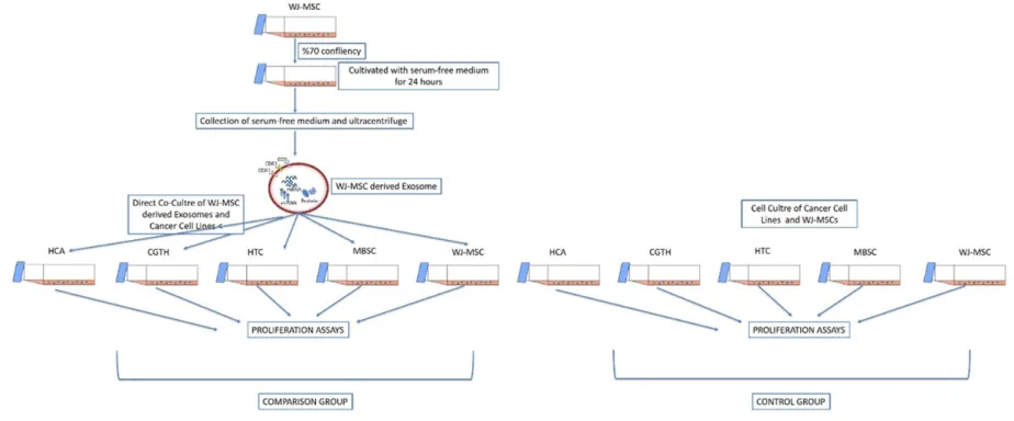
In total, four different cancer cell lines were incubated separately for 7 days, in microenvi-ronments supplied with 6 different concentra-tions (6.25, 12.5, 25, 50, 100 ug) of WJ-MSC derived exosomes and were monitored by using WST-1, Xelligence, and immunostaining with proliferation marker (PCNA). In order to deter-mine if exosomes were uptaken by cells or not, we labelled the supplied exosomes with PKH-26 (Red Fluorescent Cell Linker Kit) and exam-ined the cells under the fluorescence micr- oscope.
Four of these cell lines which are Human Colorectal Adenocarcinoma Cells (ATCC CCL-218), Human Thyroid Carcinoma Cells (ATCC CRL-1803) and Mammary Gland Adenocar- cinoma from Metastatic Site (ATCC HTB-26), were purchased from ATCC (Manassas, VA). Malign Breast Stromal Cells (MBSC) were taken
from a patient with written informed consent with the declaration of Helsinki. The study received ethical approval from the Kocaeli University, Faculty of Medicine Ethical Com- mittee (KAEK 2012/35). Additionally, the Wh- arton’s Jelly derived MSC (WJ-MSC) (PCS-500-010) was purchased from ATCC (Manassas, VA).
A comparison and a control group were set with these 5 different cell lines. The comparison group consists of cell lines co-cultured with exosomes derived from WJ-MSCs while the control group consists of cell lines without any additional treatment on cells. Overall methodol-ogy workflow was illustrated in Figure 1.
Cell lines culture
WJ-MSC cell culture: WJ-MSC cell line was
cul-tured in DMEM-F12 medium (Gibco, Thermo Fisher Scientific, Waltham, MA, USA) supple-mented with 10% fetal bovine serum (FBS), %1 streptomycin (100 mg/mL) solution at 37°C in a humidified 5% CO2 incubator. After reaching 70% to 80% confluency, adherent cells were harvested with trypsinization by 0.05% trypsin-EDTA (Gibco, Germany).
Cancer cell line culture
Four different cell lines were cultured in RPMI 1649 (Gibco, Thermo Fisher Scientific, Walth- am, MA, USA) supplemented with 10% fetal bovine serum (FBS), 2 mM L-glutamine and streptomycin (100 mg/mL) solution at 37°C in a humidified 5% CO2 incubator. After reaching 70% to 80% confluency, adherent cells were harvested with trypsinization by 0.05% trypsin-EDTA (Gibco, Germany).
Exosome isolation, characterization and label-ling: Characterized WJ-MSC were cultured with
a serum-free medium to produce a concentrat-ed and an adequate amount of exosome. Exosomes were produced in a humidified atmo-sphere containing 5% CO2 at 37°C for 24 hours then the serum-free medium was collected and centrifuged at 300 g for 5 minutes for discard-ing the remaindiscard-ing cells. Until the last step, the pellet is thrown away and the supernatant is used for the following step.
The second and the third step are designed to eliminate dead cells and cell debris by succes-sive centrifugations at increasing speeds. At
the second step, the supernatant was centri-fuged at 1000 g for 10 minutes and in the third step, the supernatant was centrifuged at 5000 g for 20 minutes. The final supernatant is ultra-centrifuged at 100,000 g for 70 minutes and collected pellet that the small vesicles that cor-respond to exosomes from the pellet.
Quantification of protein in exosomes
By the BCA protein determination method, it is shown in order to show the presence of protein in the isolated exosomes. After the exosomes were isolated, 20 µl of the resulting pellet was prepared by adding 200 µl working solution (196 µl Bicinchoninic acid + 4 µl CuSO4) and incubated at 37°C for one hour. After incuba-tion, the amount of protein was determined by spectrophotometer at 562 nm with respect to the BSA standard.
Co-culture of cancer cell lines and exosomes
Different cancer cell lines were seeded into cell culture flasks for proliferation and after reach-ing 70% to 80% confluency, WJ-MSC derived exosomes directly co-cultured on these adher-ent cells for 7 days.
Isolated exosomes were labelled with CD81, CD9 and CD63 and the characterization data were acquired with Flow Cytometry analysis. The obtained exosomes were labelled with PKH26 (PKH26 Red Fluorescent Cell Binding Kit, PKH26GL-1KT, Sigma) in order to observe whether the exosomes were localized in the co-cultured cells or not.
Proliferation tests
The proliferation effect of WJ-MSC exosomes on cancer cells was demonstrated by WST-1 and XCelligence tests. Each proliferation com-parison group repeated as triplicate.
WST-1 proliferation assay
Cancer cell lines cultivated with exosomes in 6-well temperature-sensitive culture plates, cells were tested with WST-1 assay at days 1, 3 and 7. The absorbance after the incubation period was read at 480 nm using microplate reader with the monochromatic system (Ver- saMax, Molecular Device, USA). Comparative analysis of viability and proliferation of the cell types is shown in the graph.
MSC-derived exosomes do not promote the proliferation of cancer cells in vitro
180 Int J Physiol Pathophysiol Pharmacol 2019;11(4):177-189
XCelligence proliferation assay
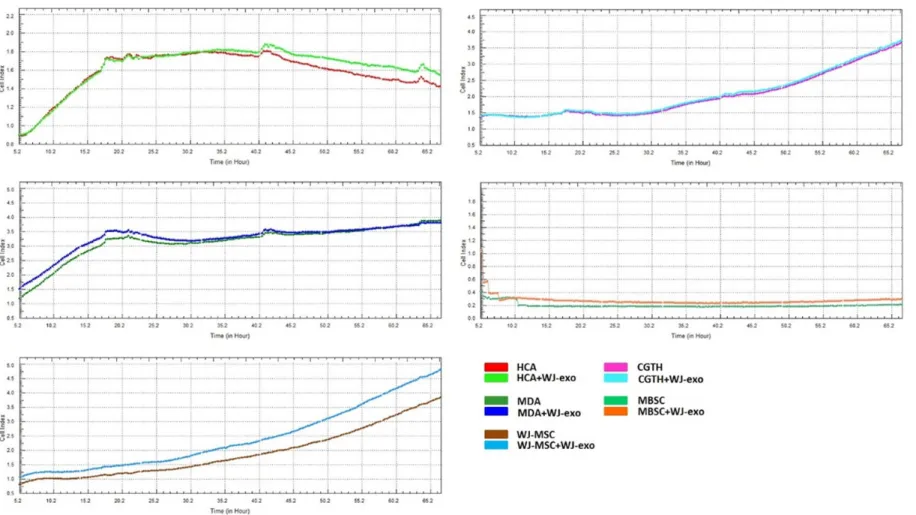
To support the WST-1 test, Xelligence was used to monitor the viability and proliferation of the cells during cell culture. Each experiment set was grouped with cancer cell line and co-cul-tured cell line with exosomes. 50 µL from each set was seeded in E96 xCELLigence plates and plate was equilibrated to 37°C. Simultaneous proliferation curves were determined by densi-ty change per time in each well. Densidensi-ty chang-es were determined with the RTCA (Real-Time Cell Analysis) (ACEA Biosciences) software.
Immunostaining by proliferation marker PCNA
Exosomes were stained by the immunoblotting of PCNA (ab29) in order to show proliferation in the cells. For immunohistochemical staining, cells were fixed with 70% methanol. After incu-bation with primer antibody for 1 hour, staining was performed by IHC kit protocol. PCNA posi-tive cells counted under the light microscope.
Statistical analysis
SPSS 10.0 (SPSS Inc, Chicago, IL, USA) soft-ware was used for statistical analyses. The data were analyzed by paired t-test and for mul-tiple sample analysis Newman-Keuls compari-son method was used. Each experiment was repeated at least three times. The level of sig-nificance was set at P<0.05 and P<0.01 it was identified as highly significant for all statistical analyses.
Results
Characterization of WJ-MSC and MSC-derived exosomes
First, MSC characterization was reviewed under the parameters which are cell surface marker staining and differentiation experiments in our previous publication [32].
We isolated exosomes from the serum-free cul-ture supernatants of MSCs to investigate the potential effects of proliferation on cancer cells. To determine whether MSC-derived exo-somes were successfully purified, we per-formed flow cytometry. As shown in Figure 2, representative markers of exosomes which are CD9, CD63 and CD81 were detected in the iso-lated exosomes 80% or higher. This means that
MSC-derived exosomes were successfully purified.
Cellular uptake of MSC-derived exosomes into cancer cells
In order to determine cellular internalization of exosomes by cancer cells, we labelled the sup-plied WJ-MSC derived exosomes with a fluores-cent dye, 26. After the incubation of PKH-labelled exosomes with cancer cell lines, exo-somes were examined under the fluorescence microscope. As shown in Figure 3, PKH26-labeled exosomes were uptaken by cancer cells.
The effect of MSC-derived exosomes on pro-liferation rate of different cancer cell lines in vitro
To evaluate the role of MSC-derived exosomes in proliferation in vitro, four different cancer cell lines were incubated separately for 7 days, in microenvironments supplied with 5 different concentrations (6.25, 12.5, 25, 50 and 100 ug) of WJ-MSC derived exosomes. The proliferation response to exosomes was assessed on can-cer cell lines by using WST-1 and XCelligence. Additionally, the proliferation markers were indicated by immunostaining.
First of all, viability and proliferation amount of HCA, CGTH, MBSC, MDA and WJ-MSCs co-cul-tured with WJ-exosomes are determined via the WST-1 test on 1st, 3rd and 7th days. A relatively significant proliferation increase was observed only on groups of WJ-MSC which were treated with 100 ug and 50 ug of WJ-MSC derived exo-somes (P<0.05, P*; P<0.01, P**; P<0.001, P***) (Figure 4). There was no significant change in the rate of proliferation in co-cultures with cancer cell lines compared to control groups.
Following the WST-1, proliferation assay for co-cultured cells and WJ-exosomes was examined via xCELLigence. A significant increase was not observed (Figure 5).
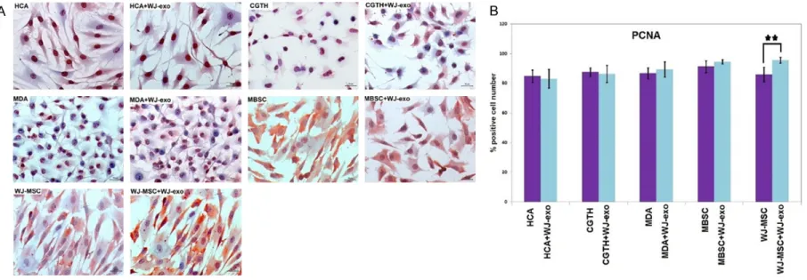
Comparing the number of PCNA positive cells between the comparison and control groups, no significant change was observed between the groups. The number of PCNA positive cells are found significantly higher in WJ-exosome treated control group when compared with
MSC-derived exosomes do not promote the proliferation of cancer cells in vitro
182 Int J Physiol Pathophysiol Pharmacol 2019;11(4):177-189
other experimental groups (P<0.05, P*; P<0.01, P**; P<0.001, P***) (Figure 6).
Discussion
MSCs are a group of progenitor cells capable of differentiating into several mesenchymal lin-eages [33] and an attractive cell source for cel-lular therapies [34]. The certain mechanism of cellular therapy action of MSCs is still doubtful [35]. The widely accepted concept is a secreto-ry or an expressed factor that reaches neigh-bouring parenchymal cells via either a para-crine effect or a direct cell-to-cell interaction promoting functional activity, survival and pro-liferation of the parenchymal cells.
Exosomes exhibit certain different characteris-tics and secreted factors according to the
source from which they are derived that are potentially associated with their biogenesis, targeting and putative immunological function [36]. That is why the sources of exosomes eval-uated for microenvironment formation are important. They provide a potential pathway short- or long-range for the cell to cell communi-cation, which may be likely to lie mechanism underlying MSCs’ tumour-suppressive proper-ties [31]. The secreted factors from the somes are known to be the roles of the exo-somes in establishing and modifying tumour microenvironments [37]. Some studies have shown that MSC-derived exosomes are one of the key elements in carcinogenesis process through cellular communication, cell growth and cell migration. These triggering paths have been elucidated by demonstrating that adi-Figure 2. Characterization of WJ-MSC derived exomes by flow cytometry.
pose-MCS derived exosomes increase tumour growth in glioblastoma cells by cellular commu-nication [29], and again Adipose-MCS derived exosomes stimulate cell migration and cell pro-liferation by inducing Wnt signalling in breast cancer cells [38]. When we had examined the other studies that put forward exosomes trig-ger cancer, we realized that cancer stem cells derived exosomes are the main character in
the carcinogenesis process. As reported by these studies, the expression of mir15a was decreased in the MM patients’ bone marrow-MSC derived exosomes rather than exosomes derived from healthy bone marrow-MSC this diminish triggered tumour growth [30]; cell growth in glioblastoma is triggered by the CLIC1 which is the cargo of the GBM-CSC-derived exo-somes [39] and angiogenesis in renal cancer is Figure 3. Fluorescence microscope images of HCA, CGTH, MBSC, MDA and WJ-MSC cells co-cultured with PKH26 labeled WJ-exosomes (red) up taking exosomes. PKH26 labeled exosomes can be seen in cytoplasm’s of all cells used in this experiment.
MSC-derived exosomes do not promote the proliferation of cancer cells in vitro
184 Int J Physiol Pathophysiol Pharmacol 2019;11(4):177-189
induced by transferring of proangiogenic fac-tors of CD105 positive-CSC-derived exosomes [40]. This is obvious that the roles of exosomes in the carcinogenesis process are determining by their sources.
Our previous study suggested that WJ-MSC is an alternative source for clinical use [41]. Con- sequently, in this study we co-cultured WJ-MSC derived exosomes and 4 different cancer cell lines then measured the proliferation amounts with WST-1 and xCelligence assays and mea-sured the common proliferation markers PCNA. We have tried to understand whether WJ-MSC derived exosomes promote proliferation of dif-ferent epithelial originated cancer cells or not. In conclusion, we observed that WJ-MSC de- rived exosomes did not cause an additional proliferation on cancer cell lines. There are other studies that supporting our result. Wu s. et. al. showed that WJ-MSC derived exosomes inhibit the cancer cell viability of bladder tumour T24 cells by cell cycle arrest and induce apop-tosis in T24 cells in vitro and in vivo by down-regulating phosphorylation of Akt protein ki- nase and upregulating cleaved caspase-3 exo-somes play anti-proliferative role on T24 cells [31]. As same as previous study, WJ- and BM-MSC derived exosomes also induced apop-tosis by inducing sub-G1 phase of malignant glioblastoma U87MG cells [29]. In addition to the apoptosis, WJ- and Bone Marrow-MSC
derived exosomes reduced the proliferation rate both in vitro and in vivo.
miRNAs are key modulators that can regulate the gene expression both during the mRNA translation and during the post translationally [42]. MSC derived exosomes are demonstrated anti-tumour [43, 44] functions by releasing mRNA and miRNA cargos for cellular communi-cation by cellular uptake [45, 46]. A few studies have shown that, MSC-derived exosomes have shuttled anti-tumorigenic miRNAs. One of them has shown that miR-124 is downregulated in glioma cells than normal tissues [47, 48]. WJ-MSC derived exosomes delivered miR-124 and decreases the migration of U87 glioblasto-ma multiform (GBM) cells [49]. Also, MSC-derived exosomes inhibited overexpressed mir-146b and reduced glioma growth in rats [50]. Another published study has shown the anti-tumour effects of exosomes can be examined by the effects of proapoptotic role, epithelium-mesenchymal transition (EMT) induction and angiogenesis modulation by miRNAs. EMT has a crucial role in cancer development and metastasis [51]. Not surprisingly, some para-crine signals have a role in cancer also play role in EMT such as WNT/β-catenin signalling, FGF and TGF-β [52]. It was reported that, bone mar-row-MSC derived exosomes induced cell death in the ovarian tumour, Kaposi’s sarcoma and
Figure 4. Viability and proliferation amounts of HCA, CGTH, MBSC, MDA and WJ-MSCs co-cultured with different
quantities of WJ-exosomes (6.25, 12.5, 25, 50, 100 ug) are determined via WST-1 test on 1st, 3rd and 7th days. A significant increase was seen on groups of WJ-MSC which were treated with 100 mg and 50 mg WJ-exosomes (P<0.05, P*; P<0.01, P**).
MSC-derived exosomes do not promote the proliferation of cancer cells in vitro
186 Int J Physiol Pathophysiol Pharmacol 2019;11(4):177-189
Figure 6. A. Immunohistochemical PCNA staining of HCA, CGTH, MBSC, MDA and WJ-MSC cells co-cultured with exosomes. The number of PCNA positive cells are higher in WJ-exosome treated group when compared with other experimental groups. B. The statistical graph of PCNA positive cells in HCA, CGTH, MBSC, MDA and WJ-MSC cells which are co-cultured with exosomes. Significant difference was observed only in WJ-exosomes treated WJ-MSC group (P<0.01, P**).
hepatoma cell lines in vitro [53]. In EMT aspect, they induced EMT markers of FGF19-FGFR in carcinomas [54]. Moreover, mir-16 is overex-pressed in breast cancer by MSC-derived exo-somes and the angiogenesis marker of VEGF is downregulated which is the target of mir-16 [55]. When we discussed all these outcomes, MSC-derived exosomes are inhibiting angio-genesis, tumour growth and inducing the proliferation.
Thus, it is well known that the tumour microen-vironment is correlated with tumorigenesis and cancer progression. In this niche, also MSCs are present and they may undergo malignant transformation. However, we cannot say that MSCs have a role in tumorigenesis. On parallel to this information, exosomes are the key com-ponents of intercellular communication with the ability to manipulate the local and systemic extracellular niche and tumour microenviron-ment [25, 56, 57]. Exosomes modulate extra-cellular niches either immune-stimulatory or inhibitory functions or both [58].
Disclosure of conflict of interest None.
Address correspondence to: Dr. Erdal Karaoz, İstinye University, Maltepe Mahallesi Edirne Çırpıcı Yolu, No. 9, Cevizlibağ-Topkapı-İstanbul 34010, Turkey. E-mail: [email protected]
References
[1] Gnecchi M, Danieli P, Malpasso G and Ciuffre-da MC. Paracrine mechanisms of mesenchy-mal stem cells in tissue repair. Methods Mol Biol 2016; 1416: 123-146.
[2] Gnecchi M, He H, Liang OD, Melo LG, Morello F, Mu H, Noiseux N, Zhang L, Pratt RE, Ingwall JS and Dzau VJ. Paracrine action accounts for marked protection of ischemic heart by Akt-modified mesenchymal stem cells. Nat Med 2005; 11: 367-368.
[3] Raposo G and Stoorvogel W. Extracellular vesi-cles: exosomes, microvesicles, and friends. J Cell Biol 2013; 200: 373-383.
[4] Valadi H, Ekström K, Bossios A, Sjöstrand M, Lee JJ and Lötvall JO. Exosome-mediated transfer of mRNAs and microRNAs is a novel mechanism of genetic exchange between ce- lls. Nat Cell Biol 2007; 9: 654.
[5] Fujita Y, Kadota T, Araya J, Ochiya T and Ku-wano K. Clinical application of mesenchymal stem cell-derived extracellular vesicle-based
therapeutics for inflammatory lung diseases. J Clin Med 2018; 7.
[6] Li T, Yan Y, Wang B, Qian H, Zhang X, Shen L, Wang M, Zhou Y, Zhu W, Li W and Xu W. Exo-somes derived from human umbilical cord mesenchymal stem cells alleviate liver fibrosis. Stem Cells Dev 2013; 22: 845-854.
[7] Lai RC, Arslan F, Lee MM, Sze NS, Choo A, Chen TS, Salto-Tellez M, Timmers L, Lee CN, El Oakley RM, Pasterkamp G, de Kleijn DP and Lim SK. Exosome secreted by MSC reduces myocardial ischemia/reperfusion injury. Stem Cell Res 2010; 4: 214-222.
[8] Salomon C, Ryan J, Sobrevia L, Kobayashi M, Ashman K, Mitchell M and Rice GE. Exosomal signaling during hypoxia mediates microvascu-lar endothelial cell migration and vasculogen-esis. PLoS One 2013; 8: e68451.
[9] Arslan F, Lai RC, Smeets MB, Akeroyd L, Choo A, Aguor EN, Timmers L, van Rijen HV, Doeven-dans PA, Pasterkamp G, Lim SK and de Kleijn DP. Mesenchymal stem cell-derived exosomes increase ATP levels, decrease oxidative stress and activate PI3K/Akt pathway to enhance myocardial viability and prevent adverse re-modeling after myocardial ischemia/reperfu-sion injury. Stem Cell Res 2013; 10: 301-312. [10] Feng Y, Huang W, Wani M, Yu X and Ashraf M.
Ischemic preconditioning potentiates the pro-tective effect of stem cells through secretion of exosomes by targeting Mecp2 via miR-22. PLoS One 2014; 9: e88685-e88685.
[11] Yu B, Gong M, Wang Y, Millard RW, Pasha Z, Yang Y, Ashraf M and Xu M. Cardiomyocyte pro-tection by GATA-4 gene engineered mesenchy-mal stem cells is partially mediated by translo-cation of miR-221 in microvesicles. PLoS One 2013; 8: e73304-e73304.
[12] Bruno S, Grange C, Deregibus MC, Calogero RA, Saviozzi S, Collino F, Morando L, Busca A, Falda M, Bussolati B, Tetta C and Camussi G. Mesenchymal stem cell-derived microvesicles protect against acute tubular injury. J Am Soc Nephrol 2009; 20: 1053-67.
[13] Gatti S, Bruno S, Deregibus MC, Sordi A, Canta-luppi V, Tetta C and Camussi G. Microvesicles derived from human adult mesenchymal stem cells protect against ischaemia-reperfusion-in-duced acute and chronic kidney injury. Nephrol Dial Transplant 2011; 26: 1474-83.
[14] He J, Wang Y, Sun S, Yu M, Wang C, Pei X, Zhu B, Wu J, Zhao W. Bone marrow stem cells-de-rived microvesicles protect against renal injury in the mouse remnant kidney model. Nephrol-ogy 2012; 17: 493-500.
[15] Tomasoni S, Longaretti L, Rota C, Morigi M, Conti S, Gotti E, Capelli C, Introna M, Remuzzi G and Benigni A. Transfer of growth factor ceptor mRNA via exosomes unravels the
re-MSC-derived exosomes do not promote the proliferation of cancer cells in vitro
188 Int J Physiol Pathophysiol Pharmacol 2019;11(4):177-189
generative effect of mesenchymal stem cells. Stem Cells Dev 2013; 22: 772-80.
[16] Zhang B, Wang M, Gong A, Zhang X, Wu X, Zhu Y, Shi H, Wu L, Zhu W, Qian H and Xu W. Huc-MSC-exosome mediated-Wnt4 signaling is re-quired for cutaneous wound healing. Stem Cells 2015; 33: 2158-2168.
[17] Payne SR and Kemp CJ. Tumor suppressor ge-netics. Carcinogenesis 2005; 26: 2031-2045. [18] Plaks V, Kong N and Werb Z. The cancer stem
cell niche: how essential is the niche in regulat-ing stemness of tumor cells? Cell Stem Cell 2015; 16: 225-38.
[19] Greaves M and Maley CC. Clonal evolution in cancer. Nature 2012; 481: 306.
[20] Ponnusamy MP and Batra SK. Ovarian cancer: emerging concept on cancer stem cells. J Ovar-ian Res 2008; 1: 4.
[21] Witsch E, Sela M and Yarden Y. Roles for growth factors in cancer progression. Physiology (Bethesda) 2010; 25: 85-101.
[22] Hall B, Andreeff M and Marini F. The participa-tion of mesenchymal stem cells in tumor stro-ma forstro-mation and their application as target-ed-gene delivery vehicles. In: Kauser K, Zeiher AM, editors. Bone Marrow-Derived Progenitors. Berlin; Heidelberg: Springer Berlin Heidelberg; 2007. pp. 263-283.
[23] Ribeiro AL and Okamoto OK. Combined effects of pericytes in the tumor microenvironment. Stem Cells International 2015; 2015: 8. [24] Bergfeld SA and DeClerck YA. Bone
marrow-derived mesenchymal stem cells and the tu-mor microenvironment. Cancer Metastasis Rev 2010; 29: 249-61.
[25] Vallabhaneni KC, Penfornis P, Dhule S, Guillon-neau F, Adams KV, Mo YY, Xu R, Liu Y, Watabe K, Vemuri MC and Pochampally R. Extracellular vesicles from bone marrow mesenchymal stem/stromal cells transport tumor regulatory microRNA, proteins, and metabolites. Oncotar-get 2015; 6: 4953-4967.
[26] Wolfson B, Eades G and Zhou Q. Roles of mi-croRNA-140 in stem cell-associated early stage breast cancer. World J Stem Cells 2014; 6: 591-7.
[27] Zhu W, Huang L, Li Y, Zhang X, Gu J, Yan Y, Xu X, Wang M, Qian H and Xu W. Exosomes de-rived from human bone marrow mesenchymal stem cells promote tumor growth in vivo. Can-cer Lett 2012; 315: 28-37.
[28] Oviedo NJ and Beane WS. Regeneration: the origin of cancer or a possible cure? Semin Cell Dev Biol 2009; 20: 557-564.
[29] Del Fattore A, Luciano R, Saracino R, Battafar-ano G, Rizzo C, Pascucci L, Alessandri G, Pes-sina A, Perrotta A, Fierabracci A and Muraca M. Differential effects of extracellular vesicles se-creted by mesenchymal stem cells from
differ-ent sources on glioblastoma cells. Expert Opin Biol Ther 2015; 15: 495-504.
[30] Roccaro AM, Sacco A, Maiso P, Azab AK, Tai YT, Reagan M, Azab F, Flores LM, Campigotto F, Weller E, Anderson KC, Scadden DT and Gho-brial IM. BM mesenchymal stromal cell-derived exosomes facilitate multiple myeloma progres-sion. J Clin Invest 2013; 123: 1542-1555. [31] Wu S, Ju GQ, Du T, Zhu YJ and Liu GH.
Mi-crovesicles derived from human umbilical cord Wharton’s jelly mesenchymal stem cells at-tenuate bladder tumor cell growth in vitro and in vivo. PLoS One 2013; 8: e61366.
[32] Dai A, Baspinar O, Yesilyurt A, Sun E, Aydemir CI, Oztel ON, Capkan DU, Pinarli F, Agar A and Karaoz E. Efficacy of stem cell therapy in am-bulatory and nonamam-bulatory children with Duchenne muscular dystrophy - Phase I-II. De-gener Neurol Neuromuscul Dis 2018; 8: 63-77. [33] Caplan AI. Mesenchymal stem cells. J Orthop
Res 1991; 9: 641-650.
[34] Conrad C and Huss R. Adult stem cell lines in regenerative medicine and reconstructive sur-gery. J Surg Res 2005; 124: 201-208. [35] Gnecchi M, Zhang Z, Ni A and Dzau VJ.
Para-crine mechanisms in adult stem cell signaling and therapy. Circ Res 2008; 103: 1204-1219. [36] Kumar D, Gupta D, Shankar S and Srivastava
RK. Biomolecular characterization of exo-somes released from cancer stem cells: possi-ble implications for biomarker and treatment of cancer. Oncotarget 2015; 6: 3280-3291. [37] Maia J, Caja S, Strano Moraes MC, Couto N
and Costa-Silva B. Exosome-based cell-cell communication in the tumor microenviron-ment. Front Cell Dev Biol 2018; 6: 18.
[38] Lin R, Wang S and Zhao RC. Exosomes from human adipose-derived mesenchymal stem cells promote migration through Wnt signaling pathway in a breast cancer cell model. Mol Cell Biochem 2013; 383: 13-20.
[39] Setti M, Osti D, Richichi C, Ortensi B, Del Bene M, Fornasari L, Beznoussenko G, Mironov A, Rappa G, Cuomo A, Faretta M, Bonaldi T, Lori-co A and Pelicci G. Extracellular vesicle-mediat-ed transfer of CLIC1 protein is a novel mecha-nism for the regulation of glioblastoma growth. Oncotarget 2015; 6: 31413-31427.
[40] Grange C, Tapparo M, Collino F, Vitillo L, Dam-asco C, Deregibus MC, Tetta C, Bussolati B and Camussi G. Microvesicles released from hu-man renal cancer stem cells stimulate angio-genesis and formation of lung premetastatic niche. Cancer Res 2011; 71: 5346-56. [41] Karaöz E, Çetinalp Demircan P, Erman G,
Güngörürler E and Eker Sarıboyacı A. Compar-ative analyses of immunosuppressive charac-teristics of bone-marrow, wharton’s jelly, and adipose tissue-derived human mesenchymal
stem cells. Turk J Haematol 2017; 34: 213-225.
[42] Fabian MR, Sonenberg N and Filipowicz W. Regulation of mRNA Translation and Stability by microRNAs. Annu Rev Biochem 2010; 79: 351-79.
[43] Du T, Ju G, Wu S, Cheng Z, Cheng J, Zou X, Zhang G, Miao S, Liu G and Zhu Y. Microvesi-cles derived from human Wharton’s jelly mes-enchymal stem cells promote human renal cancer cell growth and aggressiveness through induction of hepatocyte growth factor. PLoS One 2014; 9: e96836-e96836.
[44] Fonsato V, Collino F, Herrera MB, Cavallari C, Deregibus MC, Cisterna B, Bruno S, Romagnoli R, Salizzoni M, Tetta C and Camussi G. Human liver stem cell-derived microvesicles inhibit hepatoma growth in SCID mice by delivering antitumor microRNAs. Stem Cells 2012; 30: 1985-98.
[45] Camussi G, Deregibus MC, Bruno S, Cantalup-pi V and Biancone L. Exosomes/microvesicles as a mechanism of cell-to-cell communication. Kidney Int 2010; 78: 838-48.
[46] Christianson HC, Svensson KJ, van Kuppevelt TH, Li JP and Belting M. Cancer cell exosomes depend on cell-surface heparan sulfate pro-teoglycans for their internalization and func-tional activity. Proc Natl Acad Sci U S A 2013; 110: 17380-5.
[47] An L, Liu Y, Wu A and Guan Y. microRNA-124 inhibits migration and invasion by down-regu-lating ROCK1 in glioma. PLoS One 2013; 8: e69478-e69478.
[48] Wei J, Wang F, Kong LY, Xu S, Doucette T, Fergu-son SD, Yang Y, McEnery K, Jethwa K, Gjyshi O, Qiao W, Levine NB, Lang FF, Rao G, Fuller GN, Calin GA and Heimberger AB. miR-124 inhibits STAT3 signaling to enhance T cell-mediated im-mune clearance of glioma. Cancer Res 2013; 73: 3913-26.
[49] Sharif S, Ghahremani MH and Soleimani M. Delivery of exogenous miR-124 to glioblasto-ma multiform cells by Wharton’s Jelly mesen-chymal stem cells decreases cell proliferation and migration, and confers chemosensitivity. Stem Cell Rev 2018; 14: 236-246.
[50] Katakowski M, Buller B, Zheng X, Lu Y, Rogers T, Osobamiro O, Shu W, Jiang F and Chopp M. Exosomes from marrow stromal cells express-ing miR-146b inhibit glioma growth. Cancer Lett 2013; 335: 201-4.
[51] Heerboth S, Housman G, Leary M, Longacre M, Byler S, Lapinska K, Willbanks A and Sarkar S. EMT and tumor metastasis. Clin Transl Med 2015; 4: 6.
[52] Micalizzi DS, Farabaugh SM and Ford HL. Epi-thelial-mesenchymal transition in cancer: par-allels between normal development and tumor progression. J Mammary Gland Biol Neoplasia 2010; 15: 117-134.
[53] Bruno S, Collino F, Deregibus MC, Grange C, Tetta C and Camussi G. Microvesicles derived from human bone marrow mesenchymal stem cells inhibit tumor growth. Stem Cells Dev 2013; 22: 758-771.
[54] Shi S, Zhang Q, Xia Y, You B, Shan Y, Bao L, Li L, You Y and Gu Z. Mesenchymal stem cell-de-rived exosomes facilitate nasopharyngeal car-cinoma progression. Am J Cancer Res 2016; 6: 459-472.
[55] Lee JK, Park SR, Jung BK, Jeon YK, Lee YS, Kim MK, Kim YG, Jang JY and Kim CW. Exosomes derived from mesenchymal stem cells sup-press angiogenesis by down-regulating VEGF expression in breast cancer cells. PLoS One 2013; 8: e84256-e84256.
[56] Deregibus MC, Cantaluppi V, Calogero R, Lo Iacono M, Tetta C, Biancone L, Bruno S, Bus-solati B and Camussi G. Endothelial progenitor cell derived microvesicles activate an angio-genic program in endothelial cells by a horizon-tal transfer of mRNA. Blood 2007; 110: 2440-2448.
[57] Deregibus MC, Tetta C and Camussi G. The dy-namic stem cell microenvironment is orches-trated by microvesicle-mediated transfer of genetic information. Histol Histopathol 2010; 25: 397-404.
[58] Kahlert C and Kalluri R. Exosomes in tumor mi-croenvironment influence cancer progression and metastasis. J Mol Med (Berl) 2013; 91: 431-437.