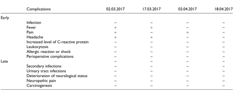
Wharton's jelly-derived mesenchymal stem cell transplantation in a patient with hypoxic-ischemic encephalopathy: a pilot study
Tam metin
Şekil
Benzer Belgeler
alternative procedures to get an initial feasible solution: l j we randomly generate 100 feasible solutions and run the heuristic starting from each, recording the best
Araştırma evreni Düzce İl Merkezi’nde sokakta çalışan çocuklardan ve onların ailelerinden oluşmaktadır. Düzce İl Em- niyet Müdürlüğü Çocuk Şubesi’nden alı-
TEMPOROMANDIBULAR DISORDERS IN SCUBA DIVERS DURING DIVING CERTIFICATION TRAINING PROGRAMME.. DALIŞ SERTİFİKASYONU EĞİTİM PROGRAMINDA SCUBA DALICILARINDA GÖRÜLEN
In conclusion, voriconazole loaded in situ gels could be offered as a promising strategy for ocular drug delivery for the treatment of fungal keratitis.. Key words: Voriconazole,
velilerden okula gelen desteğin de yeterli olmadığını göstermektedir. Her ne kadar okul içinde bir laboratuvar bulunsa da sınıflardaki öğrenci sayılarının
Both the freestream Mach number and the angle of attack are considered as random parameters and the generalized Polynomial Chaos (gPC) theory is coupled with standard
Mumun sevgili, pervânenin âfl›k oldu¤undan hareketle beyitten âfl›k olunacak sevgilinin, birçok âfl›¤› bir an- da küle çevirecek bir atefl gibi olmas› ge-
Blood urea nitrogen, creatinine, uric acid levels and tissue oxidative stress markers, total oxidant status and oxidative stress index levels significantly increased and
