DR ISIL YURDAISIK (Orcid ID : 0000-0001-8316-1229)
DR SULEYMAN HILMI AKSOY (Orcid ID : 0000-0002-2356-0268)
Article type : Original Paper
Title: The Relationship Between Lesion Density Change in Chest CT and Clinical Improvement
in COVID-19 Patients
Running title: CT Lesion Density in COVID-19
Authors: Isil Yurdaisik, Fuat Nurili, Ayse Gul Agirman, Suleyman Hilmi Aksoy
Affiliations:
Isil Yurdaisik: Department of Radiology, Istinye University, Gaziosmanpasa Medical Park
Hospital, · Istanbul, Turkey
E-mail: [email protected]
Fuat Nurili: Department of Radiology, Memorial Sloan Ketteting Cancer Center, Interventional
Radiology, New York, NY, USA e-mail: [email protected]
Ayse Gul Agirman: Department of Radiology, Dr. Siyami Ersek Thoracic and Cardiovascular
Surgery Training and Research Hospital, Istanbul, Turkey. e-mail: [email protected]
Suleyman Hilmi Aksoy: Department of Radiology, Galata University, Hisar Intercontinental
Hospital, İstanbul, Turkey e-mail: [email protected]
Address Correspondence; Isil Yurdaisik, M.D.
Tel: 0090-5327393033, Fax: 00902129793950 E-mail: [email protected]
Accepted Article
Address: Merkez Mahallesi Çukurçeşme Cad. No: 57-59 Gaziosmanpaşa - İstanbul, Turkey
The Relationship Between Lesion Density Change in Chest CT and Clinical Improvement in COVID-19 Patients
ABSTRACT
Objective: To evaluate the association of changes in chest computed tomography (CT) lesion
densities with clinical improvement in COVID-19 patients.
Methods: This was a cross-sectional analysis of hospitalized COVID-19 patients who underwent
repeated chest CT. Patients who improved clinically but showed radiological progression were included. Demographic data, presentation complaints, and laboratory results were retrieved from the electronic database of the hospital. Lesion density that was measured in Hounsfield Units (HU) was compred between admission and discharge chest CT scans.
Results: Forty patients (21males, mean age 47.4±15.1 years) were included in the analysis. The
median white blood cell count and CRP significantly decreased, whereas the median lymphocyte count significantly increased at discharge compared with the admission values. The mean density significantly reduced from admission to discharge.
Conclusion: This is the first study in the literature reporting reduction in chest CT lesion densities
correlated with clinical and laboratory improvement in COVID-19 patients.
Key words: Chest CT, Clinical improvement, COVID-19, Ground-glass opacity, Hounsfield
Units, Lesion Density
What is already known?
The radiologic appearance of the COVID-19 lung disease shows an evolution through time and somehow lags behind the clinical improvement.
More time is needed to observe the complete resolution of lung abnormalities.
If one depends solely on the chest CT characteristics for treatment change or discharge decisions, it would be misleading.
What does this study add new?
This study reports for the first time in the literature that reduction in chest CT lesion densities correlated with clinical and laboratory improvement in patients hospitalized because of COVID-19.
Chest CT density reduction might point to the future resolution of the lung lesions and may precludeunnecessarily long hospitalizations due to this disease.
This is of practical importance considering the limited inpatient bed capacities in some parts of the world.
INTRODUCTION
SARS-CoV-2 is a novel coronavirus that led to a pandemic and claimed more than 550 thousand lives worldwide as of July 2020. The virus causes Acute respiratory stress syndrome (ARDS) in about 30% of the patients.1 Although the fatality rates were lower than those of Middle East
respiratory virus and SARS-CoV, lack of an efficient treatment makes the early detection and preventive measures the foremost priority to prevent spreading. Currently, the diagnosis of the condition is most commonly based on the detection of the SARS-CoV-2 from respiratory samples via RT-PCR method. However, several studies reported the positivity rates of the test between 32% and 93% depending upon the method of sample acquisition.2 Thus, despite suggestive
clinical findings and a positive history of epidemiologic exposure to COVID-19, some patients have a negative RT-PCR test. In these patients, chest CT findings might provide supportive evidence for the diagnosis of COVID-19.3-5
On the other hand, the radiologic appearance of the COVID-19 lung disease shows an evolution through time and somehow lags behind the clinical improvement. Several studies defined chest CT stages based on the time from the symptom onset. In stage 4 (>14 days after first symptoms), 65% and 75% of patients still had ground-glass opacities and consolidation on chest CT images, respectively.2 It is clear that more time is needed to observe the complete resolution of lung
abnormalities. Thus, if one depends solely on the chest CT characteristics for treatment change or discharge decisions, it would be misleading. It was hypothesized that chest CT lesion densities might provide us with valuable information regarding the improvement of the lesions. When combined with clinical improvement of the patient, chest CT densities, measured in Hounsfield units, might serve as an adjunctive measure whether a patient is ready for discharge.
Hence, it was decided to evaluate the association of the change in the chest CT lesion densities with clinical improvement in COVID-19 patients whose chest CT lesions persisted or worsened.
Materials and Methods
Study participants, design and setting
This study was a cross-sectional analysis of COVID-19 patients who underwent repeated chest computed tomography (CT) during their hospitalization. Study subjects were diagnosed with COVID-19 by means of reverse transcription-polymerase chain reaction (RT-PCR) technique, or the diagnosis was based on compatible clinical and pulmonary radiological evidence of COVID-19. COVID-19 patients who presented to the emergency department of our pandemic hospital and were hospitalized between March 15th and April1st, 2020 were enrolled. During this period, a total
of 326 patients were hospitalized because of COVID-19. Of these patients, only 189 patients (57.9%) had a positive RT-PCR test for SARS-CoV-2.
A repeat chest CT was performed before discharge in all study subjects, irrespective of their clinical status. Patients aged below 18 years, those who had no an RT-PCR positive COVID-19 diagnosis, patients who were still staying in the hospital, those who did not show clinical improvement, patients with chest CT findings not caused by COVID-19, those who had lesions other than ground-glass opacities (GGO) that may affect density measurements, moderate to severe pleural effusion and patients with GGO that occupied a small area were excluded from the study. After the exclusion of the patients due to the aforementioned reasons, a total of 77 patients with COVID-19 remained. Of these, 37 patients were excluded because they showed clinical improvement along with radiologic improvement. The remaining 40 patients who improved clinically, but showed radiological progression were included in the analyses. The study flow-chart is seen in Figure 1.
Study protocol was approved by the local ethics committee of our hospital (2/2020.K-025). The objectives of the study were explained to all patients, and written informed consents were signed by the study participants. The study was performed in accordance with the ethical principles of the Declaration of Helsinki.
Accepted Article
Data collection
Data, including patient sex, age, presentation complaints and, laboratory results (complete blood count, basic biochemistry tests, procalcitonin, ferritin, d-dimer, and troponin I) were retrieved from the electronic database of the hospital and recorded for each participant.
Acquisition of the chest CT images
Because of the COVID-19 pandemic, every necessary measure was taken to prevent the spread of the disease via the imaging procedure. All patients who would undergo chest CT imaging were instructed to wear a face mask during the whole procedure. Disposable bed sheets were used during each examination, and all used equipment and room were thoroughly disinfected after each scan. Radiology staff wore a face mask, disposable gloves, and gowns and safety goggles during the patient admission and image acquisition.
Chest computed tomographic scans were performed with a 128-slice multi-detector CT scanner (Somatom Definition AS+ Erlangen, Germany). During each scan, the following standard parameters were used: slice thickness of 1.0 mm, reconstruction interval of 1.0-3.0 mm, tube voltage of 120 kVp, and automatic exposure controlled standard tube flow. The patients were imaged on the axial plane, in the supine position, and during end-inspiration.
Analysis of the chest CT images
All chest CT scans of the study subjects were evaluated by the same experienced radiologist. For each patient, the chest CT at hospitalization (admission chest CT) and the chest CT performed before discharge (discharge chest CT) were evaluated. Progression of the COVID-19 lung disease was assessed by comparing the extent, amount, and density of the lesions detected on the initial and repeat chest CT scans. Fusion of the lesions, development of new lesions, and/or increase in lesion density were considered as progression. On the other hand, a decrease in lesion density was accepted as absorption.
Lesion density that was measured in Hounsfield Units (HU) was compared between admission and discharge chest CT scans. This analysis was performed only in lesions with ground-glass appearance. No measurement was made in other lesions composed of consolidation, air bronchograms, pleuro-parenchymal band, air cyst etc. A fixed region of interest (ROI, mm2) was
used for the purpose of area measurements. The ROI was determined circular or oval, depending on the shape of the target lesion. Great attention was paid to that ROIs had the same size and shape as much as possible in the initial and repeat CT scans. The repeat measurements were taken from the same lesion and anatomic location of the initial chest CT scan.
Accepted Article
Statistical Analysis
The Shapiro–Wilk test was used to check the normality assumptions of the data. Normally distributed variables were presented as a mean ± standard deviation, whereas non-normally distributed variables were given as median and interquartile range. Paired Sample t-test was used for normally distributed numerical variables in two-group comparisons in paired data. For variables that were not normally distributed, the Wilcoxon signed-rank test was used. To compare paired nominal data, the McNemar's test was used. SPSS 25.0 software package (IBM, Armonk, NY, USA) was used to analyze data of the study. A p-value <0.05 was accepted as statistically significant.
Results
After exclusion criteria were applied, 40 patients (21 males (52.5%)) were included in the final analysis. The mean age of the patients was 47.4±15.1 years (range 20-70 years). Fever and cough were the most common symptoms and were present in all patients (100%). Fever, dyspnea, and cough were present at discharge evaluation in 12.5%, 17.5%, and 62.5% of the patients, respectively. The frequency of all symptoms decreased significantly at discharge evaluation compared with admission. Table 1 shows age and sex distribution, frequency of admission and discharge symptoms, and some laboratory findings of the study subjects.
The median white blood cell count significantly decreased, whereas the median lymphocyte count significantly increased at discharge compared with the admission values. At discharge evaluation, three patients had mild leucopenia while the rest of the patients had normal white blood cell counts. The median platelet count showed a significant increase at discharge compared with the admission value. The median CRP value on admission significantly reduced at discharge, from 2.45 (IQR: 1.33-4.0) to 0.25 (IQR: 0.2-0.4). There was no significant change in procalcitonin levels between the admission and discharge. The laboratory parameters of COVID-19 patients on admission and at discharge are depicted in Table 2.
The mean duration between the admission and discharge CT scans was 5.8±1.30 (min-max: 4-10) days. The mean lesion density of the ROI comprising groun-glass opacities was -292.1±177.1 (min: -579 and max: +239) on admission. On admission chest CT measurements, 2 (5%) patients had a positive lesion density value, while the rest of the patients (95%) had negative densities. At discharge chest CT, the mean density was -418.9±162.7 (min: -773 and max: -156). All patients had negative density values at discharge chest CT. The mean density significantly reduced from admission to discharge. The mean decrease in the lesion densities was found to be 129.9 ± 79.02
Accepted Article
(min-max: 11-295) HU. Three chest CT scan examples demonstrating the decrease in lung lesion densities are shown in Figures 2, 3, and 4.
DISCUSSION
The main objective of the present study was to evaluate whether the changes in the density of the chest CT lung lesions in hospitalized COVID-19 patients provide a clue for disease improvement despite persisting or worsening lung lesions. The density measurements were taken from the ground-glass opacities in each patient. All patients had significant clinical improvement throughout the hospitalization.
Increased lesion density was associated with the progression of the COVID-19 infection. In a study by Li et al.,6 increased lesion density along progression of the disease was found in 69% of
patients. In the present study, the median chest CT GGO density significantly reduced from the admission to the discharge. This might provide guidance for the practicing physician that although the number and size of the lesions were increased, the reduction in the density of the lesions were correlated with clinical improvement of the patients.
Chest CT imaging is important in the diagnosis of COVID-19. In a meta-analysis of 2738 cases, the authors reported the pooled positivity rate of COVID-19 as 89.8% with CT scans.7 On the
other hand, the sensitivity of the first RT-PCR test for COVID-19 was reported as 71%.8
Moreover, patients with false-negative RT-PCR test might still have abnormalities in chest CT imaging.4
The most common presenting lesions on chest CT of these patients, in general, include bilateral peripheral ground-glass opacities, seen along with consolidation in some cases.2 Chest CT findings
show a predictable evolution with time in COVID-19. The maximum lung involvement was reported to occur at 10 days after symptom onset.9 In the early stages, GGOs dominates, followed
by consolidation. These lesions increase in frequency during the intermediate stage (3-5 days after symptom onset).10 The late phase is generally defined as 6-12 days after the symptom onset. In
this phase, most patients still continue to have lung lesions, most common of which is GGO. Beyond 14 days is defined as the absorption stage, and 65 and 75% of COVID-19 patients have GGO and consolidation, respectively.9 In our study, the mean duration between amission and
discharge was 5.8 days. All patients had negative density values at discharge chest CT and the mean decrease in lesion density was found as 129.9 ± 79.02 HU.
A considerable number of patients still had persistent lung lesions when they were ready to be discharged based on their clinical condition. Wang and colleagues reported that 94% of their 70
Accepted Article
patients still had residual lung lesions on their discharge chest CT images.11 The most commonly
seen lesion was GGOs with a median follow-up duration of 18 days (range 5-43 days) in that study.11 In another study by Du et al. at discharge chest CT images, the most common lesion was
GGOs (in 79% of the patients). Thus, chest CT findings solely on the ground of lesion number and extensity might be misleading in the decision-making process of COVID-19 patients.
It was decided to evaluate the density in the GGO lesions because both in the early and late phases of the COVID-19, GGOs were the most commonly observed lesions. All patients included in the present study clinically improved. However, persistence or worsening of the lung lesions on chest CT images was also observed. Despite this, densities of GGOs significantly reduced in all patients at discharge compared to the admission values.
Lesion densities on chest CT images have scarcely been studied in COVID-19. Wang and colleagues 11 did a quantitative analysis of chest CT images to be able to predict the development
of ARDS in COVID19 patients. They included the volume and the density of the target lesion in their deep network analysis. The authors reported that the patients with lesion densities between -549 and -450 HU were at increased risk, while patients with lesion densities between -149 and -50 HU had a reduced risk for ARDS development. On the other hand, neither average density nor the total volume of the lesions was associated with ARDS risk in that study. Du et al.13 evaluated
chest CT findings in a cohort of ready to be discharged COVID-19 patients with the assistance of an artificial intelligence system. The authors revealed that the lesion densities reduced significantly in all patients (100%) at follow-up chest CTs that were performed after the discharge (average time 2-13 days) compared with discharge CTs. The authors actually extended the results of our results. In the present study, it was demonstrated that the density of the lesions significantly decreased from the hospital admission until the discharge. Thus, we think that lesion density decrease despite a seemingly persistent or worsening chest CT image may be a harbinger that the lungs are recovering.
On the other hand, the shortcomings of the chest CT imaging in COVID-19 patients should be taken into account. It is now well-known that chest CT images might be normal, particularly in early disease. Several studies reported that in RT-PCR-proven COVID-19, false-negative CT rates might occur up to 56% of the procedures.14, 15 Besides, chest CT findings are not COVID-19
specific and may have overlaps with many other disease processes.16, 17 Increased radiation
exposure should also be considered, especially in younger patients. However, when CT images were used as an adjunctive tool in the diagnosis and/or follow-up of COVID-19 patients, we think
Accepted Article
that examining changes in density of the lesions might provide prognostic data and might prevent unnecessary follow-up CTs to be performed to observe the complete resolution of the lesions. Sun and colleagues found significant correlations between clinical features and quantitative CT parameters, including GGO, consolidation, and total lesion score.18 Matos and coworkers also
showed that lymphocyte percentage and C-reactive protein could predict the median volume of the chest CT lesions in COVID-19.19 However, the authors did not study the relationship between
lesion density and laboratory features. In contrast, no significant correlation could be found between the admission chest CT density and presentation symptoms such as fever, dyspnea, and cough. The relatively small sample size of the current study might have caused this result.
Some limitations of the current study are worthy of mention. First, the study sample size is relatively small. Second, COVID-19 patients whose chest CT findings persisted or worsened despite clinical improvement were selected. Thus, the findings of this study can only be applied to this rather specific subpopulation of COVID-19 patients. Third, data regarding the timing of the symptom onset in COVID-19 patients were not obtained. Symptoms to admission times may differ among the patients. And symptoms and laboratory values might have changed because of this. In conclusion, despite the aforementioned limitations, this study was the first in the literature reporting that reduction in chest CT lesion densities correlated with clinical and laboratory improvement in patients hospitalized because of COVID-19. Particularly in patients whose lesions in chest CT seems persistent or worsening despite clinical and laboratory improvement, chest CT density reduction might point to the future resolution of the lung lesions and may preclude unnecessarily long hospitalizations in this patient population. This is of practical importance considering the limited inpatient bed capacities in some parts of the World.
REFERENCES
1. Huang C, Wang Y, Li X, et al. Clinical features of patients infected with 2019 novel coronavirus in Wuhan, China. Lancet 2020; 395: 497-506. 2020/01/28. DOI: 10.1016/S0140-6736(20)30183-5.
2. El Homsi M, Chung M, Bernheim A, et al. Review of chest CT manifestations of COVID-19 infection. Eur J Radiol Open 2020; 7: 100239. 2020/06/COVID-19. DOI: 10.1016/j.ejro.2020.100239. 3. Zhao W, Zhong Z, Xie X, et al. Relation Between Chest CT Findings and Clinical Conditions of Coronavirus Disease (COVID-19) Pneumonia: A Multicenter Study. AJR Am J Roentgenol 2020; 214: 1072-1077. 2020/03/04. DOI: 10.2214/AJR.20.22976.
4. Xie X, Zhong Z, Zhao W, et al. Chest CT for Typical 2019-nCoV Pneumonia: Relationship to Negative RT-PCR Testing. Radiology 2020: 200343. 2020/02/13. DOI: 10.1148/radiol.2020200343.
5. Huang P, Liu T, Huang L, et al. Use of Chest CT in Combination with Negative RT-PCR Assay for the 2019 Novel Coronavirus but High Clinical Suspicion. Radiology 2020; 295: 22-23. 2020/02/13. DOI: 10.1148/radiol.2020200330.
6. Li X, Zeng W, Li X, et al. CT imaging changes of corona virus disease 2019(COVID-19): a multi-center study in Southwest China. J Transl Med. 2020;18(1):154. Published 2020 Apr 6. doi:10.1186/s12967-020-02324-w
Accepted Article
7. Bao C, Liu X, Zhang H, et al. Coronavirus Disease 2019 (COVID-19) CT Findings: A Systematic Review and Meta-analysis. J Am Coll Radiol 2020; 17: 701-709. 2020/04/14. DOI: 10.1016/j.jacr.2020.03.006.
8. Fang Y, Zhang H, Xie J, et al. Sensitivity of Chest CT for COVID-19: Comparison to RT-PCR. Radiology 2020: 200432. 2020/02/20. DOI: 10.1148/radiol.2020200432.
9. Pan F, Ye T, Sun P, et al. Time Course of Lung Changes at Chest CT during Recovery from Coronavirus Disease 2019 (COVID-19). Radiology 2020; 295: 715-721. 2020/02/14. DOI: 10.1148/radiol.2020200370.
10. Bernheim A, Mei X, Huang M, et al. Chest CT Findings in Coronavirus Disease-19 (COVID-19): Relationship to Duration of Infection. Radiology 2020; 295: 200463. 2020/02/23. DOI: 10.1148/radiol.2020200463.
11. Wang Y, Dong C, Hu Y, et al. Temporal Changes of CT Findings in 90 Patients with COVID-19 Pneumonia: A Longitudinal Study. Radiology 2020: 200843. 2020/03/20. DOI: 12.1148/radiol.2020200843.
12. Wang Y, Chen Y, Wei Y, et al. Quantitative analysis of chest CT imaging findings with the risk of ARDS in COVID-19 patients: a preliminary study. Ann Transl Med 2020; 8: 594. 2020/06/23. DOI: 10.21037/atm-20-3554.
13. Du S, Gao S, Huang G, et al. Chest lesion CT radiological features and quantitative analysis in RT-PCR turned negative and clinical symptoms resolved COVID-19 patients. Quant Imaging Med Surg 2020; 10: 1307-1317. 2020/06/19. DOI: 10.21037/qims-20-531.
14. Li K, Fang Y, Li W, et al. CT image visual quantitative evaluation and clinical classification of coronavirus disease (COVID-19). Eur Radiol 2020; 30: 4407-4416. 2020/03/28. DOI: 10.1007/s00330-020-06817-6.
15. Zhou S, Wang Y, Zhu T, et al. CT Features of Coronavirus Disease 2019 (COVID-19) Pneumonia in 62 Patients in Wuhan, China. AJR Am J Roentgenol 2020; 214: 1287-1294. 2020/03/07. DOI: 10.2214/AJR.20.22975.
16. Sun Z, Zhang N, Li Y, et al. A systematic review of chest imaging findings in COVID-19. Quant Imaging Med Surg 2020; 10: 1058-1079. 2020/06/04. DOI: 10.21037/qims-20-564.
17. Mehrabi S, Fontana S, Mambrin F, et al. Pitfalls of Computed Tomography in the Coronavirus 2019 (COVID-19) Era: A New Perspective on Ground-Glass Opacities. Cureus 2020; 12: e8151. 2020/06/19. DOI: 10.7759/cureus.8151.
18. Sun D, Li X, Guo D, et al. CT Quantitative Analysis and Its Relationship with Clinical Features for Assessing the Severity of Patients with COVID-19. Korean J Radiol 2020; 21: 859-868. 2020/06/12. DOI: 10.3348/kjr.2020.0293.
19. Matos J, Paparo F, Mussetto I, et al. Evaluation of novel coronavirus disease (COVID-19) using quantitative lung CT and clinical data: prediction of short-term outcome. Eur Radiol Exp 2020; 4: 39. 2020/06/28. DOI: 10.1186/s41747-020-00167-0.
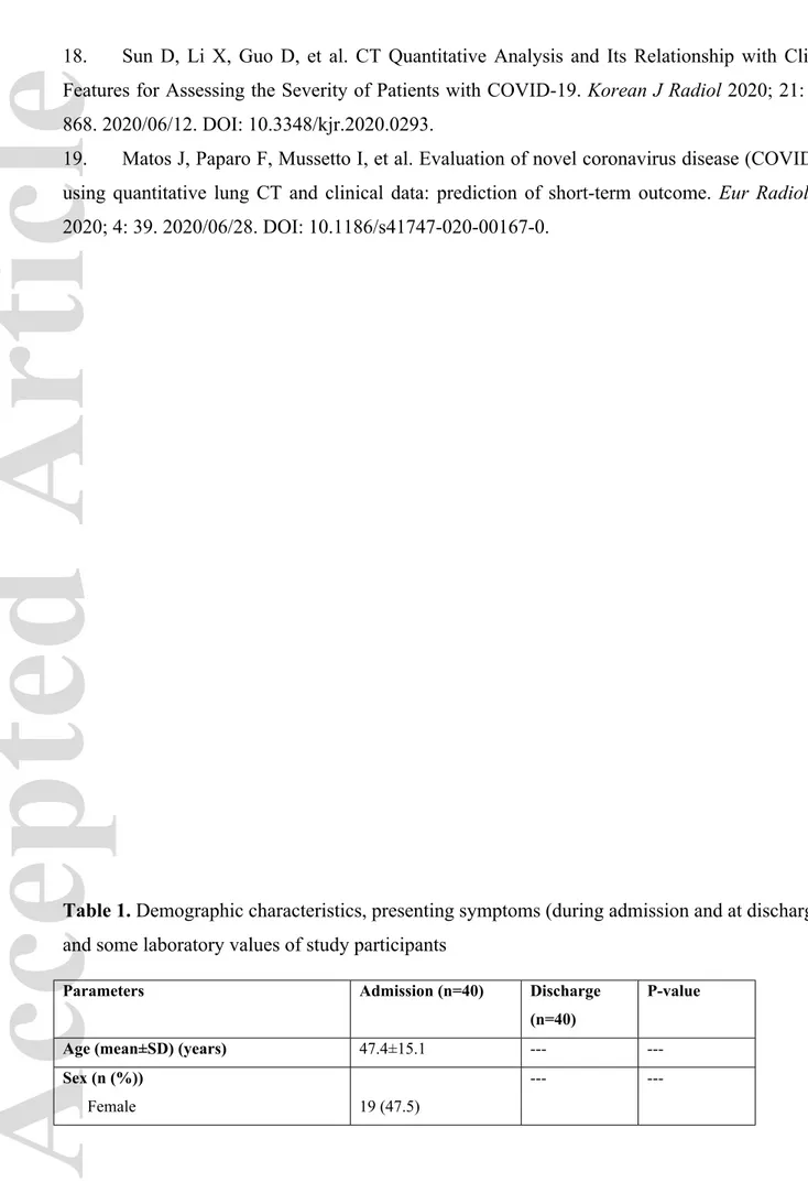
Table 1. Demographic characteristics, presenting symptoms (during admission and at discharge)
and some laboratory values of study participants
Parameters Admission (n=40) Discharge
(n=40)
P-value
Age (mean±SD) (years) 47.4±15.1 ---
---Sex (n (%))
Female 19 (47.5)
---
Male 21 (52.5) Fever (n (%)) 40 (100) 5 (12.5) <0.001* Dyspnea (n (%)) 36 (90) 7 (17.5) <0.001* Cough (n (%)) 40 (100) 25 (62.5) <0.001* Diarrhea (n (%)) 5 (12.5) 7 (17.5) 0.063* Myalgia (n (%)) 34 (85.5) 22 (5) 0.025*
Ferritin (median (IQR)) (ng/mL) 355 (259.3-452.3) ---
---Troponin (median (IQR)) 5.4 (3.4-7.95) ---
---D-dimer (median (IQR)) (ng/mL) 557 (250.5-2319.8) ---
---*Mc-Nemar test was used, IQR: Interquartile range, SD: Standard deviation
Table 2. Comparison of the changes in the lesion density (Hounsfield units) and some laboratory
values on admission and at discharge
Parameters Admission (n=40) Discharge (n=40) P-value
Lesion Density (Hounsfield units) -292.1±177.1 -418.9±162.7 <0.001*
WBC (mean±SD) (x103/µL) 7.89±3.59 5.6±1.24 <0.001*
Platelet count (mean±SD) (x103/µL) 299.6±103.4 355.7±75.5 0.002*
Lymphocyte count (median (IQR))
(x103/µL)
1.1 (0.9-1.3) 2.65 (1.83-3.75) <0.001**
spO2 (median (IQR)) (%) 86 (82.5-88) 94 (92-94) <0.001**
CRP (median (IQR)) (mg/L) 2.45 (1.33-4.0) 0.25 (0.2-0.4) <0.001**
Procalcitonin (median (IQR)) (µg/L) 0.3 (0.2-0.48) 0.2 (0.13-0.48) 0.963**
ALT (median (IQR)) (U/L) 45 (22-95.3) 44 (34-45) 0.017**
ALT: alanine aminotransferase, CRP: C-reactive protein, SD: standard deviation, spO2: oxygen saturation, WBC: White blood cell count. *Paired Samples t-test, and **Wilcoxon Signed Rank test were used.
FIGURES
Figure-1. Study flow-chart
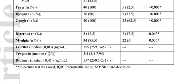
Figure 2. A 47-year-old male patient. The lesion density value decreased from -223.3 HU on
admission to -296.9 HU on discharge chest CT taken 5 days later. Newly developed lesions and size increase in the previous lesions are seen. A close anatomical location and area were used (56.8/63.7 mm2). No visible density difference is observed.
Figure 3. A 43-year-old male patient. The lesion density value was decreased from -489.3 HU on
admission to -662.9 HU on the repeat CT taken 5 days later despite the increased size and number of the lesions. No visible density difference is observed.
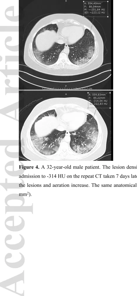
Figure 4. A 32-year-old male patient. The lesion density value was decreased from -251 HU on
admission to -314 HU on the repeat CT taken 7 days later despite the increased size and number of the lesions and aeration increase. The same anatomical location and area were used (554.4/559.8 mm2).