ARAŞTIRMA MAKALESİ (Research Article)
43
ASSESSMENT OF ANTIMICROBIAL ACTIVITY ON THE SKIN SECRETIONS OF NINE ANURAN SPECIES FROM TURKEY
Dilek AKYIL1, Arzu ÖZKARA2, Uğur Cengiz ERİŞMİŞ3
1Afyon Kocatepe Üniversitesi, Fen Edebiyat Fakültesi, Moleküler Biyoloji ve Genetik Bölümü,Afyonkarahisar, [email protected], ORCID: 0000-0002-7048-3808
2Afyon Kocatepe Üniversitesi, Fen Edebiyat Fakültesi, Moleküler Biyoloji ve Genetik Bölümü,Afyonkarahisar, [email protected], ORCID: 0000-0002-7815-5366
3Afyon Kocatepe Üniversitesi, Fen Edebiyat Fakültesi, Moleküler Biyoloji ve Genetik Bölümü,Afyonkarahisar, [email protected], ORCID: 0000-0002-6958-2016
Geliş Tarihi:25.09.2018 Kabul Tarihi: 03.07.2019
ABSTRACT
A dramatic decline of amphibians was reported for the first time about 40 years ago and then, this decline has continued at an alarming rate. Nearly one-third (32%), or 6593 amphibian species are threatened according to the most recent 2008 global assessment. Since 1980, the population size of at least 43% of amphibian species is declining and 122 amphibian species have become extinct. The causes of this declines are likely to be complex. Amphibians, like higher vertebrates, have immune systems including both adaptive and innate immunity. Innate immunity has different mechanisms that can provide an instant response nonspecifically to many pathogens. Antimicrobial peptides are the first step in these mechanisms. As each frog species has its own unique peptides, significant variation obtains among species in the number, structure and antimicrobial activity of these peptides. In our knowledge, there is no study previously performed in our country on the amphibian immune system. This study aims to evaluate the skin secretions of the nine anuran species and antimicrobial activity. Keywords: Amphibian populations, decline causes, amphibian immune system, antimicrobial peptids
TÜRKİYE’DEN DOKUZ ANURA TÜRÜNÜN DERİ SEKRESYONLARININ ANTİMİKROBİYAL AKTİVİTESİNİN DEĞERLENDİRİLMESİ ÖZ
İlk kez yaklaşık kırk yıl önce amfibilerde dramatik bir azalma rapor edilmiş ve bu düşüş endişe verici bir oranda devam etmiştir. 2008 yılı son küresel değerlendirmesine göre amfibilerin yaklaşık üçte biri (% 32) veya 6593 amfibi türü tehdit altındadır. Bu azalışın birçok karmaşık sebebi olabilir. Amfibiler omurgalılar da olduğu gibi hem doğuştan hem de kazanılmış immün sisteme sahip canlılardır. Doğal bağışıklık bireyin birçok farklı patojene karşı anlık tepki göstermesini sağlayan farklı mekanizmaları içerir. Bu mekanizmalar içerisinde antimikrobiyal peptidler ilk sırada yer alır. Her bir kurbağa türünün kendine özgü peptidleri olduğu için, bu peptidlerin sayısı, yapısı ve antimikrobiyal aktivitesindeki türler arasında önemli farklılıklar gösterir. Türkiye’de amfibi bağışıklık sistemi ile ilgili yapılmış
44
herhangi bir çalışma bulunmamaktadır. Bu çalışmada 9 amfibi türünden edilen deri sekresyonlarının antimikrobiyal aktivitelerinin belirlenmesi amaçlanmıştır.
Anahtar Kelimeler: Amfibi populasyonu, Azalma sebepleri, Amfibi immün sistemi, Antimikrobiyal peptidler
1. INTRODUCTION
Global declines of amphibian populations are a source of great concern. Many possible causes for amphibian population declines have been proposed [1]. According to researchers, deaths of amphibians are generally originate from three different factors. The first one of these factors is the abiotic factors and amphibians are interaction with this factor. People cause the change of ecosystems due to effect of abiotic factors consciously or unconsciously. The other factor is increase of number and virulance of pathogen microorganisms and organisms are adversely affected these microorganisms. Another factor is weakened immune system of organisms which live in ecosystems and organisms are under threat.
Amphibians are a transition species from the aquatic ancestors to the reptiles. Their skin is a morphologically, physiologically, and biochemically complex organ. Functions of amphibian skin are necessary for survival, including respiration, antipredator, water regulation, and antimicrobial defense, temperature control, excretion, and reproduction [2]. Their skin contains mucous and granular glands which responsible for the secretion of toxins and defences against microorganisms and predators [3,4]. The glands are usually activated by injury or stress and secrete a complex chemical mixture which vary from species to species [5, 6, 7, 8]. The skin glands of amphibians have various bioactive molecules such as peptides, steroids, proteins, alkaloids and biogenic amines that possess antibacterial and other important biological activities. According to their interactions with the environment, the compositions of the skin secretions differ among anuran groups [5, 7, 8]. These substances play different roles either in the defence against microorganisms and predators or in the regulation of physiological functions of the skin [9]. Besides, they are related to a variety of biological effects such as cytotoxic, fungicidal, bactericidal, lytic, anesthetic, neuromimetic, and phenomenal [4, 10, 11, 12, 13]. Recently, skin secretions were also noted as a rich source of multiple antimicrobial peptides effective against multidrug resistant strains[14]. Antimicrobial peptides were divided into two categories: those obtained from protein hydrolysis which called cryptides [15] and those obtained by ribosomal synthesis. Peptids which were obtained by ribosomal synthesis a significant part of innate immunity. Antimicrobial peptides constitute the first line of defense of the body [16, 17, 18]. Some antimicrobial peptides constitude by the proteolytic degradation of some proteins present within the organism. Studies on this subject is limited to a few species of frog in Turkey.
The purpose of present study is to test the antimicrobial activity of different anuran skin secretions against Gram (+), Gram (-) bacteria and Candida albicans cultures.
2. MATERIALS AND METHODS
2.1. Specimen Biodata and Secretion Harvesting
All the animal experiments were carried out in accordance with the approved guidelines with the regulations of the Turkish Department of Nature Conservation (Permit number,
DKMP-45
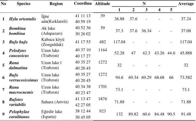
0.15.01.510.39719) and were approved by the Animal Welfare and Research Ethics Committee of Afyon Kocatepe University (permit number, AKU.0.A2.00.00-222). Adult specimens of frogs of both sexes (Bombina bombina, Rana dalmatina, Rana macrocnemis, Bufo bufo, Bufotes variabilis, Bufo verrucosissimus, Pelodytes caucasicus, Pelophylax ridibundus, Pelophylax caralitanus) obtain from different regions in Turkey (Fig1).
Figure 1 Turkey on the map of study areas, 1. Hyla orientalis (İğne ada (Kırklareli)) 2. Bombina bombina (Akgöl ( Adapazarı)) 3. Bufo bufo (Kabaca köyü (Zonguldak)) 4. Pelodytes caucasicus (Trabzon Uzungöl) ) 5. Rana dalmatina (Uzungöl (Trabzon)) 6. Bufo verrucosissimus (Uzungöl (Trabzon)) 7. Rana macrocnemis (Uzungöl (Trabzon)) 8. Bufotes variabilis (Sahara (Artvin)) 9.
Pelophylax caralitanus (Eğirdir Gölü (Isparta)). 2.2. Preparation of Skin Secretion
The frogs were washed first with tap water for the first step in the experimentand then with distilled water. Skin secretion was taken from the dorsal skin with gentle transdermal electrical stimulation [19]. Secretions were washed from the skin using deionized water and collected solutions were held in 80°C water bath for 30 min and centrifuged at 5500 rev/min for 30 min. The precipitate which constitute in centrifugation was diluted with distilled water,0.1 N HCl, 0.1 N NH4OH and 1 M phosphate buffers (pH: 4 and pH: 7)was used in the experiments [20].
2.3. Test Microorganisms and Growth Conditions
Test microorganisms (Escherichia coli, Staphylococcus aureus, Pseudomonas aeruginosa, Listeria, Micrococcus luteus, Bacillus subtilis, Klebsiella pneumoniae, Salmonella thyphi, Proteus vulgaris, Bacillus cereus, Candida albicans) were used from the culture collection of Afyon Kocatepe University, Faculty of Science and Art, Biology Department, Afyonkarahisar/Turkey. Cultures of these bacteria were grown in nutrient broth (NB) at 37°C for 24 h [21].
2.4. Determination of Antimicrobial Activity
Agar-Disc Diffusion Method were carried out in vitro antimicrobial activity studies. For antimicrobial activity Nutrient agar (NA) was preferred as the most suitable medium. 20 μl extract was inoculated into a sterile 6 mm diameter disc [22]. The turbidity of bacterial suspension was adjusted according to Mcfarland Standard Tube (0.5) with physiologic serum and suspension of the tested microorganism was spread on the solid media plates. Filter paper discs placed on the inoculated plates. These plates,
46
were incubated at 37°C for 24 h for bacteria and, at 30°C for 24 h, for yeasts [23]. The diameters of the inhibition zones were measured in millimetres. All tests were performed in duplicate. In addition, the solvent was used as negative control. 0.1 N HCl, 0.1 N NH4OH, 1 M phosphate buffers (pH: 4), 1 M phosphate buffers (pH:7) were used as positive controls. Experiments were repeated two times and results were evaluated as average values.
3. RESULTS
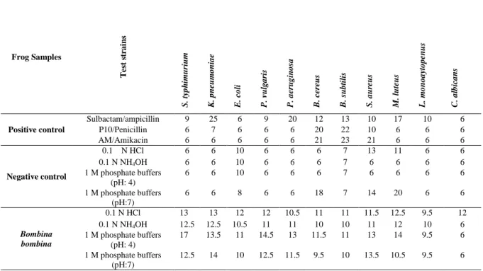
In this study, antimicrobial effects of prepared extracts on the tested microorganisms were determined by using different solvents. According to our findings, all the extracts were obtained from diffrent amphibians, exhibit antimicrobial activity effects of different extracts were obtained from the skin secretions against bacteria and yeast cultures results are given in Table 1. Adult specimens of frogs of both sexes obtain from different regions in Turkey (Table 2).
In our results show that, The highest antibacterial effect showed by 0.1 N NH4OH of Bufotes variabilis extract against Micrococcus luteus (ATCC-9341). Extracts of Bufo bufo skin secretion exhibited no effects against Pseudomonas aeruginosa and Micrococcus luteus. However the highest antibacterial effect showed against E.coli (10,5-14mm inhibition zone).The more antibacterial effect showed by 0.1 N HCL and 1M phosphate buffers (pH 7) extracts of Bufo variabilis than 0.1 N NH4OH extract and 1M phosphate buffers (pH 4). According to our findings, the highest antimicrobial activity was observed by 0.1 N HCI extract of Rana dalmatina and 1M phosphate buffers (pH 7) extracts of Rana macrocnemis against Klepsiella pneumonia.
Table 1 Antimicrobial activity effects of different extracts were obtained from the skin secretions against bacteria and yeast cultures.
Frog Samples T est st ra in s S . ty p h imu ri u m K . p n eu m o n ia e E . co li P. v u lg a ri s P. a eru g in o sa B . ce reus B . su b ti li s S . a u re u s M . lu te u s L. m o n o a yt o p en u s C . a lb ic a n s Positive control Sulbactam/ampicillin 9 25 6 9 20 12 13 10 17 10 6 P10/Penicillin 6 7 6 6 6 20 22 10 6 6 6 AM/Amikacin 6 6 6 6 6 21 23 21 6 6 6 Negative control 0.1 N HCl 6 6 10 6 6 6 7 13 11 6 6 0.1 N NH4OH 6 6 10 6 6 6 7 6 6 6 6 1 M phosphate buffers (pH: 4) 6 6 10 6 6 6 7 6 6 6 6 1 M phosphate buffers (pH:7) 6 6 8 6 6 18 7 14 20 6 6 Bombina bombina 0.1 N HCl 13 13 12 12 10.5 11 11 11.5 12.5 9.5 12 0.1 N NH4OH 12.5 12.5 10.5 11 11 10 10 11 12 10 6 1 M phosphate buffers (pH: 4) 17 13.5 11 14.5 13 11.5 11 13 14 9.5 6 1 M phosphate buffers (pH:7) 12.5 14 10 12.5 11.5 9.5 10 13.5 10.5 9.5 6
47
Rana dalmatina 0.1 N HCl 9.5 14.5 11 13 11 10 9.5 10.5 12 10.5 12 0.1 N NH4OH 6 6 11 6 10 6 6 6 6 6 9 1 M phosphate buffers (pH: 4) 6 6 11 6 6 6 6 6 6 9.5 6 1 M phosphate buffers (pH:7) 7.5 14 10.5 10 9.5 9 10.5 10 6 11.5 8.5 Rana macrocnemis 0.1 N HCl 14 13.5 12 13.5 13.5 10.5 10.5 14.5 11 11 10.5 0.1 N NH4OH 12 13.5 13 12.5 6 14 11.5 13.5 12 7.5 6 1 M phosphate buffers (pH: 4) 9 10 9.5 6 6 11 6 9.5 6 6 6 1 M phosphate buffers (pH:7) 12.5 14.5 11 12 13.5 8 9.5 9.5 6 8.5 6 Bufo bufo 0.1 N HCl 11.5 16 14 10.5 6 9 9 9 6 8.5 6 0.1 N NH4OH 9 12 11 9 6 6.5 10 7.5 6 8 6.5 1 M phosphate buffers (pH: 4) 6 6 11.5 6 6 8 6.5 6 6 6 8.5 1 M phosphate buffers (pH:7) 9 6 10.5 9.5 - 8.5 8.5 8 6 9 9 Bufotes variabilis 0.1 N HCl 11 11 10.5 11 12.5 11 11 9.5 15.5 9 12.5 0.1 N NH4OH 6 6 9 6 8.5 6 6 6 6 6 6 1 M phosphate buffers (pH: 4) 6 6 10 6 6 6 6 6 6 7.5 9 1 M phosphate buffers (pH:7) 10 6 8.5 10 10 9 9 11.5 6 8 6 Bufo verrucosissimus 0.1 N HCl 8.5 11.5 11 12 12 9.5 9.5 10.5 10.5 12 11 0.1 N NH4OH 6 8 13.5 6 6 6 6 6 6 6 6 1 M phosphate buffers (pH: 4) 6 6 11.5 6 6 6 6 6 6 6 6 1 M phosphate buffers (pH:7) 8.5 8 8 11 10 9.5 7.5 9.5 6 10 6 Pelodytes caucasicus 0.1 N HCl 9 11 10 11.5 10.5 9.5 10 10 11 10.5 10 0.1 N NH4OH 6 6 11.5 6 6 6 6 6 6 6 7 1 M phosphate buffers (pH: 4) 6 6 12 6 6 9 6 6 6 11 6 1 M phosphate buffers (pH:7) 9.5 9.5 11.5 10 8.5 8.5 10 9.5 10 9.5 6.5 Pelophylax ridibundus 0.1 N HCl 11 6 6 10 6 8.5 8.5 9.5 7 12 6 0.1 N NH4OH 10 9 6 10 6 12 10 11 13.5 11 6 1 M phosphate buffers (pH: 4) 12 9 8 11.5 6 15 13.5 11.5 10.5 11 6.5 1 M phosphate buffers (pH:7) 11 8 6 9.5 6 11 11 10.5 8.5 8 6 Pelophylax caralitanus 0.1 N HCl 9.5 10 11 11.5 12 9 9 9 12.5 10 11 0.1 N NH4OH 6 9 12.5 8 6 6 10 6 8.5 6 6 1 M phosphate buffers (pH: 4) 6 6.5 12.5 6 6 6 8.5 6 6 7.5 6 1 M phosphate buffers (pH:7) 10.5 6 10.5 11 10.5 10.5 9.5 11 10.5 9 648
Table 2 Adult specimens of frogs of both sexes obtain from different regions in Turkey.
4. DISCUSSION
The skin of amphibians is an extraordinarily rich source of antimicrobial peptides (AMPs). The first AMP was detected in the skin of the European frog Bombina variegata 42 years ago [24]. Since that time, several hundreds of different antimicrobial peptides have been obtained from the skin of amphibians belonging to the families Bombinatoridae, Hylidae, Hyperoliidae, Leiopelmatidae, Leptodactylidae, Myobatrachidae, Pipidae, and Ranidae [28]. AMPs belong to a large group of linear amphipathic helical peptide which are cationic, containing a flexible number of positively charged residues and hydrophobic regions. These properties supply them with an ability to bind to membrane lipids and/or negatively charged molecules and disrupt the membrane structure (Rollins-Smith et al. 2005).
In general, the antimicrobial activity of frog skin peptides is determined with some bacterial and fungal strains. These include the Gram-negative Escherichia coli and Pseudomonas aeruginosa, the Gram-positive Staphylococcus aureus, and the yeast Candida albicans [24].
The antimicrobial activity of bombinin-like peptides or bombinins H obtain from Bombina sp. can be distinguished on the basis of their cytolytic properties. Bombinins were found to be active against Gram-positive [G(+)] (Bacillus megaterium, Staphylococcus aureus) and Gram-negative [G(-)] (Escherichia coli, Yersinia pseudotuberculosis, Pseudomonas aeruginosa) bacteria as well as against Candida albicans [25]. According to our findings, skin secretion from Bombina bombina was No Species Region Coordina
te
Altitude N Average
1 2 3 4 5
1 Hyla orientalis İğne ada(Kırklareli) 41 11 13 39 36.88 37.6 - - - 37.24 40 59 19 2 Bombina bombina Ak lake (Adapazarı) 40 52 36 39 37.3 37.6 36.34 - - 37.08 30 26 02
3 Bufo bufo Kabaca köyü
(Zonguldak) 41 17 53 482 117.04 - - - - 117.04 4 Pelodytes caucasicus Uzun lake (Trabzon) 40 37 10 1164 52.28 47 42.3 43.26 44.6 45.888 40 17 27 5 Rana dalmatina Uzun lake l (Trabzon) 40 35 27 1272 32 - - - 32 40 20 45 6 Bufo verrucosissimus Uzun lake (Trabzon) 40 35 27 1272 94.6 69.34 69.29 68.68 66 73.582 40 20 45 7 Rana macrocnemis Uzun lake (Trabzon) 40 34 38 1701 73.1 - - - - 73.1 40 23 47 8 Bufotes
variabilis Sahara (Artvin)
41 13 47 1876 71.88 - - - - 71.88 42 27 05 9 Pelophylax caralitanus Eğirdir lake (Isparta) 38 12 44 923 132 89.82 60.6 84.48 90.5 91.48 30 45 05
49
observed antimicrobial activities against both G(-) and G(+) bacteria. The highest antibacterial effect showed by 1M phosphate buffer (pH4) of Bombina bombina skin extract against Proteus vulgaris, Salmonella typhimurium (NRRLB-4420) and Bacillus cereus (ATCC-11778). Only 0.1N HCI extracts of skin secretions showed antiyeast effects. The results of our study are similar to the other literatures. Dülger et al. [26] investigated antimicrobial activity of skin secretions from Bufo viridis. In this study, antimicrobial activity was determined with Escherichia coli, Staphylococcus aureus, Klebsiella pneumoniae, Pseudomonas aeruginosa, Proteus vulgaris, Bacillus cereus, Mycobacterium smegmatis, Listeria monocytogenes, Micrococcus luteus, Candida albicans, Rhodotorula rubra, and Kluyveromyces fragilis. Park et al. [27] report a novel antimicrobial peptide named buforin I and II purified from the stomach of Bufo bufo gargarizans, an Asian toad, which has been used as a wound-healing agent in traditional Korean medicine. Both buforin I and buforin II displayed strong antimicrobial activities against a broad spectrum of bacteria including, Bacillus subtilis, Staphylococcus aureus, Streptococcus mutans, Streptococcus pneumoniae, Escherichia coli, Serratia sp., Pseudomonas putida, and Salmonella typhimurium. Furthermore, Candida albicans, Saccharomyces cerevisiae and Cryptococcus neoformance were also killed.
In our results show that, the highest antibacterial effect was observed by 0.1 N NH4OH of Bufotes variabilis extract against Micrococcus luteus (ATCC-9341). Extracts of Bufo bufo skin secretion exhibited no effects against Pseudomonas aeruginosa and Micrococcus luteus. However the highest antibacterial effect showed against E.coli (10,5-14mm inhibition zone).The more antibacterial effect showed by 0.1 N HCL and 1M phosphate buffers (pH7) extracts of Bufotes variabilis than 0.1 N NH4OH extract and 1M phosphate buffers (pH4).
Frogs which belong to family Ranidae represent a important source of antimicrobial peptides [28]. Çevikbas [29] reported that skin secretion of Rana ridibunda determines antibacterial activity at different levels. However, Afsar et al. [20] showed that, skin secretions of Rana macrocnemis against the yeast cultures show more antimicrobial activity than that of the bacterial cultures. According to our results, the highest antimicrobial activity was observed by 0.1 N HCI extract of Rana dalmatina and 1M phosphate buffers (pH7) extracts of Rana macrocnemis against Klepsiella pneumoniae. The highest antibacterial effect showed by 1M phosphate buffers (pH 7) Rana dalmatina extract against Listeria monoaytopenus (ATCC-7644); 0.1 N NH4OH and 1M phosphate buffer (pH 7) of Rana macrocnemis skin extract against Pseudomonas aeruginosa. The highest antimicrobial activity was observed by 0.1 N HCI extract of Ranamacrocnemis skin extract against Stapylococus aureus (MRRL-B 767).1 M phosphate buffer (pH4) of Pelophylax ridibunda skinextract against Bacillus subtilis (NRS-744). Sensitivity of the microorganisms to the different agents changes from strain to strain [30]. According to our results, all the extracts of skin secretion, were obtained from diffrent anurans, exhibit antimicrobial activity. The present study has demonstrated that antimicrobial activity of skin secretions of varries at both the generic and ecological.
In conclusion, amphibians are the first group of organism forming a connecting link between land and water. Moreover, they have to fight to adopt a different conditions of environment and laden with pathogenic microbes. Therefore, theywere endowed with an excellent chemical defense system composed of pharmacological and antimicrobial peptides [31]. New peptides have been found that could inspire the design of substance prevent or treat infections. Peptide-based antibiotics are considered a potential solution to the growing problem of resistance to traditional antibiotics. In the development of AMPs as therapeutics, peptides obtained from amphibian skin and their synthetic analogues will play a very important role.
50
ACKNOWLEDGMENTSThis research was supported by Project no. Tubitak 113Z139. Ethical endorsement was ratified by the Ethical Committee of Afyon Kocatepe University and the Turkish Department of Nature Conservation (Permit number, DKMP-51039719).
REFERENCES
[1] Rollins-Smith, L. A, Doersam J. K, Longcore J. E, Taylor S. K, Shamblin J. C, Carey C. and Zasloff M. A., (2002), Antimicrobial peptide defenses against pathogens associated with global amphibian declines, Developmental- Comparative Immunology, 26(1), 63-72.
[2] Clarke, B.T., (1997), The natural history of amphibian skin secretion, their normal functioning and potentialmedical applications, Biological Reviews, 72, 365-379.
[3] Barra, D. and Simmaco. M., (1995), Amphibian skin: a promising resource for antimicrobial peptides, Trends Biotechnology, 13, 205- 209.
[4] Guo, W., Ao, M., Li, W., Wang, J., Yu, L. and Naturforsch, Z., (2012), Major biological activities of the skin secretion of the chinese giant salamander, Andrias davidianus, Zeitschrift fur Naturforsehung C. Journal of Biosciences, 67, 86-92.
[5] Gomes, A., Giri, B., Saha, A., Mishra, R., Dasgupta, S. C., Debnath, A. and Gomes, A., (2007), Bioactive molecules from amphibian skin: Their biological activities with reference to therapeutic potentials for possible drug development, Indian Journal of Experimental Biology, 45, 579-593.
[6] Siano, A., Gatti, P. I., Imaz, M. S., Zerbini, E., Simonetta, A. C., Lajmanovich, R. and Tonarelli, G. G., (2014), A comparative study of the biological activity of skin and granular gland secretions of Leptodactylus latrans and Hypsiboas pulchellus from Argentina, Records of Natural Products, 8, 128-135.
[7] Libério, M., Bastos, I. M. D., Junior, O. R. P., Fontes, W., Santana, J. M. and Mariana, S. C., (2014), The crude skin secretion of the pepper frog Leptodactylus labyrinthicus is rich in metallo and serine peptidases, Plos one, https://doi.org/10.1371/journal.pone.0096893.
[8] Artika, I. M., Pinontoan, S. and Kusrini, M. D., (2015), Antibacterial activity of skin secretion of bleeding toad Leptophryne cruentata and Javan tree frog Rhacophorus margaritifer, American Journal of Biochem Biotechnology, 11(3), 127-131.
[9] Stebbins, R. C. and Cohen, N. W., (1995), A natural history of amphibians, Princeton University Press, New Jersey.
[10] Apponyi, M. A., Pukala, T. L., Brinkworth, C. S., Maselli, V. M., Bowie, J. H., Tyler, M. J., Booker, G. W., Wallace, J. C., Carver, J. A., Separovic, F., Doyle, J. and Llewellyn, L. E., (2004), Host-defence peptides of Australian anurans: structure, mechanism of action and evolutionary significance, Peptides, 25, 1035-1054.
51
[11] Daly, J. W., Spande, T. F. and Garraffo, H. M., (2005), Alkaloids from amphibian skin: a tabulation of over eighthundred compounds, Journal of Natural Products, 68, 1556- 1575. [12] Giangaspero, A., Sandri, L. and Tossi, A., (2001), Amphipathic alpha helical antimicrobial
peptides, European Journal of Biochemistry, 268, 5589- 5600.
[13] Woodley, S. K., (2010), Pheromonal communication in amphibians, Journal of Comparative Physiology A, Neuroethology, Sensory, Neural and Behavioral Physiology, 196, 713-727. [14] Calderon, L. A., Silva, A. A. E., Ciancaglini, P. and Guerino, S. R., (2011), Antimicrobial
peptides from Phyllomedusa frogs: from biomolecular diversity to potential nanotechnologic medical applications, Amino Acids, 40, 29–49.
[15] Ueki, N., Someya, K., Matsuo, Y., Wakamatsu, K. and Mukai, H., (2007), Cryptides: functional cryptic peptides hidden in protein structures, Biopolymers, 88, 190–198.
[16] Devine, D. A., (2003), Antimicrobial peptides in defence of the oral and respiratory tracts, Molecular Immunology, 40, 431–443.
[17] Levy, O., (2004), Antimicrobial proteins and peptides: anti-infective molecules of mammalian leukocytes, Journal of Leukocyte Biology, 76, 909–25.
[18] Marshall, S. H. and Arenas, G., (2003), Antimicrobial peptides: a natural alternative to chemical antibiotics and a potential for applied biotechnology, Electronic Journal of Biotechnology, 6, 271–84.
[19] Zhou, M., Chen, T., Walker, B. and Shaw, C., (2006), Pelophylaxins: Novel antimicrobial peptide homologs from the skin secretion of the Fukien gold-striped pond frog, Pelophylax plancyifukienensis Identification by ‘‘shotgun’’ cDNA cloning and sequence analysis, Peptides, 27(1), 36-41.
[20] Afsar, B., Afsar, M. and Kalyoncu, F., (2011), Antimicrobial activity in the skin secretion of brownfrog, Rana macrocnemis (Boulenger, 1885) collectedfrom Turkey, Scientific Research and Essays, 6(5), 1001-1004.
[21] Oskay, M. and Sarı, D., (2007), Antimicrobial screening of some Turkish medicinal plants, Pharmaceutical Biology, 45, 176-181.
[22] Solak, M. H., Kalmis, E., Saglam, H. and Kalyoncu, F., (2006), Antimicrobial activity of two wild mushrooms Clitocybe alexandri (Gill.) Konr. and Rhizopogon roseolus (Corda) T.M. Fries collected from Turkey, Phytotherapy Research, 20, 1085-1087.
[23] Collins, C. M., Lyne, P. M. and Grange, J. M., (1989), Microbiological Methods, Six Edition, Butterworths & Co Ltd London, p. 416.
[24] Rinaldi, A.C., (2002), Antimicrobial peptides from amphibian skin: an expanding scenario, Current Opinion in Chemical Biology, 6, 799–804.
52
[25] Simmaco, M., Kreil, G., Barra D., (2009), Bombinins, antimicrobial peptides from Bombina species, Biochimica et Biophysica Acta, 1788, 1551–1555.
[26] Dülger, B., Ugurtas, I. H., Sevinc, M., (2004), Antimicrobial activity in the skin secretion of Bufo viridis (Laurenti, 1768), Asiatic Herpetology Research, 10, 161-163.
[27] Park, C. B, Kim, M. S., Kim, S. C., (1996), A Novel Antimicrobial peptide from Bufo bufo gargarizans, Biochemical and Biophysic Research Community, 218, 408–413.
[28] Conlon, J. M., (2008), Reflections on a systematic nomenclature for antimicrobial peptides from the skins of frogs of the family Ranidae, Peptides, 29, 1815–1819.
[29] Cevikbas A., (1978), Antibacterial activity in the skin secretion of the frog Rana ridibunda, Toxicon, 16, 195-197.
[30] Cetin, T. E., Gurler, N., (1989), Bakterilerin antibiyotiklere duyarlilik deneyinin yapılması, Kukem, 12, 2-5.
[31] Boman, H. G., (1991), Antibacterial peptides: key components needed in immunity, Cell, 65(2), 205–207.