• Sonuç bulunamadı

Mastocytosis

N/A
N/A
Protected

Academic year: 2021

Share "Mastocytosis"

Copied!
10
0
0

Yükleniyor.... (view fulltext now)

Tam metin

(1)

R E V I E W

İMMÜNOLOJİ

119

Mastocytosis

Reyhan GÜMÜŞBURUN1 , Uğur Hacı MUŞABAK2 , Sevim BAVBEK1

ABSTRACT

Mast cell activation diseases are common as either atopic diseases or rarely occur as mastocytosis and mast cell activation syndrome. Mastocytosis is a clonal disorder characterized by proliferation and accumulation of mast cells in various tissues, particularly the skin and bone marrow. Mast cell activation syndrome is characterized by systemic symptoms ranging from flushing and abdominal cramps to anaphylaxis secondary to mast cell degranulation. The aim of this review was to increase the awareness of the clinical features, classification, diagnosis, and treatment approaches of systemic mastocytosis. In addition, the role of mast cell activation syndrome in the differential diagnosis will be discussed.

Keywords: Mast cell, mastocytosis, mast cell activation syndrome (MCAS), triptase, anaphylaxis

1 Department of Immunology and Allergy Diseases, Ankara University, School of Medicine, Ankara, Turkey 2 Department of Immunology and Allergy, Başkent University, Ankara Hospital, Ankara, Turkey

INTRODUCTION

Mast cells (MC) are an important part of the immune system and the main effector cells of the allergic processes. As progenitor cells originating from precursor cells in the bone marrow, they migrate from the bone marrow to peripheral tissues where they complete their maturation. They have a widespread tissue distribution, but are mainly present at interfaces such as the skin, respiratory mucosa, and gastrointestinal tract with potential entry sites between the host and the external environment. MCs interact with the microenvironment by being activated by stimuli acting on various receptors on the surface and release biologically active mediators in response (Figure 1) (1). Structurally, MCs are designed to detect and respond to triggers of internal or external stress or danger. Therefore, a certain amount of MC activation may be physiologic or even necessary to maintain normal homeostasis on a daily basis. However, MC proliferation (mastocytosis and monoclonal mast cell activation syndrome) or MC activation disproportionately from the perceived hazard (anaphylaxis) may cause this physiological process to become disease (2).

Mast cell activation syndrome (MCAS) was used primarily in 2010 as an umbrella term to describe all clinical signs and symptoms secondary to MC degranulation, rather than a specific diagnosis. The diagnostic criteria were recently approved by an international consensus (Table I) and classified into 3 subgroups (Table II) (3,4). The signs and symptoms of MCAS are necessary to fulfill the three diagnostic criteria.

Mastocytosis identifies a heterogeneous group of diseases characterized by the proliferation and accumulation of neoplastic MCs in one or more systems. It was first described by Nettleship and Tay in 1869 as a skin disease, and the first case of systemic mastocytosis (SM) was reported in 1949 (5,6). Between 1991 and 2000, some clinical and laboratory parameters with distinct diagnostic and / or prognostic effects were identified and used to develop subtype-specific criteria. These criteria were the basis for the first World Health Organization (WHO) classification published in 2001. However, new prognostic parameters and more effective treatments were found and the WHO classification of mastocytosis was updated. In the 2016 WHO classification, at least one major and one minor criteria or at least three minor criteria are sufficient for the diagnosis of mastocytosis and seven different subgroups are defined (Table III, IV) (7,8).

Corresponding Author: Reyhan GÜMÜŞBURUN * reyhangumusburun@gmail.com

ORCID: Reyhan GÜMÜŞBURUN / 0000-0002-0274-5917, Uğur Hacı MUŞABAK / 0000-0003-1511-7634, Sevim BAVBEK / 0000-0002-7884-0830

Received: 25/12/2018 • Accepted: 18/07/2019 Online Published: 04/09/2019

(2)

Figure 1. Mast cell mediators. Figure 1. Mast cell mediators

Activators

Bacteria, fungi, virus, allergen, drug, cytokine, peptide, toxin

Mast Cell

Prostaglandin D2 / E2 Leukotriene B4 / C4, PAF

1, IL2, 3, IL4, 5, 6, 8, 9, IL-10, IL-12, IL-13, IL-16, IL-17, IL- 33, IFN-type1 / 2, TNF-α, TGF-β, VEGF, NGF, SCF, FGF, GM-CSF, nitric oxide, complement factor C3 and C4

Histamine, serotonin, dopamine, polyamine, cathepsin, β hexozaminidase, β glucuronidase, β galactosidase, aryl sulfatase, cathepsin, chymase, tryptase, carboxypeptidase, granzyme B, matrix metalloproteinase, heparin, chondrin sulfate, TNF-α, IL-4/8 / 15, RANTES, eotaxin, MCP-1/3/4, TGF-β, VEGF, NGF, SCF, FGF, CTRH, endorphin, endothelium-1, suptance P, VIP, Major basic protein.

Preformed

Neoformed

Neosynthesized

Table I. Diagnostic criteria for mast cell activation syndrome. 1. Typical signs and symptoms of mast cell degranulation

(affect at least two organ systems) Ø Ø Skin: redness, itching, urticaria, angioedemaCardiovascular: hypotension, palpitations

Ø Respiratory: wheezing, throat swelling, shortness of breath Ø GI: diarrhea

Ø Nazo-ocular: itching 2. Objective evidence of mediator release Ø Increased serum tryptase

Ø Increased 24-hour urinary histamine metabolites (methylhistamine)

Ø Increased 24-hour urinary prostaglandins (prostaglandin D2; 11β platelet-derived growth factor 2α)

3. Response to therapy that blocks mast cell mediator activity Ø H1- and H2-blockers, Leukotriene antagonists, Cromolyn sodium, Ketotifen, Aspirin

Table II. Mast cell activation syndrome classification.

1. Primary Ø Cutaneous mastocytosis (CM)( urticaria pigmentosa, maculopapular cutaneous mastocytosis (MPCM)) Ø Systemic mastocytosis (SM) (indolent(ISM), aggressive(ASM), associated with a hematologic non-mast cell

lineage disease (SM-AHNMD), mast cell leukemia(MCL) Ø Mast cell sarcoma (MCS)

Ø Mastocytoma

Ø Monoclonal mast cell activation syndrome (MMAS)

2. Secondary Ø IgE-mediated hypersensitivity reactions (eg, food, insect, drug-induced anaphylaxis) Ø Drugs (eg, vancomycin, opioids, taxanes)

Ø Mast cell hyperplasia (associated with chronic infections, neoplasia, and autoimmune conditions possibly due to an excess of stem cell factor; these reactive states are infrequently the cause of mast cell activation disorders) 3. Idiopathic Ø Idiopathic Anaphylaxis (IA)

(3)

121

Asthma Allergy Immunol 2019;17:119-128 Epidemiology

Although all types of mastocytosis are rare, the exact prevalence is not known and no difference in incidence has been observed between the sexes (9). Mastocytosis in chil-dren is often seen as cutaneous mastocytosis (CM) in the first year of life and the majority regress in adolescence (10, 11). In adults, indolent systemic mastocytosis (ISM) is seen most commonly, and the second most frequent is SM with an associated hematologic (non-MC lineage) neoplasm (SM-AHN). Also, cutaneous forms of the disease constitute less than 5% of cases and usually persist into SM (12).

DIAGNOSIS Clinical Evaluation

Clinical features that may lead to a suspicion of mast cell activation (MCA) are either pruritus, flushing, urti-caria, angioedema, nasal congestion, wheezing, bron-chospastic cough, headache, fatigue, lethargy, concentra-tion problems, diarrhea, gastric hyperacidity, abdominal cramping, nausea, vomiting, hypotension, tachycardia due to degranulation of MC mediators with the effect of various factors or end-organ dysfunction such as hypersplenism, Table III. Diagnostic criteria for systemic mastocytosis.

Major SM

criterion Multifocal dense infiltrates of MCs (≥15 MCs in aggregates) in BM biopsies and/or in sections of other extracutaneous organ(s) Minor SM

criteria

1. > 25% of all MCs are atypical cells (type I or type II) on BM smears or are spindle-shaped in MC infiltrates detected on sections of visceral organs

2. KIT point mutation at codon 816 in the BM or another extracutaneous organ 3. MCs in BM or blood or another extracutaneous organ exhibit CD2 and/or CD25

4. Baseline serum tryptase level > 20 ng/mL (in case of an unrelated myeloid neoplasm, item is not valid as an SM criterion)

B findings

1. Bone marrow biopsy showing >30% infiltration by MCs (focal, dense aggregates) and/or serum tryptase level >200 ng/mL.

2. Signs of dysplasia or myeloproliferation in non-MC lineages, but insufficient criteria for definitive diagnosis of a hematopoietic neoplasm (AHNMD), with normal or slightly abnormal blood counts.

3. Hepatomegaly without impairment of liver function, and/or palpable splenomegaly without hypersplenism, and/or lymphadenopathy on palpation or imaging.

C findings

1. Bone marrow dysfunction manifesting as cytopenia (absolute neutrophil count <1.0 x 10⁹/L, hemoglobin <10 g/dL, or platelets <100 x 109/L), but no obvious non-MC hematopoietic malignancy.

2. Palpable hepatomegaly with impairment of liver function, ascites, and/or portal hypertension. 3. Skeletal involvement with large osteolytic lesions and/or pathological fractures.

4. Palpable splenomegaly with hypersplenism.

5. Malabsorption with weight loss due to gastrointestinal mast cell infiltrates.

Systemic mastocytosis is diagnosed when there is at least 1 major and 1 minor or at least 3 minor criteria (6).

Table IV. WHO classification and diagnosis of mastocytosis 2016.

Classification Diagnosis

Cutaneous mastocytosis (CM)

Ø Maculopapular cutaneous mastocytosis (MPCM) Ø Diffuse cutaneous mastocytosis (DCM)

Ø Mastocytoma of skin (cutaneous mastocytoma)

More than 15 mast cells in skin biopsy aggregates Absence of SM criterion

Systemic mastocytosis (SM)

Ø Indolent systemic mastocytosis (ISM) Ø Smoldering systemic mastocytosis (SSM) Ø Aggressive systemic mastocytosis (ASM)

Ø SM with an associated hematologic (non-MC lineage) neoplasm (SM-AHN)

Ø Mast cell leukemia (MCL)

The presence of SM criteria and

Ø Lack of C and B findings, absence of other clonal hematological disease findings

Ø The absence of signs of C and other findings of clonal hematological disease with the presence of 2 or more B signs Ø The presence of signs of C with the absence of mast cell

leukemia characteristics

Ø Presence of other hematological mast cell manifestations Ø More than 20% mast cell presence in bone marrow

Mast cell sarcoma (MCS) undifferentiated Ø An extracellular organ infiltration with undifferentiated mast cell with a destructive growth pattern

(4)

pathologic bone fracture, bile acid malabsorption, and cytopenia, which is caused by tissue infiltration of MCs in patients with aggressive disease (3, 4). Chronic urticaria, angioedema, and upper airway swelling are almost never associated in patients with mastocytosis but can be a fea-ture of patients with secondary [immunoglobulin (Ig)-E and non-IgE-mediated] and idiopathic mast cell activation syndromes (2). MCA symptoms may be chronic or epi-sodic in patients with nonclonal MCAS (13).

Therefore, every symptom and sign that the patient describes should be evaluated in detail to determine whether it is compatible with MCA and triggers should be determined. Triggers can be insect stings (snakes, jellyfish, bees), drugs (narcotics, radiocontrast, analgesics and antibiotics), physical factors (hot, cold, strong scrubbing, friction, UV light, exercise), invasive procedures (endoscopy, biopsy, surgery), psychovegetative agents (fear, stress), alcohol, spicy foods, and infections.

Characteristic skin lesions including urticaria pigmen-tosa, plaque, nodule, mastocytoma, and telangiectasia can develop. In patients with CM, the formation of erythema and urticaria-like lesions as a result of the mechanical stimulation of the skin and spreading of the mediators re-leased from MCs into the adjacent cells is typical for these patients (Darier sign) (Figure 2) (14). An experienced phy-sician can detect CM with a characteristic appearance of maculopapular cutaneous mastocytosis (MPCM). Howev-er, in the presence of doubt, a biopsy should be performed and evaluated.

The prevalence of atopy in patients with mastocytosis is not different from the healthy population, but the incidence of anaphylaxis is higher in mastocytosis. Anaphylaxis in adults with mastocytosis is most commonly seen with bee stings (19-53%), followed by foods (3-16%) and

drugs (5-9%), and the cumulative anaphylaxis prevalence is between 22-49%. Although alcohol, exercise, and temperature changes are potential triggers of anaphylaxis, they are mainly active as cofactors in patients with SM. Thirty-nine percent of anaphylaxis cases are idiopathic anaphylaxis in SM (15). Therefore, in all patients with hypotensive anaphylaxis, those with systemic reactions to hymenoptera and those with anaphylaxis that cannot be explained by IgE-mediated reasons, clonal mast cell disease should be considered (2). Alvarez-Twose et al. proposed a predictive model for clonal MC disorders based on clinical and laboratory findings for patients presenting with anaphylaxis without CM in a study from Spain (REMA). According to this, scores ≥2 can predict the presence of clonal MCAS with 92% sensitivity and 81% specificity (Table V) (16,17).

Laboratory Investigations

Laboratory tests should be performed to determine the presence of MC mediators or to evaluate abnormalities due to MC tissue infiltration. The primary evaluations for bone marrow and liver are complete blood count, peripheral smear, and routine liver function tests. Serum tryptase is the most specific indicator of mast cell load and activation (18). The basal tryptase level should be checked in the patient’s normal healthy state or at least 24 hours after the event and >11.4 ng/mL is considered high in most diagnostic laboratories (19). Primary mast cell disorder, chronic eosinophilic leukemia, myelodysplastic syndromes, acute leukemias, chronic renal failure, and other hematologic disorders that cause bone marrow suppression can cause the elevation of basal tryptase concentrations. In addition, familial tryptase elevations have been described, but it is not clear whether tryptase elevation is caused by MCA in these families (20). Serum tryptase should be measured from 15 minutes to 4 hours

Figure 2. Darier sign in a patient with cutaneous mastocytosis.

Picture . Darier sign in patient with cutaneous mastocytosis

Picture . Darier sign in patient with cutaneous mastocytosis

(5)

123

Asthma Allergy Immunol 2019;17:119-128

after symptom onset to provide evidence that MCA is responsible for the patient’s symptoms. The increase in tryptase should be at least 20% + 2 ng/mL above baseline to be considered significant as MCA (19). Elevated baseline tryptase values >20 ng/mL indicate that a patient is at increased risk for severe reaction (21, 22).

Normal urinary N-methyl-histamine is highly specific for mast cell activation but can be affected by diet or bacterial contamination. The normal range is 30 to 200 mcg/gm creatinine (Cr) (13).

Urinary prostaglandin D2 or metabolites are increased in patients with mast cell activation but not specific to mast cells. In patients with SM, urinary excretion of 11β PGF2α is above 3500 ng/24 hours (reference range <1000 ng/24 hours) (23). Urinary leukotriene E4 is increased in patients with mast cell activation. Increased urinary concentrations of LTE4 have been reported in several conditions, such as aspirin-exacerbated respiratory disease and anaphylactic reactions (24). The reference range among Cr normal volunteers was measured as <104 pg/ mg (25). Plasma interleukin (IL)-6 levels in patients with SM were correlated with disease severity and progression, serum tryptase concentrations and symptom severity. IL-6 concentrations higher than 2.5 pg/mL indicate severe disease (13).

Bone marrow biopsy should be performed in patients with basal tryptase concentrations above 20 ng/mL in ad-dition to typical symptoms of MC degranulation, in adults with CM, especially in the case of unexplained hypotensive attacks, flushing or anaphylaxis, gastrointestinal abnor-malities such as unexplained malabsorption or diarrhea, peripheral blood abnormalities such as cytopenia, unex-plained liver dysfunction, organomegaly, osteoporosis or pathologic bone fractures, and REMA scores ≥2 (26, 27).

The most important factor in MC growth is the stem cell factor, which leads to the overexpression of MCs and giving rise to mutations to the receptor to which they bind (27). The mutation of the KIT gene, which encodes the stem cell factor receptor, is a minor criterion in the diagnosis of SM and is usually screened in peripheral blood and bone marrow. If the most common KIT D816V mutation is detected in peripheral blood, bone marrow biopsy should be performed to evaluate other diagnostic criteria. However, if the KIT D816V mutation is not detected in peripheral blood and clinical suspicion is high, bone marrow biopsy should be performed. Bone marrow aspiration and biopsy are performed to evaluate CD117, immunoglobulin E, CD 25-positive cells by flow cytometry, mutation analysis (D816V BMT), and immunochemical assays involving tryptase, CD117, CD2, CD25, and antibodies. Patients should be referred to centers specialized in mastocytosis for diagnosis when possible because these diseases are rare and there are low numbers of abnormal MCs in bone marrow. Histologic evaluation of additional organs is generally not recommended because the bone marrow is almost always involved and the importance of increased MCs in other tissues is uncertain (3). Bone lesions are visualized using bone densitometry and the presence of organomegaly or acid is displayed by ultrasonography (USG), computed tomography (CT), and magnetic resonance (MRI) in SM.

Steps to the Diagnosis of Mastocytosis

As a first step, the baseline serum tryptase level should be measured in all patients with suspected MCA. MCAS should be considered when there is a response to treatment that interferes with MCA symptoms, at least two organ systems affecting MCA symptom findings, and high serum total tryptase or other MC mediators (histamine, PGD2) are detected. IgE-related conditions Table V. REMA scoring model.

Variable Score

Gender Male +1

Female -1

Clinical symptoms

Lack of urticaria and angioedema +1

Urticara and/or angioedema -2

Presyncope and/orsyncope +3

Serum basal tryptase < 15 ng/ml -1

(6)

that may cause MCA, and drugs or underlying diseases that may cause MC hyperplasia should be investigated for secondary MCAS. For patients with low clinical suspicion for SM or for patients with basal tryptase levels normal or less than 20.0 ng/mL, or for patients with no suggestive skin lesions, the D816V KIT mutation should be screened first in peripheral blood (19). If the result is negative, it may eliminate the need for bone marrow biopsy, but bone marrow biopsy is indicated if the result is positive (19). The presence of characteristic skin lesions in adults, unexplained pathologic fractures, osteoporosis, diffuse skeletal anomaly, cytopenia, organomegaly, unexplained recurrent anaphylaxis, and basal tryptase levels greater than 20.0 ng/mL in patients with MCA are generally considered indications for bone marrow biopsy (19, 28). The bone marrow biopsy confirms the diagnosis of SM, MMAS, and SM-AHNMD or excludes these diseases but leads to the diagnosis of idiopathic MCAS (Figure 3) .

Differential Diagnosis

Mastocytosis is diagnosed clinically, pathologically, and genetically based on the WHO criteria. MCAS and

anaphylaxis are the leading causes of histamine increase in urine and serum tryptase concentrations with similar clinical findings. Monoclonal MCAS describes patients with MCA symptoms who do not meet the exact diagnostic criteria of SM, but have MC clonality (15). Idiopathic MCAS has been proposed to identify idiopathic disorders that meet three of the MCAS diagnostic criteria, but the diagnosis of idiopathic anaphylaxis is more appropriate if these signs and symptoms meet the criteria for anaphylaxis. In anaphylaxis, tryptase and urine histamine concentrations increase during the acute event, but the basal tryptase concentration is normal. In the case of unexplained, frequently recurring anaphylaxis, basal tryptase values should be evaluated for SM.

Other differential diagnoses that have similar clinical findings to mastocytosis do not elevate serum tryptase and histamine in urine because they do not cause MCA, include endocrine disorders such as carcinoid syndrome, pheochrocytoma, thyroid medullary carcinoma, acute hypoglycemia, acute hypothyroidism, and adrenal insufficiency; gastrointestinal disorders such as Zolinger-Ellison syndrome, vasoactive intestinal peptide secreting

Figure 3. Diagnostic algorithm.

MCAS: Mast cell activation

syndrome,

SM: Systemic mastocytosis, MMAS: Monoclonal mast

cell activation syndrome,

SM-AHNMD: Associated

hematologic non-mast cell lineage disease.

Figure 2. Diagnostic algorithm. MCAS: Mast cell activation syndrome, SM: Systemic mastocytosis, MMAS:

• Mast cell activation symptoms

• Elevation of Mast cell mediator

• Response to anti-mediator therapy MCAS

• Investigate IgE-related triggering factors in mast cell activation (drug, food, insect)

• Assess for underlying diseases that could cause mast cell hyperplasia (chronic

infection, cancer, autoimmunity)

Secondary MCAS

SM WHO diagnostic criteria

• Characteristic lesions (MPCM, DCM, Mastocytoma)

• Unexplained pathological fracture, osteoporosis,

• Common skeletal anomaly (lytic, sclerotic lesion)

• Cytopenia, organomegaly

• REMA score ≥ 2

• High serum basal tryptase

• Unexplained recurrent anaphylaxis

Screen for D816V KIT mutation in peripheral blood and bone marrow

SM-AHNMD

1 major and 1 minor criteria Or 3 minor criteria 1 or 2 minor criteria SM MMAS Idiopathic MCAS

(7)

125

Asthma Allergy Immunol 2019;17:119-128

tumors, food-related reactions, irritable bowel syndrome, eosinophilic esophagitis, and gastroenteritis; cardiac diseases such as Kounis syndrome, postural orthostatic hypotension, myocardial infarction, syncope due to aortic stenosis, and endomyocarditis; neurological disorders such as epilepsy, central nervous system tumors, and intoxication; psychological disorders; and hereditary or acquired angioedema, pemphigus vulgaris, acute lupus vulgaris, acutely toxic dermatosis diseases, and drug-related adverse effects (28, 29). Furthermore, chronic eosinophilic leukemia, primary myelofibrosis, reactive mastocytosis, which is a similar sign of bone marrow due to increased MC infiltration and dermato fibroma; psoriasis; atopic dermatitis; and nevi because of similar skin biopsy findings due to increased MC infiltration should be considered in the differential diagnosis.

Treatment

There is generally no curative treatment in MCAS and mastocytosis, but avoidance of general and specific triggers is very important in reducing the risk of anaphylaxis. Therefore, sudden heat exchange, and excessive heat and cold exposure should be avoided, air conditioning and warm water should be preferred for bathing, exposure to heavy exercise and intense UV light should be reduced, and mastocytomas should be avoided by strong rubbing and friction. Anxiolytic or relaxation techniques for stress and anxiety or both should be considered. If drugs such as non-steroidal anti-inflammatory drugs (NSAIDs), opioids, radiocontrast media, and anesthetics have to be used, tolerable drugs should be preferred. However, if the tolerance is unknown but the drug should be used, a drug provocation test should be performed and prophylactic anti-mediator therapy should be considered if necessary.

Premedication regimens are preferred to be used for major procedures are a combination of anti-H1, anti-H2 histamine receptor drugs, a leukotriene (LT) receptor blocker and 0.5 mg/kg prednisone; and a combination of anti-H1 and anti-H2 histamine receptor drugs for small procedures. Alcohol; spices; strawberries; tomatoes; cheese; seafood; and smoked, canned, and fermented foods containing high amounts of histamine should be avoided. Insect repellents can be used to prevent insect stings and bites, perfumed lotions should be avoided, and light long-sleeved shirts and long trousers should be worn (26, 13). Venom immunotherapy is considered a life-saving treatment for patients with venom allergy and should be administered lifelong.

All patients with mastocytosis and MCAS with history of anaphylaxis, hypotension, presyncope, and mastocyto-sis with elevated tryptase should be prescribed 2 doses of intramuscular epinephrine autoinjectors and should be informed about how and when to use them. H1 antihista-mines, H2 antihistaantihista-mines, and glucocorticoids may be giv-en as adjunctive therapies in the treatmgiv-ent of anaphylaxis. Intravenous fluid should be applied in all cases of anaphy-laxis. Due to the lack of data on drug anaphylaxis, it is not possible to present clear recommendations at present (15). The mainstays of treatment of MCAS and SM symp-toms are blockade of mediator receptors (H1 and H2 anti-histamines, LT receptor blockade), inhibition of mediator synthesis (aspirin, zileuton), mediator release (sodium cro-molyn), tricyclic antidepressants (amitriptyline, doxepin), prochlorperazine, PUVA or narrow-band ultraviolet B, anti-IgE therapy, or a combination of these approaches (Table VI) (19). Intercalary urinary MC mediator levels and serum tryptase concentrations can be used (13). Table VI. Symptomatic treatment.

Symptom Treatment

Itching, flushing, urticaria, angioedema, dermatographism H1 or H2 blockers, Leukotriene Antagonists, Aspirin, Ketotifen Diarrhea, abdominal cramps, nausea, vomiting H2 blockers, Cromolyn sodium, Proton pomp inhibitors, Leukotriene

Antagonists, Ketotifen

Headache, poor concentration and memory H1 or H2 blockers, Cromolyn sodium, Ketotifen Presenkop, syncope, tachycardia H1 or H2 blockers, Corticosteroids, Omalizumab

Wheezing H1 or H2 blockers, Leukotriene Antagonists, inhaled and systemic

corticosteroids, Omalizumab

Itching, blockage in the nose H1 blockers (Topical or systemic), Topical Corticosteroids, Topical Cromolyn sodium

Anaphylaxis Epinephrine (intramuscular), H1 or H2 blockers, Corticosteroids, Omalizumab.

(8)

The starting regimen should include once or twice-daily non-sedating H1-receptor antagonists, which can be increased up to 4 times without anecdotal evidence of severe adverse effects except for sedation and mucosal dryness. Leukotriene antagonists and non-sedating anti-H1 receptor antagonists can also be used as second-line and add-on therapies. H2-receptor antagonists can be combined if gastrointestinal symptoms are present. Sodium cromolyn is suggested in patients at 100 mg daily and escalated over 8 weeks to 800 mg divided as 200 mg 4 times daily, taken on an empty stomach before meals and at bed time. Severe symptoms may require further escalation to 1000 to 1200 mg daily with skin, gastrointestinal, and neuropsychiatric symptoms (30). Patients with severe refractory symptoms can benefit from the addition of a glucocorticoid. It is recommended that aspirin should be given by gradual updosing schedule even if the patient has tolerated aspirin or other NSAIDs previously (13, 31). Especially recently, there are reports that omalizumab has prevented or even completely eliminated life-threatening anaphylaxis attacks and MC mediator symptoms in mastocytosis and MCAS (32-35). Omalizumab is also used to increase tolerance to venom immunotherapy and to reduce adverse effects (36-38). Benign forms of mastocytosis may turn into aggressive forms, and complications (bone loss, hepatosplenomegaly, cytopenia) should be closely monitored. Although there is no definitive guidelines about the frequency of bone mineral density (BMD) measurements, some studies recommended 18 months or 24 months (13, 39). Treatment mainstays include adequate calcium and vitamin D intake, weight-bearing exercise, and the use of appropriate medications (bisphosphonates, rank ligand inhibitors, and interferon alpha 2α).

Another treatment approach is to prevent MC proliferation and survival (cladribine, INF-α, tyrosine kinase inhibitors, allogenic hemopoietic stem cell transplantation) (7, 17). These cytoreductive therapies are required for recurrent anaphylaxis conditions despite other treatments in ISM and SSM and other advanced forms of SM, such as ASM, SM-AHN, and MCL. Traditionally used IFN-α and cladribine have been shown to control mast cell activation episodes and are highly effective agents (7, 2). The IFN-α effect is independent of the c-kit mutation, and has been used successfully in imatinib-resistant systemic mastocytosis. It can be used in high doses (10 million units [MU] 3/week) alone or in lower doses in combination with prednisolone (1.5 MU 3/week). Cladribine (2CdA) is a purine analog with activity against both resting and

actively dividing cells. Cladribine is administered at a dosage of 0.13-0.17 mg/kg/day in a 2-hour intravenous infusion over a period of 5 consecutive days, and repeated every 4 to 12 weeks for up to 9 courses, and its efficacy and toxicity has been investigated. However, many patients develop resistance to cladribine or poly-chemotherapy (poly-CT). Therefore, in recent years, new treatment concepts have been developed and tested in clinical studies and clinical applications (7).

Midostaurin, a multikinase inhibitor with activity against D816V KIT, has been used for mast cell cytoreduction and reducing the symptoms of mast cell activation (40). The prototypic tyrosine kinase inhibitor imatinib, which has activity against wild-type KIT and platelet-derived growth factor receptor, is highly successful in controlling symptoms, and the control can be prolonged. In vitro, imatinib does not reduce mast cell activation; therefore, it is not recommended for the treatment of non-clonal mast cell diseases and mast cell activation syndromes or patients with mastocytosis carrying the D816V KIT mutation, which confers resistance to this drug (41, 42). Masitinib, an inhibitor of wild-type (but not D816V-mutated) KIT and LYN, has been reported to be an effective and well-tolerated agent for the treatment of severely symptomatic indolent or smouldering systemic mastocytosis (43). Ibrutinib, a Bruton tyrosine kinase inhibitor, has no effect on non-specific non-IgE-mediated mast cell activation. More data are needed on the negative effects on antibody-mediated immune function, especially before considering the treatment of IgE-mediated disease (2). Avapritinib, a potent and highly specific oral inhibitor of KIT activation loop mutants including D816V, is well tolerated; there was no discontinued treatment because of drug-related adverse reactions in a phase 1 study in patients with advanced SM (44). Allogeneic hematopoietic stem cell transplantation (HSCT) appears to be an important approach in patients with advanced SM, including MCL responding to initial therapy. The results of transplanted patients appear to be positive in non-transformed patients with ASM and SM-AHN compared with those with overt MCL (45). It is appropriate to choose patients who are young and have no severe comorbid disease because the risk of death associated with transplantation is very high. Therefore, HSCT may not be an option in many patients. These patients should be directed to centers with expertise in this area because they have an aggressive clinical course with end-organ damage and survival is significantly reduced.

(9)

127

Asthma Allergy Immunol 2019;17:119-128 REFERENCES

1. Da Silva EZM, Jamur MC, Oliver C. Mast Cell Function: A new vision of an old cell. J Histochem Cytochem 2014;62(10):698-738.

2. Akin C. Mast cell activation syndromes. J Allergy Clin Immunol 2017;140 (2):349-55.

3. Akin C, Valent P, Metcalfe DD. Mast cell activation syndrome: Proposed diagnostic criteria. J Allergy Clin Immunol 2010;126:1099-104.

4. Valent P, Akin C, Arock M, Brockow K, Butterfield JH, Carter MC, et al. Definitions, criteria and global classification of mast cell disorders with special reference to mast cell activation syndromes: A consensus proposal. Int Arch Allergy Immunol 2012;157:215-25.

5. Nettleship E, Tay W. Rare forms of urticaria. Br Med J 1869;2:323-4.

6. Ellis JM. Urticaria pigmentosa; A report of a case with autopsy. Arch Pathol (Chic) 1949;48(5):426-35.

7. Valent P, Akin C, Metcalfe DD. Mastocytosis: 2016 updated WHO classification and novel emerging treatment concepts. Blood 2017;129(11):1420-7.

8. Arber DA, Orazi A, Hasserjian R, Thiele J, Borowitz MJ, Le Beau MM, et al. The 2016 revision to the World Health Organization classification of myeloid neoplasms and acute leukemia. Blood 2016; 127(20): 2391-405.

9. Méni C, Bruneau J, Georgin-Lavialle S, Le Saché de Peufeilhoux L, Damaj G, Hadj-Rabia S, et al. Paediatric mastocytosis: A systematic review of 1747 cases. Br J Dermatol 2015;172:642-51. 10. Wolff K, Komar M, Petzelbauer P. Clinical and histopathological

aspects of cutaneous mastocytosis. Leuk Res 2001;25:519-28. 11. Escribano L, Garcia-Montero A, Sanchez-Muñoz L, Teodosio

C, Alvarez‐Twose I, Jara‐Acevedo M, et al. Diagnosis of adult mastocytosis: Role for bone marrow analysis. In: Kottke-Marchant K, Davis B (eds). Laboratory Hematology Practice. London: Wiley-Blackwell, 2012: 388-98.

12. Horny HP, Sotlar K, Sperr WR, Valent P. Systemic mastocytosis with associated clonal haematological non-mast cell lineage diseases: A histopathological challenge. J Clin Pathol 2004;57:604-8.

13. Castells M, Butterfield J. Mast Cell Activation Syndrome and Mastocytosis: Initial treatment options and long-term management. J Allergy Clin Immunol Pract 2019;7(4):1097-106. 14. Rotolo A, Familiari U, Nicoli P, Cilloni D, Saglio G, Guerrasio A. Systemic mastocytosis: An intriguing disorder, hematology-science and practice. In: Lawrie C (ed). Hematology-Science and Practice. In Tech 2012:467-85.

15. Bonadonna P, Lombardo C, Zanotti R. Mastocytosis and allergic diseases. J Investig Allergol Clin Immunol 2014;24(5): 288-97. 16. Alvarez-Twose I, González de Olano D, Sánchez-Muñoz L,

Matito A, Esteban-López MI, Vega A, et al. Clinical, biological, and molecular characteristics of clonal mast cell disorders presenting with systemic mast cell activation symptoms. J Allergy Clin Immunol 2010;125:1269-78.

17. Gümüsburun R, Kendirlinan R, Altiner S, Toprak SK, Kuzu I, Heper A, et al. Is mastocytosis really rare as thought before? 3 case experience. EAACI Congress; 2017 June 17-21; Helsinki, Finland; 2017: p.448-50.

18. Schwartz LB. Diagnostic value of tryptase in anaphylaxis and mastocytosis. Immunol Allergy Clin North Am 2006;26:451-63. 19. Picard M, Giavina-Bianchi P, Mezzano V, Castells M. Expanding

spectrum of mast cell activation disorders: Monoclonal and idiopathic mast cell activation syndromes. Clin Ther 2013;35(5):548-62.

20. Lyons JJ, Sun G, Stone KD, Nelson C, Wisch L, O’Brien M, et al. Mendeial n inheritance of elevated serum tryptase associated with atopy and connective tissue abnormalities. J Allergy Clin Immunol 2014; 133:1471-4.

21. Aberer E, Savic S, Bretterklieber A, Reiter H, Berghold A, Aberer W. Disease spectrum in patients with elevated serum tryptase levels. Australas J Dermatol 2015;56:7-13.

22. Fellinger C, Hemmer W, Wohrl S, Sesztak-Greinecker G, Jarisch R, Wantke F. Clinical characteristics and risk profile of patients with elevated baseline serum tryptase. Allergol Immunopathol (Madr) 2014;42:544-52.

23. Divekar R, Butterfield J. Urinary 11beta-PGF2alpha and N-methyl histamine correlate with bone marrow biopsy findings in mast cell disorders. Allergy 2015;70:1230-8.

24. Denzlinger C, Haberl C, Wilmanns W. Cysteinyl leukotriene production in anaphylactic reactions. Int Arch Allergy Immunol 1995;108:158-64.

25. Lueke AJ, Meeusen JW, Donato LJ, Gray AV, Butterfield JH, Saenger AK. Analytical and clinical validation of an LC-MS/ MS method for urine leukotriene E4: A marker of systemic mastocytosis. Clin Biochem 2016;49: 979-82.

26. González-de-Olano D, Matito A, Orfao A, Escribano L. Advances in the understanding and clinical management of mastocytosis and clonal mast cell activation syndromes. F1000Res. 2016;5:2666.

27. Feger F, Ribadeau DA, Leriche L, Valent P, Arock M. Kit and c-kit mutation in mastocytosis: A short overview with special reference to novel molecular and diagnostic concepts. Int Arch Allergy Immunol 2002;127:110-4.

28. Theoharides TC, Valent P, Akin C. Mast cells, mastocytosis, and related disorders. N Engl J Med 2015;373(2):163-72.

29. Valent P. Mast cell activation syndromes: Definition and classification. Allergy 2013;68(4):417-24.

30. Horan R, Sheffer AL, Austen KF. Cromolyn sodium in the management of systemic mastocytosis. J Allergy Clin Immunol 1990;85:852-5.

31. Wong JT, Nagy CS, Krinzman SJ, MacLean JA, Bloch KJ. Rapid oral challenge-desensitization for patients with aspirin-related urticaria-angioe-dema. J Allergy Clin Immunol 2000;105:997-1001.

32. Broesby-Olsen S, Vestergaard H, Mortz CG, Jensen B, Havelund T, Hermann AP, et al. Omalizumab prevents anaphylaxis and improves symp-toms in systemic mastocytosis: Efficacy and safety observations. Allergy 2018; 73:230-8.

(10)

33. Lieberoth S, Thomsen SF. Cutaneous and gastrointestinal symptoms in two patients with systemic mastocytosis successfully treated with omalizumab. Case Rep Med 2015;2015:903541. 34. Chen M, Kim A, Zuraw B, Doherty TA, Christiansen S. Mast cell

disorders: Protean manifestations and treatment responses. Ann Allergy Asthma Immunol 2018;121:128-30.

35. Jagdis A, Vadas P. Omalizumab effectively prevents recurrent refractory anaphylaxis in a patient with monoclonal mast cell activation syndrome. Ann Allergy Asthma Immunol 2014;113:115-6.

36. Sokol KC, Ghazi A, Kelly BC, Grant JA. Omalizumab as a desensitizing agent and treatment in mastocytosis: A review of the literature and case report. J Allergy Clin Immunol Pract 2014;2(3):266-70.

37. Castells MC, Hornick JL, Akin C. Anaphylaxis after hymenoptera sting: Is it venom allergy, a clonal disorder, or both? J Allergy Clin Immunol Pract 2015; 3:350-5.

38. da Silva EN, Randall KL. Omalizumab mitigates anaphylaxis during ultrarush honey bee venom immunotherapy in monoclonal mast cell activation syndrome. J Allergy Clin Immunol Pract 2013;1:687-8.

39. Rossini M, Zanotti R, Orsolini G, Tripi G, Viapiana O, Idolazzi L, et al. Prevalence, pathogenesis, and treatment options for mastocytosis-related osteoporosis. Osteoporosis Int 2016;27:2411-21.

40. Gotlib J, Kluin-Nelemans HC, George TI, Akin C, Sotlar K, Hermine O, et al. Efficacy and safety of midostaurin in advanced systemic mastocytosis. N Engl J Med 2016;374:2530-41. 41. Pardanani A, Elliott M, Reeder T, Li CY, Baxter EJ, Cross NC, et

al. Imatinib for systemic mast-cell disease. Lancet 2003;362:535-6.

42. Musto P, Falcone A, Sanpaolo G, Bodenizza C, Carella AM. Inefficacy of imatinib-mesylate in sporadic, aggressive systemic mastocytosis. Leuk Res 2004;28:421-2.

43. Lortholary O, Chandesris MO, Bulai Livideanu C, Paul C, Guillet G, Jassem E, et al. Masitinib for treatment of severely symptomatic indolent systemic mastocytosis: A randomised, placebo-controlled, phase 3 study. Lancet 2017;389:612-20. 44. DeAnelo DJ, Quiery AT, Radia D, Drummond MW, Gotlib J,

Robinson WA, et al. Clinical activity in a phase 1 study of Blu-285, a potent, highly-selective inhibitor of KIT D186V in advanced systemic mastocytosis (AdvSM). Blood 2017;130(Suppl 1):2. 45. Ustun , Reiter A, Scott BL, Nakamura R, Damaj G, Kreil S, et al.

Hematopoietic stem-cell transplantation for advanced systemic mastocytosis. J Clin Oncol 2014;32(29):3264-74.

Şekil

Table I. Diagnostic criteria for mast cell activation syndrome. 1.  Typical signs and symptoms of mast cell degranulation
Table IV. WHO classification and diagnosis  of mastocytosis 2016.
Figure 2. Darier sign in  a patient with cutaneous  mastocytosis.
Figure 3. Diagnostic  algorithm.

Referanslar

Benzer Belgeler

Unlike SC-FDMA (Single Carrier Frequency Division) multiple access, only DFT (Discrete Fourier Transform) spread spectrum and FBMC-OQAM (filter bank multi-carrier and offset

The inclusion criteria were as follows: 1) Patients above 18 years of age with painful mandibular movement and with a maximal mouth opening (MMO) of less than 35 mm.

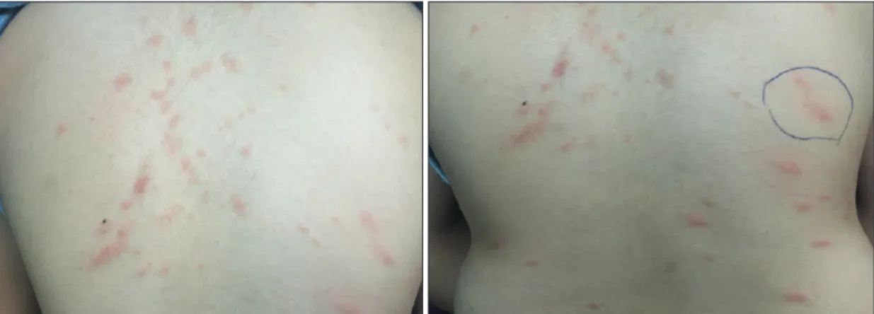
Observed mucocutaneous signs were classified as infectious dermatosis (mucocutaneous candidiasis, paronychia, tinea versicolor), inflammatory dermatoses (asteatotic eczema,

In addition to these, EMB should be considered if a patient has HF symp- toms accompanied with heart blocks or new ventricular arrhyth- mias &gt;3 weeks, rapidly progressive HF

In fact, we were still be able to show the increased aortic stiffness in metabolic syndrome patients compared to healthy controls despite the fact that aortic stiffness might

The study of post-stress test troponin T readings, in patients with stable angina pectoris and with negative stress test result, may be of great help in detecting especially

Cooperation status of the patient, advanced age, bleeding disorders, presence of underlying diseases (such as cirrhosis, ascites, or malignancy), biopsy experience of the

In 10 of 33 patients in the metabolic group, vitamin B12 deficiency was detected and its rate compared to the total patient number was 2.5%.. Parathyroid adenoma was iden- tified