Introduction
Androgenetic development (all paternal inheritance) in fish can be triggered by fertilising gamma, X-rays or UV irradiated eggs with normal spermatozoa, but the resultant embryos show inviable abnormality due to haploidy.
Viable diploid androgens can be produced by doubling the paternal chromosome set through suppression of the first cleavage using physical shocks such as temperature and pressure or by fertilisation of inactivated eggs with diploid spermatozoa from tetraploid males.
The applicable potential of androgenesis involves the rapid establishment of inbred lines for breeding programmes and research purposes (1,2), sex control using anticipated super male (YY) in the male heterogametic species (3,4), production of a nucleo-cytoplasmic hybrid between different species and recovery of genotypes from cryopreserved sperm, particularly for those which are facing extinction or the threat of contamination by hybridisation (1,5-7).
Haploid androgenesis has been induced using 60Co in the loach (Misgurnus anguillicaudatus) (1), flounder (Pleuronectes flesus) (2), masu salmon (Oncorhynchus masou) (8), rainbow trout (Oncorhynchus mykiss) (9) and brook trout (Salvelinus fontinalis) (10). Briedis and Elinson (11) induced haploid androgenetics in fertilised frog (Rana pipiens) eggs using pressure and deuterium oxide (D2O) to inhibit male pronucleus movement by the disruptive effects of microtubule-specific agents on pronuclear movement.
Studies with amphibians (12,13) showed that the transparency of the amphibian egg and the fact that the egg pronucleus is oriented toward the animal pole after fertilisation facilitated treatments with UV. However, the opacity of some fish eggs and the failure of the egg nucleus to demonstrate any particular orientation before or after fertilisation may present problems owing to the poor penetrance of UV (3,6). Despite these disadvantages, UV light has been successfully used in the irradiation of eggs from the white sturgeon (Acipenser
Optimisation of UV Treatment Duration to Induce Haploid
Androgenesis in the Nile tilapia (Oreochromis niloticus L.)
‹smihan KARAYÜCEL, Sedat KARAYÜCEL
University of Ondokuz May›s, Faculty of Fisheries, 57000 Sinop – TURKEY
Received: 16.11.2001
Abstract: The optimum UV duration times of eggs were examined in order to develop a simple and safe method for inducing
androgenetic development in the Nile tilapia, Oreochromis niloticus. The yield of androgenetic haploid O. niloticus to the pigmentation stage was 18.53 ± 5.3% (relative to controls) with an optimal UV irradiation dose of 540 Jm-2(at 150 µWcm-2) for 6 min. Most embryos developing after fertilisation with normal spermatozoa showed abnormal morphology and a haploid number of chromosomes (n = 22). The success of oocyte denucleation was also assessed by using the recessive “blond” skin pigmentation character.
Key Words: Androgenesis, chromosome manipulations, Nile tilapia, Oreochromis niloticus
Nil Tilapyas›nda (Oreochromis niloticus L.) Haploid Androgenesis Üretimi ‹çin UV Muamele Süresinin Optimizasyonu
Özet: Nil tilapyas›nda, Oreochromis niloticus, androgenetik geliflmeyi sa¤lamak için, basit ve güvenli metot gelifltirmek amac›yla,
yumurtalar›n optimum UV ›fl›nlar›na maruz b›rak›lma süreleri incelendi. Optimum UV irradizasyon dozu olan 540 Jm-2 de (150 µWcm-2 de) 6 dakika muamele, pigmentasyon safhas›nda % 18,53 ± 5,3 (kontrol grubuna oranla) androgenetik haploid O. niloticus üretmifltir. Normal spermatozoa ile döllenmeden sonra geliflen birçok embriyo abnormal morfoloji ve haploid kromozom say›s› göstermifltir. Yumurtalar›n çekirdekten mahrumiyetleri resesif ‘’sar›’’ deri pigmentasyon karakteri kullan›larak da de¤erlendirilmifltir. Anahtar Sözcükler: Androgenesis, kromozom manipulasyonlar›, Nil tilapyas›, Oreochromis niloticus
transmontanus) (14), common carp (Cyprinus corpio) (15,16), Nile tilapia (4), loach Misgurnus anguillicaudatus (17) and African catfish (Clarias gariepinus) (18). Furthermore, UV irradiation results in no residual fragments, in contrast to gamma irradiation, and it is easy to use anywhere, inexpensive and safer to apply (1,3,4,19,20). Therefore, optimisation of the intensity and the duration of irradiation is the first step for the successful production of haploid androgens. In the present study, we examined the optimum UV duration time of eggs in order to develop a simple and safe method for inducing androgenetic development in the Nile tilapia.
Materials and Methods
Origin of fish stock and their maintenance The O. niloticus brood stock used for this study were descended from an electrophoretically tested, pure stock of the Tilapia Reference Collection maintained at the Institute of Aquaculture, University of Stirling, Scotland (21). The origin of blond fish is described by Scott et al. (22), McAndrew et al. (23) and Hussain (24).
All fish were reared in recirculating freshwater systems. Lighting in all the systems was adjusted by an automatic timer to 12 h light and 12 h dark. The water temperature was maintained at 28 ± 1ºC. Individual female broodstock were kept in partitioned glass tanks of 120 cm X 44 cm X 30 cm. All tanks were aerated by airstones coupled to a low-pressure blower unit. All fish were fed with commercial trout feed (Trouw Aquaculture Nutrition, Russhive, UK) three times a dayad libitum.
Fish breeding, stripping and fertilisation of eggs Under aquarium conditions, mature females of O. niloticus spawn at approximately 2-6 week intervals. Females which are ready to spawn have a swollen urogenital papilla and show pre-spawning behaviour such as nest building and cleaning. After anaesthetising the female, the eggs were collected by applying gentle downward pressure with the fingers from below the pectoral fin to the genital opening of the fish. The eggs were collected in a clean, sterile Petri dish (100 mm in diameter) and were washed carefully with water from the recirculating system several times until ovarian fluid and any blood were removed. Then the eggs were sub-divided into a number of batches, as the experimental design required. Milt was also stripped from males in a similar way to egg collection using a glass capillary tube to collect
the milt, which was then put into a clean 1.5 ml microtube and stored at 4 ºC until use. Milt contaminated with water and urine was rejected.
Eggs were fertilised in vitro by mixing the milt with “dry” eggs and then 10-20 ml of aquarium water was added. The fertilised eggs were left in the Petri dish for 2-30 min for water hardening, washed, and transferred to a recirculated system for further development.
The embryos in each batch were checked and counted at four development stages: morula 6-8 h after fertilisation (a.f.); pigmentation 45-50 h a.f; hatching 80-90 h a.f. and yolk sac resorption 9-11 days a.f. Survival was calculated as (Number of embryos surviving at a given development stage / total number of eggs) x 100.
UV irradiation of eggs
UV irradiation of eggs was carried out according to Myers et al. (4). A 254 nm UV lamp (Ultra-Violet Products, San Gabriel, California) mounted on a camera copy stand was used for irradiation. UV treatments were standardised by placing 4 ml of unfertilised eggs (150-250 eggs) in a vial with enough filtered water to bring the total volume of eggs and water to 14-15 ml. The eggs in water were then poured into a glass Petri dish (75 mm in diameter) which was then placed on a stirrer. The distance between the lamp and Petri dish was adjusted to provide a dose of 150 µW/cm-2 using a radiometer (Ultra-Violet Products, San Gabriel, California).
All treated and untreated batches of eggs were incubated identically in a recirculated system.
Determination of ploidy
Fish metaphase chromosome spreads were prepared from newly hatched or one-day-old post-hatched larvae according to the original procedures described by Kligerman and Bloom (25), Chourrout and Itskovich (26) and Chourrout (20).
Experimental design for optimisation of UV duration time
Optimisation of UV duration time was carried out by irradiating six batches of eggs with UV light for 2, 4, 6, 8, 10 or 12 min and fertilising with sperm from blond tilapia males. This colour pattern was first reported by Scott et al. (22) and can be used as a visual marker to indicate the successful production of the haploid androgenetic fish because of a recessive “blond” skin pigmentation marker. A portion of the eggs was retained
as a control group and fertilised with sperm from the same blond male. Four different females and one blond male were used for this experiment.
Statistical analyses
Since the egg quality of each spawn varied greatly within and between females, the survival of each treatment was always calculated relative to the survival of their corresponding diploid control group. When the survival rate of the control group was less than 30%, that particular batch of eggs was not included (4). The data from the results of morula stages were transformed to arc-sine for statistical analyses and normality was tested by the Anderson-Darling Normality test and a test for homogeneity of variance was applied (27). Only the results of the morula stages were tested by one-way ANOVA since they were normally distributed. The other non-parametric data for pigmentation, hatching and yolk sac resorption stages, which included many zero values, were transformed to square root and tested by the Kruskal-Wallis Test (27,28). The results were presented as mean and standard error of mean ( ± SE). All statistical analyses were performed by Minitab 9.2 software.
Results
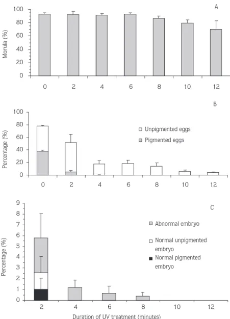
The effect of UV exposure for 2, 4, 6, 8, 10 and 12 min on the percentage of morula, the percentage of pigmented and unpigmented embryo and the percentage of abnormal embryos in the presumptive haploid “blond” androgenetic Nile tilapia are presented in the Table and Figure 1. All measurements are in relation to untreated controls.
At the morula stage, all the levels of fertilisation of the treatment groups and controls were quite similar and there were no significant differences between them (P > 0.05). As seen in Figure 1A, the fertilisation levels decreased with increased UV exposure time.
At the pigmentation stage, the survival of average pigmented (38.29 ± 1.18%) and unpigmented embryos (39.56 ± 0.97%), were not significantly different in the control group indicating that the females used in these experiments were heterozygous for the blond locus (Figure 1B). For 2 and 4 min UV duration, pigmented embryos were observed at a survival rate of 5.51 ± 1.93% and 0.22 ± 0.22%, respectively. Although the highest survival of unpigmented embryos (46.03 ± 13.6%) was obtained for 2 min UV duration, these
treatments yielded pigmented embryos showing only partial success with oocyte denucleation. A 6 min UV exposure time with a survival rate of 18.53 ± 5.3 % provided the best survival amongst the treatments giving only blond embryos. The survival rates of the treatment groups declined with increasing UV exposure time.
At the hatching stage, there were no significant differences between normal developed pigmented and unpigmented embryos with a survival rate of 32.14 ± 1.08% and 35.48 ± 2.10%, respectively, in the control group (P > 0.05) (Figure 1C). In the 2 min treatment group, survival rates of 1.01 ± 1.01%, 1.52 ± 1.52% and 3.24 ± 2.24% were observed in normally developed pigmented and unpigmented embryos and abnormal embryos, respectively. Only abnormal embryos were produced in 4, 6 and 8 min treatments while 10 and 12 min UV duration did not result in any hatched embryos. There were no significant differences between the treatments in terms of abnormality (P > 0.05). None of the UV treated embryos, including pigmented embryos, in the 4, 6, 8, 10 and 12 min treatments survived for more than a few days post-hatching.
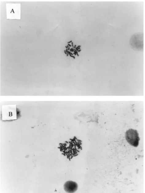
Analysis of some of the embryos by karyological examination (Figure 2) showed a typical single set of chromosomes (n = 22) (29).
Discussion
The present study indicates that UV irradiation successfully inactivated the nuclear DNA in Nile tilapia eggs. The yields of viable denucleated eggs to the pigmentation stage varied between 4.76 and 30.52% (relative to the controls) with a mean of 18.53 ± 5.3% for 6 min UV duration with a total dose of 540 Jm-2. The yield is comparable with that of 22.9 ± 1.6% in the Nile tilapia (4), 22% in the loach (30) and 22.5 ± 2.8% in the muskellungen (Esox masquinongy) (31). In the common carp, an optimal dose of 2500 Jm-2 produced 53.9% surviving haploids at hatching as well as a few biparental diploids (16). Bongers et al. (18) were able to produce higher numbers of androgenetic haploids (81% to hatching, relative to control) in African catfish using an optimum UV dose of 1250 Jm-2. Arai et al. (30) successfully produced 22% hatched androgenetic haploids in loach with a dose of 750 Jm-2. The yield of haploid androgenetic muskellunge was 22.5 ± 2.8% with optimal UV irradiation doses of 620-1320 Jm-2 (31). Marengoni and Onoue (32) obtained survival rates of
Table. The effect of UV exposure (at 150 µWcm-2) for 2, 4, 6, 8, 10 and 12 min on the morula, pigmentation and hatching of presumptive “blond” androgenetic haploid Nile tilapia, O. niloticus. % Relative control data are in parentheses. Common superscripts in the same column signify means which are not significantly different. *: Females were heterozygous for blond gene, R: Relative to controls.
Pigmentation stage Hatching stage
UV dose Experiment Morula Unpigmented Pigmented Normal developed Normal developed Abnormal (min) no. embryos embryos embryos pigmented embryos unpigmented embryos embryos
0 1* 98.02 37.62 39.60 30.20 30.69 0.99 2* 92.26 40.07 35.35 33.67 34.01 1.35 3* 90.94 38.51 37.54 34.30 36.57 1.29 4* 91.12 42.06 40.65 30.37 40.65 2.80 Mean 93.08 ± 1.67a 39.56 ± 0.97b 38.29 ± 1.18c 32.14 ± 1.08b 35.48 ± 2.10b 1.61 ± 0.41a 2 1* 96.66 (98.61) 63.59 (82.35) 6.28 (8.13) 2.51 (4.06) 3.77 (6.09) 5.86 (9.47) 2* 70.91 (76.86) 33.33 (44.20) 6.67 (8.84) 0.00 0.00 2.17 (3.51) 3* 88.44 (97.26) 12.54 (16.49) 0.32 (0.43) 0.00 0.00 0.00 4* 87.50 (96.03) 33.98 (41.08) 3.85 (4.65) 0.00 0.00 0.00 Mean 85.88 ± 5.40 35.86 ± 10.50 4.28 ± 1.50 0.63 ± 0.63 0.94 ± 0.94 2.00 ± 1.38 Mean (R) 92.19 ± 5.14a 46.03 ± 13.6b 5.51 ± 1.93b 1.01 ± 1.01a 1.52 ± 1.52a 3.24 ± 2.24a 4 1* 93.31 (95.19) 11.48 (14.87) 0.00 0.00 0.00 1.44 (2.32) 2* 78.41 (84.99) 20.60 (27.31) 0.66 (0.88) 0.00 0.00 1.66 (2.41) 3* 80.91 (89.00) 4.37 (5.75) 0.00 0.00 0.00 0.00 4* 87.50 (96.03) 19.68 (23.80) 0.00 0.00 0.00 0.00 Mean 85.03 ± 3.36 14.03 ± 3.82 0.17 ± 0.17 0.00 0.00 0.78 ± 0.45 Mean (R) 91.30 ± 2.62a 17.93 ± 4.83ab 0.22 ± 0.22a 0.00 0.00 1.18 ± 0.69a 6 1* 92.55 (94.42) 14.36 (18.60) 0.00 0.00 0.00 0.00 2* 82.74 (89.68) 23.02 (30.52) 0.00 0.00 0.00 1.80 (2.61) 3* 82.24 (90.43) 3.62 (4.76) 0.00 0.00 0.00 0.00 4* 89.73 (98.48) 16.73 (20.23) 0.00 0.00 0.00 0.00 Mean 86.82 ± 2.56 14.43 ± 4.04 0.00 0.00 0.00 0.45 ± 0.45 Mean (R) 93.25 ± 2.02a 18.53 ± 5.3ab 0.00 0.00 0.00 0.65 ± 0.65a 8 1* 81.10 (82.74) 7.20 (9.32) 0.00 0.00 0.00 0.00 2* 72.32 (78.39) 19.72 (26.15) 0.00 0.00 0.00 1.04 (1.50) 3* 79.83 (87.78) 4.04 (5.31) 0.00 0.00 0.00 0.00 4* 87.96 (96.53) 14.38 (17.39) 0.00 0.00 0.00 0.00 Mean 80.30 ± 3.20 11.34 ± 3.53 0.00 0.00 0.00 0.26 ± 0.26 Mean (R) 86.36 ± 3.89a 14.54 ± 4.62a 0.00 0.00 0.00 0.38 ± 0.38a 10 1* 68.96 (70.35) 4.51 (5.84) 0.00 0.00 0.00 0.00 2* 79.33 (85.98) 3.39 (4.49) 0.00 0.00 0.00 0.00 3* 65.58 (72.11) 1.31 (1.72) 0.00 0.00 0.00 0.00 4* 81.93 (89.91) 9.35 (11.30) 0.00 0.00 0.00 0.00 Mean 76.45 ± 5.93 4.64 ± 1.70 0.00 0.00 0.00 0.00 Mean (R) 79.59 ± 4.91a 5.83 ± 2.01a 0.00 0.00 0.00 0.00 12 1* 68.12 (69.50) 3.18 (4.12) 0.00 0.00 0.00 0.00 2* 74.83 (81.11) 5.86 (7.77) 0.00 0.00 0.00 0.00 3* 31.10 (34.20) 0.79 (1.04) 0.00 0.00 0.00 0.00 4* 86.40 (94.82) 2.94 (3.56) 0.00 0.00 0.00 0.00 Mean 65.11 ± 11.96 3.19 ± 1.04 0.00 0.00 0.00 0.00 Mean (R) 69.91 ± 12.98a 4.12 ± 1.38a 0.00 0.00 0.00 0.00
57.6% and 55.8%, and 57.1% and 56.0% (relative to controls) in androgenetic haploid O. aureus and O. niloticus, respectively, at total UV doses of 594 and 693 Jm-2, respectively.
By using the recessive “blond” skin pigmentation character in spermatozoa, it was possible to assess whether oocyte denucleation was successful. All haploid embryos showed non-pigmentation under optimal UV irradiation of 5-8 min whereas 2 min and 4 min UV irradiation produced some pigmented embryos. The blond colour variant was also used successfully by Myers et al. (4) in the production of androgenetic haploid tilapia and they also observed some pigmented embryos in the same UV irradiation treatments as the present study.
Pigmented embryos were observed in the 2 and 4 min UV treatments and blond embryos showed aberrant development. Therefore, to ensure that host eggs are totally denucleated, the UV treatment should be at least 5 min to 8 min at 150 µWcm-2or a total dose of between 450 Jm-2 and 720 Jm-2. Myers et al. (4) reported that variable sensitivity to UV irradiation from species to species could be explained by differences in the thickness, composition and optical qualities of egg chorion, egg size and shape, and the relative position of the female pronucleus.
It was concluded that for successful haploid induction, the UV duration time has to be at least 5 min which will ensure total degradation of the egg nucleus.
0 20 40 60 80 100 0 2 4 6 8 10 12 Morula (%) A 0 20 40 60 80 100 0 2 4 6 8 10 12 Percentage (%) Unpigmented eggs Pigmented eggs B 0 1 2 3 4 5 6 7 8 9 2 4 6 8 10 12
Duration of UV treatment (minutes)
Percentage (%) Abnormal embryo Normal unpigmented embryo Normal pigmented embryo C
Figure 1. Percentage of embryos observed at morula (A), pigmentation (B) and hatching (C) of presumptive “blond” androgenetic haploid Nile tilapia, O. niloticus, (% relative to the diploid control) subjected to 150 µWcm-2intensity.
Figure 2. Metaphase chromosome spread of A. androgenetic haploid (N=22) and B. diploid (N=44) embryos of O. niloticus.
References
1. Ihssen, P.E., McKay, L.R., McMillan, I., Phillips, R.B.: Ploidy manipulation and gynogenesis in fishes. Trans. Am. Fish. Soc. 1990; 119: 698-717.
2. Purdom, C.E.: Radiation-induced gynogenesis and androgenesis in fish. Heredity. 1969; 24: 431-444.
3. Thorgaard, G.H.: Chromosome set manipulation and sex control in fish. In: Fish Physiology (eds. Hoar, W.S., Randall, D.J. and Donaldson, E.M.). Vol. IX-B. Academic Press, New York. 405-434; 1983.
4. Myers, J.M., Penman, D.J., Basavaraju, Y., Powell, S.F., Baoprasertkul, P., Rana, K.J., Bromage, N., McAndrew, B.J.: Induction of diploid androgenetic and mitotic gynogenetic Nile tilapia (Oreochromis niloticus L.). Theor. Appl. Genet. 1995; 90: 205-210.
5. Stoss, J.: Fish gamete preservation and spermatozoan physiology. In: Fish Physiology (eds. Hoar, W.S., Randall, D.J and Donaldson, E.M.). Vol 9(B) Academic Press, New York. 305-350; 1983. 6. McAndrew, B.J., Rana, K.J., Penman, D.J.: Conservation and
preservation of genetic variation in aquatic organisms. In: Recent Advances in Aquaculture (Eds. R.J. Roberts and J. Muir). Vol IV, Blackwell Sci. Publ. 295-337; 1993.
7. Mair, G.C.: Chromosome-set manipulation in tilapia - techniques, problems and prospects. Aquaculture. 1993; 111: 227-244. 8. Arai, K., Onozato, H., Yamazaki, F.: Artificial androgenesis induced
with gamma irradiation in masu salmon, Oncorhynchus masou. Bull. Fac. Fish. Hokkaido Univ. 1979; 30: 181-186.
9. Parsons, J.E., Thorgaard,G.H.: Production of androgenetic diploid rainbow trout. J. Heredity. 1985; 76: 177-181.
10. May, B., Henley, K.J., Krueger, C.C., Gloss, S.P.: Androgenesis as a mechanism for chromosome set manipulation in brook trout (Salvelinus fontinalis). Aquaculture. 1988; 75: 57-70.
11. Briedis, A., Elinson, R. P.: Suppression of male pronuclear movement in frog eggs by hydrostatic pressure and deuterium oxide yields androgenetic haploids. J. Exp. Zool. 1982; 222: 45-57.
12. Gillespie, L.L., Armstrong, J.B.: Production of androgenetic diploid axolotls by suppression of first cleavage. J. Exp. Zool. 1980; 201: 423-425.
13. Gillespie, L.L., Armstrong, J.B.: Suppression of first cleavage in the Mexican axolotl (Ambystoma mexicanum) by heat shock or hydrostatic pressure. J. Exp. Zool. 1981; 218: 441-445. 14. Kowtal, G.V.: Preliminary experiments in induction of polyploidy,
gynogenesis, and androgenesis in the white sturgeon, Acipenser transmontanus Richardson. In: Proceedings of World Symposium on Selection, Hybridization and Genetic Engineering in Aquaculture (Ed. K. Tiews). Bordeaux, Vol. II, Heenemann, Berlin. 317-324; 1987.
15. Bongers, A.B.J., Eding, E.H., Richter, C.J.J.: Androgenesis in common carp, Cyprinus carpio L. Proceedings of International Workshop on Genetics in Aquaculture and Fisheries Management. 1993; 73-76.
16. Bongers, A.B.J., Veld, E.P.C., Abo Hashema, K., Bremmer, I.M., Eding, E.H., Komen, J., Richter, C.J.J.: Androgenesis in common carp (Cyprinus carpio L.), using UV-irradiation in a synthetic ovarian fluid and heat shocks. Aquaculture. 1994; 122: 119-132. 17. Arai, K., Masaoka, T., Suzuki, R.: Optimum conditions of UV ray irradiation for genetic inactivation of loach eggs. Nippon Suisan Gakkaishi. 1992; 58 (7): 1197-1201.
18. Bongers, A.B.J, Nguenga, D., Eding, E.H., Richter, C.J.J.: Androgenesis in the African catfish, Clarias gariepinus. Aquat. Liv. Res. 1995; 8: 329-332.
19. Chourrout, D.: Pressure induced retention of second polar body and suppression of first cleavage in rainbow trout: production of all-triploids, all-tetraploids, and heterozygous and homozygous diploid gynogenetics. Aquaculture. 1984; 36: 111-126.
20. Chourrout, D.: Techniques of chromosome manipulation in rainbow trout: a new evaluation with karyology. Theor. Appl. Genet. 1986; 72: 627-632.
21. McAndrew, B.J., Majumdar, K.C.: Tilapia stock identification using electrophoretic markers. Aquaculture. 1983; 30: 249-261. 22. Scott, A.G., Mair, G.C., Skibinski, D.O.F., Beardmore, J.A.: “Blond”
a useful genetic marker in the Oreochromis niloticus L. Aquacult. Fish. Manage. 1987; 18: 159-165.
23. McAndrew, B.J., Roubal, F.R., Roberts, R.J., Bullock, A.M., McEwen, I.M.: The genetics and histology of red, blond and associated colour variants in Oreochromis niloticus. Genetica. 1988; 76: 127-137.
24. Hussain, M.G.: Genetic manipulation studies in Oreochromis niloticus L. Ph. D. Thesis, Institute of Aquaculture, University of Stirling, U.K. 1994.
25. Kligerman, A.D., Bloom, S.E.: Rapid chromosome preparation from solid tissues of fishes. J. Fish. Res. Board Can. 1977; 34 : 266-269.
26. Chourrout, D., Itskovich, J.: Three manipulations permitted by artificial insemination in tilapia: induced diploid gynogenesis, production of all triploid population and intergeneric hybridisation. In: International Symposium on Tilapia in Aquaculture. (Eds. L. Fishelson and Z. Yaron) Nazareth, Israel. 246-255; 1983. 27. Sokal, R.R., Rohlf, F.J.: Introduction to Biostatistics. W.H.
Freeman and Company. 1987.
28. Gardiner, W.P.: Statistics for the Bioscience. Prentice Hall Europe, Great Britain. 1997.
29. Majumdar, K.C., McAndrew, B.J.: Relative DNA content of somatic nuclei and chromosomal studies in three genera, Tilapia, Sarotherodon, and Oreochromis of the tribe Tilapiini (Pisces, Cichlidae). Genetica. 1983; 68: 165-188.
30. Arai, K., Ikeno, M., Suzuki, R.: Production of androgenetic diploid loach Misgurnus anguillicaudatus using spermatozoa of natural tetraploids. Aquaculture. 1995; 137: 131-138.
31. Lin, F., Dabrowski, K.: Androgenesis and homozygous gynogenesis in muskellunge (Esox masquinongy): evaluation using flow cytometry. Mol. Rep. Dev. 1998; 49: 10-18.
32. Marengoni, N.G., Onoue, Y.: Ultraviolet-induced androgenesis in Nile tilapia, Oreochromis niloticus (L.) and hybrid Nile X blue tilapia, O. aureus (Steindachner). Aqua. Res. 1998; 29: 359-366.