Detection of exfoliative toxin in Staphylococcus spp. isolated from
skins of healthy dogs and dogs with skin lesions
*H. Kaan MUSTAK1, Ömer M. ESENDAL2
1Central Veterinary Control and Research Institute, Breeding Disease Laboratory, 2Department of Microbiology, Faculty of
Veterinary Medicine, University of Ankara, Ankara.
Summary: The aim of this study was to determine the presence of exfoliative toxin in Staphylococcus species isolated from nose and skins of 50 healthy dogs and skin lesions of 50 dogs at different race, age and gender; and to investigate the nasal carriage of Staphylococcus species in dogs. A total of 135 Staphylococcus intermedius (67.5 %), 12 S. aureus (6 %), 2 S. chromogenes (1 %) and 1 S. capitis (0.5 %) were isolated from 200 skin and nose samples. No bacterial growth was observed in 50 (25 %) samples. Among the isolated staphylococci, exfoliative toxin was not determined in 2 S. chromogenes and 1 S. capitis isolates but it was determined phenotypically in 54 S. intermedius isolates and both phenotypically and genotypically in 12 S. aureus isolates. As a result; it was determined that S. intermedius and S. aureus strains isolated in this study produced exfoliative toxin and because of their high isolation rates the opinion of their role in the etiology of canine pyoderma and importance of nasal carriage was supported. In this study the Staphylococcus spp. isolated from both skin and nasal mucosa samples of the same dog were compared for the first time and also it was determined that S. aureus strains isolated from dog skin produced exfoliative toxin for the first time.
Key words: Dog, exfoliative toxin, isolation, pyoderma, Staphylococcus spp.
Sağlıklı ve deri lezyonlu köpeklerin derilerinden izole edilen stafilokok türlerinde eksfoliatif toksin varlığının belirlenmesi
Özet: Bu çalışmada farklı ırk, yaş ve cinsiyetteki 50 sağlıklı ve 50 deri lezyonu bulunan köpeğin burun ve derilerinden izole edilen stafilokok türlerinde eksfoliatif toksin varlığının belirlenmesi, izole edilen stafilokok türlerinin köpeklerdeki burun boşluğunda etken taşıyıcılığının araştırılması amaçlandı. Toplanan 200 deri ve burun örneğinden 135’inde S. intermedius (% 67.5), 12’sinde S.
aureus (% 6), 2’sinde S. chromogenes (% 1) ve 1’inde S. capitis (% 0.5) saptanırken 50 (% 25) örnekte üreme olmadığı görüldü.
İzole edilen stafilokoklardan, 2 S. chromogenes ve 1 S. capitis izolatında eksfoliatif toksin varlığı belirlenemedi ancak 54 S.
intermedius izolatının fenotipik olarak, 12 S. aureus izolatının ise hem fenotipik hem de genotipik olarak eksfoliatif toksin ürettiği
saptandı. Sonuç olarak, bu çalışmada izole edilen S. intermedius ve S. aureus’ların eksfoliatif toksin sentezleyebildikleri gösterilmiş olup yüksek izolasyon oranlarından dolayı kanin piyodermaların etiyolojisinde rol aldıkları ve burun boşluğunda etken taşıyıcılığının önemli olduğu görüşü desteklendi. Bu çalışmada ilk defa, aynı köpeğin hem derisinden hem de burun mukozasından örnek alınarak üreyen stafilokok türleri karşılaştırılmış ve yine ilk defa bu çalışmada, köpek derisinden izole edilen S. aureus suşlarının eksfoliatif toksin sentezlediği belirlenmiştir.
Anahtar sözcükler: Eksfoliatif toksin, izolasyon, köpek, piyoderma, Staphylococcus spp.
* Summarized from the PhD dissertation of the first author. The study was carried out in accordance with the ethical rules and
recommendations.
Introduction
Staphylococci are commensally found on mucosal surfaces and skin. However, they also emerge as causative agents of various diseases in animals (8). S.
aureus (7, 9), S. schleiferi (4, 17), S. epidermidis (27), S. hyicus, S. xylosus, S. simulans (18) and S. intermedius
(23) are the most important Staphylococcus species isolated from skin of dogs. Within the Staphylococcus species, S. intermedius is the most frequently isolated species from both healthy dogs and dogs with skin lesion. Previous studies have shown that oral cavity,
nasal cavity, perineum region and anus are the reservoir zones for S. intermedius (3, 5, 7).
Staphylococci have various exoproteins in order to colonize and cause disease in their hosts. S. aureus is the mostly investigated species among the Staphylococci and its exfoliative toxin within pathogenicity factors, plays a vital role in the etiology of Staphylococcal Scalded Skin Syndrome (SSSS), characterized by peeling of the superficial layer of the skin. S. aureus produces four antigenically distinct exfoliative toxin serotypes. Exfoliative toxin A (ETA) and B (ETB) are the serotypes
responsible from SSSS that is seen in humans. ETA which has 26 950 Da molecular mass and 246 aminoacid is a heat-stable toxin, whereas ETB is heat-labile and has 27 274 Da molecular mass and 246 aminoacid (14). Furthermore, S. aureus produces exfoliative toxin types of C (ETC) and D (ETD). ETC which is heat-labile and has a molecular mass of 27 kDa has been isolated from a horse with phlegmone by Sato et al. (25). In addition to this, ETD was found during comparative studies on genomes of S. aureus strain TY114 and N315 (21, 30).
S. intermedius is known as the causative agent of
canine pyoderma which is usually a superficial pyoderma characterized by erythema, vesicle formation, yellowish exudates, yellow-brown crusts and loss of the horny layer of the epidermis (7, 9). Such clinical findings resemble those of SSSS in humans and of exudative epidermitis (EE) in pigs (6, 19, 29). Therefore, it has been speculated that S. intermedius also produces an exfoliative toxin as the virulence factor in the pathogenesis of canine pyoderma (6, 29).
Depending on similar clinical findings in SSSS and canine pyoderma, Terauchi et al. (29) proposed that S.
intermedius may have a toxin similar to that of S. aureus
exfoliative toxin and they achieved to isolate S.
intermedius exfoliative toxin (SIET) which has a
molecular mass of 30 kDa and serologically distinct from ETA, ETB and ETC. Studies up to now showed that within the staphylococci isolated from animals only S.
intermedius from dogs, and S. chromogenes (24) and S. hyicus (13) from pigs produced exfoliative toxin.
In this study, the aim was to determine the presence of exfoliative toxin in Staphylococcus species isolated from nose and skins of healthy dogs and dogs with skin lesions; and to investigate the nasal carriage of
Staphylococcus species in dogs.
Materials and Methods
Bacterial strains: In this research, 100 skin and 100
nasal swab samples were collected from 50 healthy dogs (with no skin lesion) and 50 dogs with skin lesion (suspect of pyoderma) by applying swabs on nasal mucosa and skin. Swab samples were plated on blood agar-base containing 5-7 % ovine blood at 37°C for 24 h. Gram positive, coccoid bacteria were subjected to catalase, coagulase and DNase tests. Catalase-positive-bacteria were evaluated with Microbact Staphylococcal 12S Identification System (Oxoid MB 1561). The bacteria were then suspended in 20 % glycerol and stored at -80°C until used. ETA and ETB positive S. aureus strains and SIET positive S. intermedius strain were obtained from Prof. Dr. Christoph Lammler, Giessen University, Germany.
Isolation of the exfoliative toxin: Staphylococcus
strains cultured on Heart Infusion Agar were suspended in Dulbecco’s phosphate buffered saline without CaCl2
and MgCl2 at a concentration of 109 CFU/ml. A 2 ml
portion of this suspension was inoculated in 200 ml of TY broth [Yeast extract 10 g, Trypticase 17 g, NaCl 5 g, K2HPO4 2.5 g, distilled water 1000 ml (pH= 7.1 ± 0.2)
(12)] and cultured at 37°C for 18 h with shaking at 75 oscillations/min. After the incubation, 40 ml of bacterial culture was centrifuged at 8000 rpm for 20 min at 4°C and the culture supernatant was passed through a membrane filter. Then the culture filtrates were lyophilized and dissolved in 10 mM Tris-HCl buffer (pH 7.5). The solution was designated as concentrated culture filtrate (24, 26, 28, 29).
Inoculation of experimental animals: In order to
determine the presence of exfoliative toxin in-vivo and to understand the biologic effect of the toxin, one-day-old conventional chickens [White Leghorn (Ross 308)] were used. Five hundred micrograms of concentrated culture filtrate was injected subcutaneously into the abdominal region of chickens. The exfoliative activity was regarded as positive when the Nikolsky signs described as peeling off the normal appearing skin surface by rubbing with fingertips were detectable within 3 h of injection (19, 29).
Cell culture assay: In order to show the rounding
effects of isolated exfoliative toxin on Vero cells, cell culture inoculation was performed as described previously (22). A 100 µl portion of each culture filtrate was diluted and added to Vero cell monolayers in 96-well microplates. After incubation at 37°C for 48 h, the wells were examined microscopically.
Histopathological examination: Skin samples of
experimental chickens were processed routinely and examined histopathologically (16).
Sodium dodecyl sulphate/polyacrylamid gel electrophoresis (SDS/PAGE): The SDS/PAGE was
performed at room temperature by the method of Laemmli (15). In order to evaluate the protein bands appeared on the gel, 18-105 kDa molecular weight standarts were used and the bands were compared with the band magnitudes of previous studies.
Polymerase chain reaction (PCR): Extraction of
DNA from isolated staphylococcus species was carried out by the method of Ardıç et al. (1, 2). Primers (MGV BIOTECH, Germany) used to amplify the specific segments of 119 and 200 bp of ETA and ETB genes, respectively, are shown in Table 1. PCR total reaction volume was 50 µl and the final concentration: 50 mM potassium chloride-10 mM Tris chloride (ph 8.3); 1.5 mM magnesium chloride; 0.01 % gelatine; 200 µM for each dATP, dCTP, dGTP, dTTP; 10 pmol for each primer and 2.5 U DNA polymerase (Taq polymerase, Fermentas, Latvia) was prepared. DNA amplification consisted of 30 cycles was carried out with the following thermal cycling profile: an initial denaturation at 94°C for 2 min, annealing at 55°C for 2 min, and extension at 72°C for 1 min (11). Amplified products were resolved by 1.5 % agarose (Prona, EU) gel electrophoresis at 90 V for 60 min.
Table 1. Primers that specifically amplify exfoliative toxin genes of S. aureus (11).
Tablo 1. S. aureus eksfoliatif toksin genlerini spesifik olarak amplifiye eden primerler (11).
Primers Sequence (5’-3’) product PCR (bp)
eta
ETA-1
ETA-2 CTA GTG CAT TTG TTA TTC AA TGC ATT GAC ACC ATA GTA CT 119
etb
ETB-1 ETB-2
ACG GCT ATA TAC ATT CAA TT TCC ATC GAT AAT ATA CCT AA
200
Results
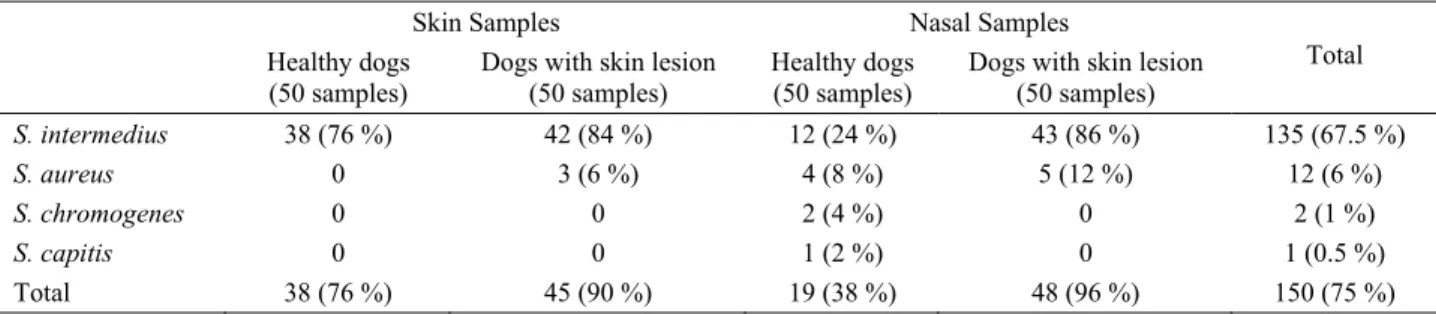
Isolation and identification: Staphylococcus species
that were isolated from nares and skin of healthy dogs and dogs with skin lesions are shown in Table 2 with
isolation ratios. Skin and nasal isolation results of
Staphylococcus species that were isolated from healthy
dogs and dogs with skin lesions are comparatively shown in Figure 1.
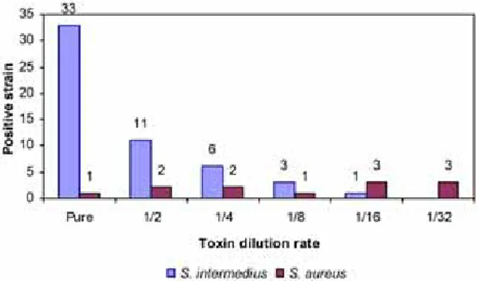
Cell culture assay: It was determined that all of the
examined culture filtrates of 54 S. intermedius and 12 S.
aureus had toxic effects on Vero cells. On the other hand,
culture filtrates of S. chromogenes and S. capitis had no effects on the same cells (Table 3). Toxic effects of bacteria determined on cell culture are shown in Figure 2 according to their toxin dilution steps. The basic microscobic finding of exfoliative toxin in culture filtrate was determined as the lysis of the cells. In the process of cell lysis, main toxicity step was noted as caryorexis (disintegration of the nucleus) and hyperchromatic cell development. Intracytoplasmic granulation and vacuolisation of cells were also seen in this period.
Table 2. Staphylococcus species isolated from skin and nasal samples and isolation ratios. Tablo 2. Deri ve burun örneklerinden izole edilen stafilokok türleri ve izolasyon oranları.
Skin Samples Nasal Samples
Healthy dogs
(50 samples) Dogs with skin lesion (50 samples) Healthy dogs (50 samples) Dogs with skin lesion (50 samples)
Total S. intermedius 38 (76 %) 42 (84 %) 12 (24 %) 43 (86 %) 135 (67.5 %) S. aureus 0 3 (6 %) 4 (8 %) 5 (12 %) 12 (6 %) S. chromogenes 0 0 2 (4 %) 0 2 (1 %) S. capitis 0 0 1 (2 %) 0 1 (0.5 %) Total 38 (76 %) 45 (90 %) 19 (38 %) 48 (96 %) 150 (75 %)
Table 3. Methods used for determination of exfoliative toxin production in isolated Staphylococcus species and obtained data. n, strain; -, not done.
Tablo 3. İzole edilen stafilokok türlerinde, eksfoliatif toksin üretiminin belirlenmesi için kullanılan yöntemler ve elde edilen veriler. n, suş; -, yapılmadı
Applied method
Animal assay Histopathology Cell culture PCR
Positive Negative Bacteria Isolation region Positive Negative Positive Negative Positive Negative
eta etb eta & etb
Lesion (n=42) 42 0 42 0 42 0 0 0 42 Skin Healthy (n=4) 4 0 4 0 4 0 0 0 4 Lesion (n=4) 4 0 4 0 4 0 0 0 4 S. intermedius Nares Healthy (n=4) 4 0 4 0 4 0 0 0 4 Lesion (n=3) 0 3 - - 3 0 0 3 3 eta Skin Healthy (n=0) - - - - Lesion (n=5) 0 5 - - 5 0 0 5 5 eta S. aureus Nares Healthy (n=4) 0 4 - - 4 0 0 4 4 eta Lesion (n=0) - - - - Skin Healthy (n=0) - - - - Lesion (n=0) - - - - S. chromogenes Nares Healthy (n=2) 0 2 0 2 0 2 0 0 2 Lesion (n=0) - - - - Skin Healthy (n=0) - - - - Lesion (n=0) - - - - S. capitis Nares Healthy (n=1) 0 1 0 1 0 1 0 0 1
Following this, apoptic particles were seen on the hyperchromatic cell surface due to chromosome fragmentation and finally seperation of cells from the colony and cell lysis were determined.
Figure 1. Number of animals according to the Staphylococcus species isolated from skin and nose. Skin/Nares isolation combinations: a, S. intermedius/S. intermedius; b, S.
intermedius/No growth; c. S. intermedius/S. capitis; d, S. intermedius/S. chromogenes; e, S. intermedius/S. aureus; f, No
growth/No growth; g, No growth/S. intermedius; h, S. aureus/S.
intermedius; i, No growth/S. aureus.
Şekil 1. Deri ve burundan izole edilen stafilokok türlerine göre hayvan sayıları. Deri/Burun izolasyon kombinasyonları: a, S.
intermedius/S. intermedius; b, S. intermedius/Üreme yok; c. S. intermedius/S. capitis; d, S. intermedius/S. chromogenes; e, S. intermedius/S. aureus; f, Üreme yok/Üreme yok; g, Üreme
yok/S. intermedius; h, S. aureus/S. intermedius; i, Üreme yok/S.
aureus.
Figure 2. The toxic effects of diluted culture filtrates of 12 S.
aureus and 54 S. intermedius isolated from healthy dogs and
dogs with skin lesions on cell cultures.
Şekil 2. Sağlıklı ve deri lezyonlu köpeklerden izole edilen 12 adet S. aureus ve 54 adet S. intermedius’un kültür filtratı sulandırmalarının hücre kültürü üzerindeki toksik etkileri.
Histological examination: Typical subcorneal
splitting of the epidermis was accepted as the positive result. Histological results of isolated species are shown in Table 3.
SDS/PAGE: Specific bands of 30 kDa and 27 kDa
were observed in 54 S. intermedius and 12 S. aureus isolates, respectively.
PCR: After agarose gel electrophoresis specific
bands (200 bp size) were detected only for etb in 12 S.
aureus template DNA (Table 3).
Discussion and Conclusion
It is necessary to understand the role of Staphylococci in canine pyoderma because of gaining resistance to antibiotics and transmission to humans from their own pets. Isolation of exfoliative toxin frequently from Staphylococci isolated from dog skin and nares showes the toxin may be effective on canine pyoderma like SSSS seen in humans either because of its importance to understand the mechanism of exfoliative toxin and its target tissue exactly.
In this study, the Staphylococcus spp. isolated from both skin and nasal mucosa samples of the same dog were compared for the first time. S. intermedius was isolated from both skin and nasal mucosa of 36 dogs with skin lesions but only in 10 of healthy dogs. However, according to the results shown in Figure 1, it was seen that the highest isolation rate belonged to this combination (combination a). Statistical evaluation of the results have shown that the rate of Staphylococcus isolation from nasal mucosa of dogs with skin lesion was 37,5 times higher than the isolation rates from nasal mucosa of healthy dogs and the difference was found statistically significant (P < 0.001). This result puts forward the hypothesis that the increase of S. intermedius amount in nasal mucosa for any reason has an effect on skin infections in dogs due to Staphylococcus spp. When compared with other Staphylococcus species, higher isolation rates of S. intermedius from nasal mucosa of healthy dogs and dogs with skin lesion supports the opinion of Devriese and De Pelsmaecker (3) that nasal region is the main reservoir for S. intermedius.
In this study, 54 S. intermedius, 12 S. aureus, 2 S.
chromogenes and 1 S. capitis were isolated from skin and
nasal mucosa of 50 healthy dogs and 50 dogs with skin lesions which were investigated for the presence of exfoliative toxin. There was no evidence for S.
chromogenes and S. capitis that they produce exfoliative
toxin individually but all S. intermedius isolates were shown to produce exfoliative toxin with phenotypic methods such as animal assay, cell culture (different dilutions), SDS-PAGE and histophatologic investigation. However, genotypic evaluation of the S. intermedius isolates were found to be negative for eta and etb genes since the primers used for PCR were specific for S.
aureus. Therefore, it is clear that a new specific primer
design is necessary for determination of S. intermedius
siet gene.
It has been well documented that S. aureus isolated from human SSSS patients can produce ETA and ETB but it has not been cleared up that whether animal isolates could produce ETA and ETB or not. In this research, 12 S. aureus isolates gave negative results in animal assays but positive results in cell cultures. Similarly, negative results were also observed in previous investigations in which exfoliation effect of S.
aureus were found to be positive (determination of
Nikolsky sign) only in one day old suckling mice (10, 12, 13, 20). An observation of a 27 kDa protein band in SDS-PAGE and 200 (etb) bp band in PCR products on agarose gel electrophoresis showed the presence of ETB at protein and gene level, but, no evidence was found for the presence of ETA in 12 S. aureus strains. According to the results, it was determined for the first time that S.
aureus strains isolated from the skin of dogs produced
exfoliative toxin.
Also it can be said that S. intermedius is the most frequently isolated Staphylococci from skin of dogs and an increase in the isolation rate of S. intermedius from nasal mucosa of dogs with skin lesion indicates that nasal carriage is an important factor for staphylococcal skin infections.
References
1. Ardıc N, Özyurt M, Sareyyüpoğlu B, Haznedaroğlu T (2005): Investigation of erythromycin and tetracycline
resistance genes in methicillin-resistant staphylococci. Int
J Antimicrob Agents, 26, 213-218.
2. Ardıc N, Sareyyüpoğlu B, Özyurt M, Haznedaroğlu T, Ilga U (2006): Investigation of aminoglycoside modifying
enzyme genes in methicillin-resistant staphylococci.
Microbiol Res, 161, 49-54.
3. Devriese LA, De Pelsmaecker K (1987): The anal region
as a main carrier site of Staphylococcus intermedius and Streptococcus canis in dogs. Vet Rec, 121, 302-303.
4. Frank LA, Kania SA, Hnilica KA, Wilkes RP, Bemis DA (2003): Isolation of Staphylococcus schleiferi from
dogs with pyoderma. J Am Vet Med Assoc, 222, 451-454.
5. Hartmann FA, White DG, West SEH, Walker RD, Deboer DJ (2005): Molecular characterization of
Staphylococcus intermedius carriage by healthy dogs and comparison of antimicrobial susceptibility patterns to isolates from dogs with pyoderma. Vet Microbiol, 108,
119-131.
6. Hesselbarth J, Flachsbarth MF, Amtsberg G (1994):
Studies on the production of an exfoliative toxin by Staphylococcus intermedius. J Vet Med B, 41, 411-416.
7. Hill PB, Moriello KA (1994): Canine pyoderma. J Am Vet Med Assoc, 204, 334-340.
8. Hoekstra KA, Paulton RJL (2002): Clinical prevalence
and antimicrobial suspceptibility of Staphylococcus aureus and Staph. intermedius in dogs. J Appl Microbiol, 93,
406-413.
9. Ihrke PJ (1987): An overview of bacterial skin disease in
the dog. Br Vet J, 143, 112-118.
10. Johnson AD, Spero L, Cades JS, de Cicco BT (1979):
Purification and characterization of different types of exfoliative toxin from Staphylococcus aureus. Infect
Immun, 24, 679-684.
11. Johnson WM, Tyler SD, Ewan EP, Ashton FE, Pollard DR, Rozee KR (1991): Detection of genes for
enterotoxins, exfoliative toxins, and toxic shock syndrome toxin 1 in Staphylococcus aureus by the polymerase chain reaction. J Clin Microbiol, 29, 426-430.
12. Kapral FA, Miller MM (1971): Product of
Staphylococcus aureus responsible for the scalded-skin syndrome. Infect Immun, 4, 541-545.
13. Ladhani S (2003): Understanding the mechanism of
action of the exfoliative toxins of Staphylococcus aureus.
FEMS Immunol Med Microbiol, 39, 181-189.
14. Ladhani S, Joannou CL, Lochrie DP, Evans RW, Poston SM (1999): Clinical, microbial and biochemical
aspects of the exfoliative toxins causing staphylococcal scalded skin syndrome. Clin Microbiol Rev, 12, 224-242.
15. Laemmli UK (1970): Cleavage of structural proteins
during the assembly of the head of bacteriophage T4.
Nature (London), 227, 680-685.
16. Luna LG (1968): Manual of Histologic Staining Methods
of The Armed Forces Institute of Pathology 3rd edition.
New York: The Blakiston Division, McGraw-Hill Book Company.
17. May ER, Hnilica KA, Frank LA, Jones RD, Bemis DA (2005): Isolation of Staphylococcus schleiferi from healthy
dogs and dogs with otitis, pyoderma, or both. J Am Vet
Med Assoc, 227, 928-931.
18. Medleau L, Long RE, Brown J, Miller WH (1986):
Frequency and antimicrobial susceptibility of Staphylococcus species isolated from canine pyodermas.
Am J Vet Res, 47, 229-231.
19. Melish ME, Glasgow LA (1970): The staphylococcal
scalded skin syndrome: development of an experimental model. N Engl J Med, 282, 1114-1119.
20. Piemont Y, Monteil H (1983): New approach in the
separation of two exfoliative toxins from Staphylococcus aureus. FEMS Microbiol Lett, 17, 191-195.
21. Prěvost G, Couppiě P, Monteil H (2003): Staphylococcal
epidermolysins. Curr Opin Infect Dis, 16, 71-76.
22. Sabini L, Torres C, Demo M, Sutil S, Lara L (2001):
Effect of staphylococcus toxins isolated from dairy cow milk on vero cell monolayers. Rev Latinoam Microbiol,
43, 13-18.
23. Sasaki A, Shimizu A, Kawano J, Wakita Y, Hayashi T, Ootsuki S (2005): Characteristics of Staphylococcus
intermedius isolates from diseased and healthy dogs. J Vet
Med Sci, 67, 103-106.
24. Sato H, Hirose K, Terauchi R, Abe S, Moromizato I, Kurokawa S, Maehara N (2004): Purification and
characterization of a novel Staphylococcus chromogenes exfoliative toxin. J Vet Med B, 51, 116-122.
25. Sato H, Matsumori Y, Tanabe T, Saito H, Shimizu A, Kawano J (1994): A new type of staphylococcal
exfoliative toxin from a Staphylococcus aureus strain isolated from a horse with phlegmon. Infect Immun, 62,
3780-3785.
26. Sato H, Tanabe T, Kuramoto M, Tanaka K, Hashimoto T, Saito H (1991): Isolation of exfoliative toxin from
Staphylococcus hyicus subsp. hyicus and its exfoliative activity in the piglet. Vet Microbiol, 27, 263-275.
27. Sentürk S, Özel E, Sen A (2005): Clinical efficacy of
rifampicin for treatment of canine pyoderma. Acta Vet
Brno, 74, 117-122.
28. Sugai M, Inoue S, Hino T, Kuwabara M, Hong Y, Miyake Y, Suginaka H (1990): Purification of
staphylococcal exfoliative toxin by high pressure liquid chromatography. Zentralbl Bakteriol, 273, 5-11.
29. Terauchi R, Sato H, Hasegawa T, Yamaguchi T, Aizawa C, Maehara N (2003): Isolation of exfoliative
toxin from Staphylococcus intermedius and its local toxicity in dogs. Vet Microbiol, 94, 19-29.
30. Yamaguchi T, Nishifuji K, Sasaki M, Fudaba Y, Aepfelbacher M, Takata T, Ohara M, Komatsuzawa H, Amagi M, Sugai M (2002): Identification of the
Staphylococcus aureus etd pathogenecity island which encodes a novel exfoliative toxin, ETD, and EDIN-B.
Infect Immun, 70, 5835-5845.
Geliş tarihi: 29.06.2009 / Kabul tarihi: 20.11.2009
Corresponding Adress
Dr. H. Kaan Müştak
Central Veterinary Control and Research Institute, Breeding Disease Laboratory,
06020 Etlik, Ankara, Turkey e-mail: [email protected]