Short Communication / Kısa Bilimsel Çalışma
Necrolytic migratory erythema in a dog
Devrim Şebnem TÜFENK
1, Thierry OLIVRY
21
Pet Health Veterinary Clinic, Istanbul/Turkey; 2Department of Clinical Sciences, Center of Comparative Medicine and Translational Research, College of Veterinary Medicine, North Carolina State University, Raleigh, NC, USA.
Summary: We report here a case of Necrolytic Migratory Erythema (NME) seen in a dog with cirrhosis in which octreotide therapy succeeded. Histopathologic examination of skin revealed NME. Blood parameters and liver cytology result, USG and CT examination of liver and pancreas indicated a severe cirrhosis, however any mass or tumor couldn’t be found on pancreas at the time. In this case, the skin disease was believed to be due to a chronic hepatopathy (cirrhosis) associated with secondary hyperglucagonemia. Octreotide, high level of protein diet, fatty acids and zinc supplement were used, and this provided temporary relief of signs. Neurologic symptoms related to liver failure, profuse vomiting, weight loss and apparent pain were eventually observed and the patient was euthanized. Postmortem examination including gross necropsy and microscobic evaluation of skin, liver and pancreas tissues confirmed our diagnosis.
Key words: dog, Necrolytic Migratory Erythema.
Bir köpekte nekrolitik migratuar eritem
Özet: Bu çalışmada octreotide tedavisinin başarıyla sonuçlandığı, sirozlu bir köpekteki Nekrolitik Migratuar Eritem (NME) olgusu sunulmaktadır. Derinin histopatolojik incelemesi NME’i göstermiştir. Kan parametreleri, karaciğer biyopsi sonucu, karaciğer ve pankreasın ultrasonografik ve bilgisayarlı tomografik incelemeleri şiddetli siroz varlığına işaret etmiş, bununla beraber pankreasta herhangi bir kitle ya da tümör bulunamamıştır. Hastada görülen deri hastalığının, sekonder hipergliseminin eşlik ettiği kronik hepatopati (siroz) sonucu oluştuğu düşünülmüştür. Octreotide, yüksek protein diyeti, yağ asitleri ve çinko kullanılmış ve böylelikle semptomlarda geçici bir düzelme sağlanmıştır. Sonunda karaciğer yetmezliğine bağlı nörolojik semptomlar, tekrarlanan kusma, kilo kaybı ve belirgin ağrı görülmüş ve hasta ötanazi edilmiştir. Deri, pankreas ve karaciğer dokularının makroskobik ve mikroskobik değerlendirmelerini kapsayan postmortem muayene sonuçları tanımızı doğrulamıştır.
Anahtar sözcükler: köpek, Nekrolitik Migratuar Eritem.
Necrolytic migratory erythema is a dermatopathy characterized some histopathological findings associated liver or pancreas. It was reported in humans, dogs, cats and black rhinoceroses (3). The disease is also known as superficial necrolytic dermatitis (SND), hepatocutaneous syndrome (HCS), metabolic epidermal necrosis (MEN) and diabetic dermatopathy (4, 7). However, some authors describe superficial necrolytic dermatitis as a progressive, debilitating, cutaneous disorder commonly associated with hepatic abnormalities. The term of hepatocutaneous syndrome (HCS) describes the condition in which a concurrent hepatopathy is present and the terms necrolytic migratory erythema (NME) and metabolic epidermal necrolysis (MEN) describe the cutaneous disease (10). In 1986, Walton et al. describe a unique dermatosis in four diabetic dogs which had the clinical and histological features of NME. They felt that, until more detailed information was available, the syndrome
should be referred as a canine diabetic dermatosis (12). As the disease in dogs is clinically, histologically and causally similar to NME of humans, we prefer the use of the NME terminology for the canine disease. The objective of this study is to report success of octreotide therapy in a case of NME due to chronic hepatopathy (cirrhosis) in a dog.
The cause of NME is not completely explained. However there are multiple theories about cause of disease. One of them is raised plasma glucagon level in some NME patients (17). In dogs, hyperglucagonemia has been recognized in diabetes mellitus, pancreatitis and chronic hepatic insuffiency (12). According to Torres and Caywood (1997), NME is directly associated to plasma glucagon level. In NME patients, excision of glucagon secreting tumour or administration of somatostatin or somatostatin analogs make the skin lesions to be regressed (17). The other theory is effects of hypoalbuminemia.
The hypoalbuminemia appears in chronic diseases like cirrhosis and malabsorbtion syndrome is a significant finding in nonglucagonemic patients. As is known, one of physiologic roles of albumin is to transport essential aminoacids and zinc (3). Zinc (cofactor) and essential amino acids (especially linoleic acid)(precursor) enhance amount of arachidonic acid that plays an important role in proliferation stage of skin (11). Another theory is that in nonglucagonemic patients, the increasement of glucagon- like hormones producing acts in NME pathogenesis. This theory supports enteroglucagon or the other peptid hormones alter aminoacid metabolism with their gluconeogenetic effects (16).
A 12.5-year-old, female, spayed, West Highland White Terrier was presented to North Carolina State University, Veterinary Teaching Hospital, Dermatology Service with a complaint on pododermatitis, erythema and erosion progressing to ventral abdomen for a year.
In our clinic, during dermatological examination, dry, brittle, misshapen nails; severe stained paws due to excessive licking and biting were determined. Paw pads were also had cracks on edges and interdigital area was moist with exudate, erythematous and crusty. Erythema, erosion, moistness, fissures, crusting and exudate were observed on ventral tail base, perineum, perivulvar area, perianal area, groin and ventral abdomen. These areas were painful to touch. Hyperpigmentation was also present on inguinal region. There was also epidermal collarette on ventrum. Severely pruritus was present particularly on paws and perineum.
Skin cytology was performed from lesions on interdigital areas. It revealed coccoid bacteria and
Malassezia spp. Skin biopsies were done from different
areas (foot, thigh, ventrum). Most samples contained diffuse parakeratotic hyperkeratosis, which often extends into follicular infundibula. There was mostly irregular epidermal hyperplasia with patchy laminar hydropic degeneration of the upper stratum spinosum. In some areas, epidermal separation occured through the zone of hydropic change and merges with erosion or ulcer. The deep stratum spinosum and basal layer were diffusely hyperbasophilic, with hyperplastic cells characterized by prominent nucleoli and enlarged nuclei and more basophilic cytoplasm. The combination of these above changes created multifocal areas of the epidermis with a red (parakeratotic), white (hydropic change) and blue (hyperbasophilia) pattern known very characteristic for NME. The parakeratotic crust often merged with neutrophilic exudates that contain multifocal aggregates of bacterial cocci. Within the superficial dermis, were patchy areas of moderate edema and moderate infiltrates of perivascular and interstitial lymphocytes and plasma cells with fewer neutrophils in areas of erosion and ulceration (Figure 1).
Figure 1. Histopathologic appearance of foot pad. Diffuse parakeratotic hyperkeratosis and laminar hydropic degeneration of the stratum spinosum (H&E stain; x250)
Şekil 1. Ayak tabanının histopatolojik görünümü. Yaygın parakeratotik hiperkeratoz ve stratum spinosumun laminar hidropik dejenerasyonu (H&E boyama; x250)
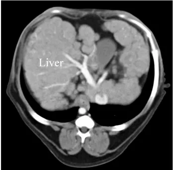
Figure 2. Computed tomography. Transvers section of abdomen. In this view, the liver is smaller than normal and multiple nodules in liver are seen.
Figür 2. Bilgisayarlı tomografi. Karın boşluğunun transversal kesiti. Bu görüntüde karaciğerin normale göre küçük olduğu ve karaciğerdeki çok sayıda noduler yapı görülüyor.
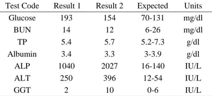
On serum chemistry profile, serum glucose, alkaline phosphatase, alanine transaminase and creatine kinase increased (Table 1). Serum insulin was 1127 pmol/L as serum glucose 8.1 pmol/L. Insulin/glucose ratio was 139 (normal range: 14-43). Mild raise of serum bilirubin (0.3 mg/dL) was seen.
Abdominal ultrasound was performed. The liver was irregularly margined with several hypoechoic
nodules in both the left and right liver ranging in size from 3.4 mm-6.1mm. The left and right liver lobes were small and retracted from the stomach separated by more mesentery than normal. Hyperechoic septations were identified in both sides of the liver as with fibrosis/cirrhosis. These changes were more consistent with an chronic hepatitis and cirrhosis. The left pancreatic lobe was ultrasonographically normal. The right pancreatic lobe was heterogeneously hyperechoic almost isoechoic to surrounding mesentery measuring 9.9 mm wide. The pancreatic body was not identified. The changes in the right pancreas were consistent with fatty infiltrate and degenerative change as with fibrosis. A pancreatic mass or nodule were not identified.
After about 3 weeks, the patient was presented to clinic for a scheduled Computed Tomography (CT) scan. On CT scan, the pancreas was normal and a nodule, such as a glucagonoma was not identified. The liver was small as it did not surround the gallbladder and had extremely undulating and nodular margins. As the contrast medium diffuses through the hepatic parenchyma, a course knobbly texture with homogeneously isoattenuating parenchyma was evident. These findings were consistent with cirrhosis and severe chronic liver disease (Figure 2). Ultrasound guided liver aspiration was done. Liver was of low cellularity with marked hemodilution on a clear background. Moderate numbers of dense clusters of hepatocytes with round nucleus, coarse chromatin, one distinct blue round nucleolus and abundant pink-blue cytoplasm were seen. Occasionally these hepatocytes had larger N:C ratio. Occasinal slightly damaged neutrophils were noted within the background, which were proportionate to the hemodilution. No etiologic agents or evidence of neoplasia were noted.
The complete blood cell count was performed. While normal erythrocyte and mean corpuscular hemoglobin concentration (MCHC) levels were seen, decreased hemoglobine (13.5 g/dl); mean corpuscular volume (MCV) (55.4 FL); mean corpuscular hemoglobine (MCH) (17.7 pg) were revealed. Serum chemistry profile was performed. It revelaed hyperglycemia and elevation of ALP, gamma-glutamyltransferase (GGT) and ALT was observed (Table 1).
We believed the cause of NME in this patient to be chronic liver disease thanks to persistently elevation of liver values, results of CT and USG and USG guided liver aspiration and the patient was believed to have NME associated with an extrapancreatic glucagonoma with secondary hyperinsulinemia to compensate for the chronic neoglucogenesis when we had considered histopathological findings, elevation of serum insulin level and atypical USG results for NME. Glucagon serum levels could not be determined at that time.
Table 1: Serum biochemistry parameters belong to 1st and 4th weeks of treatment.
Tablo 1: Tedavinin 1. ve 4. haftasına ait serum biyokimyasal parametreleri.
Test Code Result 1 Result 2 Expected Units
Glucose 193 154 70-131 mg/dl BUN 14 12 6-26 mg/dl TP 5.4 5.7 5.2-7.3 g/dl Albumin 3.4 3.3 3-3.9 g/dl ALP 1040 2027 16-140 IU/L ALT 250 396 12-54 IU/L GGT 2 10 0-6 IU/L
BUN: blood urea nitrogen, TP: total protein, ALP: alkaline phosphatase, ALT: alanine transaminase, GGT: gamma-glutamyltransferase.
BUN: kan üre nitrojen, TP: total protein, ALP: alkalin fosfataz, ALT: alanin transaminaz, GGT: gama-glutamiltransferaz.
To prevent excessive itching and treat secondary bacterial and fungal infections cefpodoxime (75 mg SID orally), terbinafine (90 mg SID orally) and Hexadene shampoo which had been used by referring veterinarian were continued to be used till all diagnostic test process was completed. After that, treatment with glucagon antagonist octreotide (2 µg/kg subcutaneously twice daily ) was started. To increase the patient’s appetite, as an appetite stimulator, mirtazapine (0.5 mg/kg orally once daily for 14 days) was given. Hill’s z/d switched into Hill’s n/d diet. A supplement of fatty acids and zinc was started. A topical lotion containing hydrocortisone (%1) and chlorhexidine containing shampoo was recommended to use if the lesions flare up.
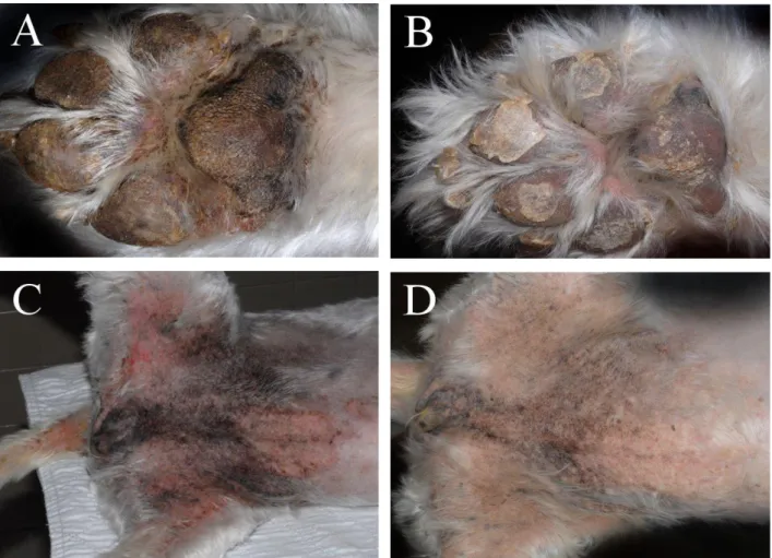
After last medications and diet, the appetite was increased, movelessness was over and the skin lesions regressed (Figure 3). However, about two months later, there had been a rapid degradation of its mental status (neurologic symptoms, such as “staring into space” related to hepatic encephalopathy), profuse vomiting, weight loss and apparent pain. This indicated likely liver failure and it was euthanized. The first skin problem had been seen 19 months ago before she passed away. Octreotide treatment was used at 15th month of disease. Our patient survived approximately 5 months after our treatment had started.
On gross necropsy; the liver was diffusely friable and distorted by myriad 0.1-2.0 cm, raised, light brown, nodules with an intervening greatly depressed, gray, thickened capsular surface in areas of with bands of fibrosis (Figure 4). The grossly observed crusting skin lesions and advanced liver disease were consistent with the reported hepatocutaneous syndrome variant of necrolytic migratory erythema. The pancreas was dissected out completely and no glucagonoma was discovered. On microscobic examination; on toe pad margin, the epidermis was diffusely, irregular hyperplastic
and diffusely overlain by alternating parakeratotic hyperkeratosis (red) with a decreased to absent granular layer that had mild hydropic change (white), and increased deep epidermal basophilia (blue). The superficial dermis contained a mild, perivascular infiltrate of lymphocytes with occasional exocytosis. In addition to the previously described changes, there were foci with a wave of orthokeratotic hyperkeratosis underlying the parakeratotic change, with a more prominent granular layer, and less basophilia (resolving lesion) on toe pad.
As noted grossly the liver was dramatically nodular. Nodules were composed of well-differentiated, but poorly organized hepatic cords lined by plump hepatocytes with minimal biliary component. The nodules were separated by a compressed zone of increased fibrous connective tissue with an increased density of embedded biliary profiles and admixed with hemorrhage, fibrin, and pigment-laden macrophages. Vacuolation of hepatocytes was minimal. Diffusely, there was decreased zymogen granules with a mild focus of lobular hyperplasia on nine slides that belong to pancreas.
Figure 4. Multiple nodules in liver that observed in necropsy. Şekil 4. Nekropside karaciğerde gözlenen multiple nodüller. Figure 3. A) Appearance of a front footpad with hyperkeratosis, crusting and interdigital erythema before octreotide treatment. B) Appearance of lesions after 14 days of octreotide treatment. C) Markedly hyperpigmentation and erythematous crusting dermatitis on ventrum, perivulvar and perianal regions before octreotide treatment. D) Appearance of lesions after 14 days of octreotide treatment. Şekil 3. A) Ön ayaktaki hiperkeratoz, kabuklanma ve interdigital kızarıklığın octreotide tedavisinden önceki görünümü. B) 14 günlük octreotide tedavisinden sonra lezyonların görünümü. C) Karında, vulva ve anüs çevresinde belirgin hiperpigmentasyon ve eritematöz kabuklu dermatitisin octreotide tedavisindan önceki görünümü. D) 14 günlük octreotide tedavisinden sonra lezyonların görünümü.
The syndrome is generally seen in dogs between 4-16 years of age. NME is most common in West Highland White, Jack Russell, Scottish and Yorkshire Terriers, Shetland Sheepdogs and Cocker Spaniels (3, 15). In this study, the patient was 12,5-year-old, West Highland White Terrier.
NME in dogs is mostly associated with hepatopathies, glucagon secreting tumors are rarely seen (3). Most of hepatic diseases are idiopathic. However it was reported that copper storage disease, ingestion of mycotoxin, long-term administration of anticonvulsant drugs (such as primidone) cause NME (3, 8). In this case, any tumor couldn’t be found on pancreas on CT and USG. In consideration of all diagnostic procedures’ results, the reason of NME in our patient was determined as a chronic liver disease. On the other hand, as far as we learnt from pet owner, although our patient had no systemic illness, long-term medical treatment was never used. So it made us think mostly the chronic liver disease might have been “idiopathic”.
Hyperkeratosis and fissures on paw-pads, exudative- ulcerative dermatitis, bullas and vesicular lesions (3, 10), hyperpigmentation are mostly seen in dogs in NME (2). Lesions are observed on paw-pads; mucocutaneous junctions and the regions exposed to pressure and friction (3). In this case similar lesions and lesion localization were determined. Paronychia was reported in a case (6). In this dog, onychomalacia and onychodystrophy were observed on claws. Lethargy, anorexia and weight loss are commonly observed generalized symptoms in NME cases (3). In this case, in addition to them, neurologic symptoms, profuse vomiting and apparent pain were observed.
In a research study involved in 35 dogs with MEN-HCS at University of Pennsylvania, elevated bile acids (100%), ALP (100%), ALT (86%) and Diabetes mellitus (43%), hypoalbuminemia (29%) were determined (3). In our case serum glucose, ALP, ALT and CK values increased. Anemia seems to be a commom feature of the syndrome in dogs (1). In this case,
microcytic-normochromic anemia was determined. The
ultrasonographic view is liken to Swiss cheese or honeycomb (9). In our case, USG exam results match diffusely the hepatic parenchyma was more hypoechoic than normal. The liver didn’t have the typical honeycomb appearance that would be expected with HCS.
Glucagon is not a hormone that is routinely measured in dogs and there are also biologically active metabolites that may not be detected by all assays (7). Glucagon secretion can increase threefold without significant increases in peripheral plasma glucagon concentrations (14). In this case plasma glucagon level was not measured. However USG and CT examination showed us that there was not any mass or tumor in
pancreas. As it’s known, in dogs, hyperglucagonemia has been recognized in diabetes mellitus, pancreatitis and chronic hepatic insuffiency (12). We diagnosed it as chronic hepatopathy (cirrhosis) associated with secondary hyperglucagonemia.
Fatty acid supplements can be given to patient to collaborate with zinc supplement (3). A supplement of fatty acids and zinc were given to our patient. Some authors think corticosteroids are contraindicated in NME patients (3). However, in recent years, low dose topical hydrocortisone was started to use succesfully in NME patients in human medicine (5). On the basis of this information, low-strength hydrocortisone lotion (%1) was used.
Oberkirchner et al. (2010) administered Octreotide at 2 µg/kg subcutaneously twice daily to a dog with glucagonoma and within 10 days of starting treatment, the skin lesions had markedly improved and the patient no longer exhibited signs of lameness or pain when walking. In the same way octreotide treatment succeeded and general condition, skin lesions and lameness of dog apparently recovered in 14 days following octreotide treatment in this study.
We saw in here, particularly skin lesions can be regressed and by the way the quality of life can be enhanced with octreotide and supportive therapy not only in pancreatic glucagonoma patients but also in a chronic hepatopathy case.
References
1. Allenspach K, Arnold P, Glaus T, Hauser B, Wolff C, Eberle C, Komminoth P (2000): Glucagon-producing
neuroendocrine tumour associated with hypoaminoacidemia and skin lesions. J Small Anim Pract, 41, 402-406.
2. Bond R, McNeil PE, Evans H, Srebernik N (1995):
Metabolic epidermal necrosis in two dogs with different underlying diseases. Vet Rec, 136, 466-71.
3. Byrne KP (1999): Metabolic epidermal
necrosis-hepatocutaneous syndrome. Vet Clin N Am-Small 29,
1337-55.
4. Godfrey DR, Rest JR (2000): Suspected necrolytic
migratory ervthema associated with chronic hepatopathy in a cat. J Small Anim Pract, 41, 324-28.
5. Griffing GT (2013): Hyperglucagonemia treatment &
management. Erişim tarihi: 18.06.2014. Erişim adresi:
http://www.emedicine.medscape.com/article/121575-treatment.
6. Gross TL, O'Brien TD, Davies AP, Long RE (1990):
Glucagon-producing pancreatic endocrine tumors in two dogs with superficial necrolytic dermatitis. J Am Vet Med
Assoc, 197, 1619-22.
7. Gross TL, Song MD, Havel PJ, Ihrke PJ (1993):
Superficial Necrolytic Dermatitis (Necrolytic Migratory Erythema) in Dogs. Vet Pathol, 30, 75-81.
8. Henricks PM (1987): Dermatitis associated with the use
9. Hill PB, Auxilia ST, Munro E, Genovese, L, Silkstone MA, Kirby B (2000): Resolution of skin lesions and
long-term survival in a dog with superficial necrolytic dermatitis and liver cirrhosis. J Small Anim Pract, 41,
519-523.
10. March PA, Hillier A, Weisbrode SE, Mattoon JS, Johnson SE, DiBartola SP, Brofman PJ (2004):
Superficial Necrolytic Dermatitis in 11 Dogs with a History of Phenobarbital Administration (1995-2002). J
Vet Intern Med, 18, 65-74.
11. Marsh KA, Ruedisueli FL, Coe SL, Watson TGD (2000): Effects of zinc and linoleic acid supplementation
on the skin and coat quality of dogs receiving a complete and balanced diet. Vet Dermatol, 11, 277-284.
12. Miller WH, Scott DW, Buerger RG, Shanley KJ, Paradis M, McMurdy MA, Angarano DW (1990):
Necrolytic Migratory Erythema in Dogs: A Hepatocutaneous Syndrome. J Am Anim Hosp Assoc, 26, 573-580.
13. Oberkirchner U, Linder KE, Zadrozny L, Olivry T (2010): Successful treatment of canine necrolytic
migratory erythema (superficial necrolytic dermatitis) due to metastatic glucagonoma with octreotide. Vet Dermatol,
21, 510-516.
14. Outerbridge CA (2010): Hepatocutaneous Syndrome. 112. In: SJ Ettinger, EC Feldman. (Eds), Textbook of Veterinary Internal medicine, 7th ed, Elsevier Saunders, St. Louis, Missiouri, USA.
15. Outerbridge CA, Marks SL, Rogers QR (2002): Plasma
amino acid concentrations in 36 dogs with histologically confirmed superficial necrolytic dermatitis. Vet Dermatol,
13, 177-186.
16. Torres S, Johnson K, McKeever P, Hardy R (1997):
Superficial necrolytic dermatitis and a pancreatic endocrine tumour in a dog. J Small Anim Pract, 38,
246-50.
17. Torres SMF, Caywood DD, O’Brien TD, O’Leary TP, McKeever PJ (1997) : Resolution of Superficial Necrolytic
Dermatitis Following Excision of a Glucagon-Secreting Pancreatic Neoplasm in a Dog. J Am Anim Hosp Assoc,
33, 313-319.
18. Walton DK, Center SA, Scott DW, Collins K (1986):
Ulcerative Dermatosis Associated with Diabetes Mellitus in the Dog: A Report of Four Cases. J Am Anim Hosp
Assoc, 22, 79-88.
Geliş tarihi: 21.04.2014 / Kabul tarihi: 20.02.2015
Address for correspondence:
Dr. Devrim Şebnem Tüfenk Pet Health Veterinary Clinic, B. Beykoz Istanbul-Turkey e-mail: [email protected]