Ankara Üniv Vet Fak Derg, 60, 155-156, 2013
Short Communication / Kısa Bilimsel Çalışma
A case of Leucocytozoon dubreuili in a starling (Sturnus vulgaris)
Ozlem OZMEN1, Ramazan ADANIR2, Mehmet HALIGUR1, Bayram Ali YUKARI21University of Mehmet Akif Ersoy, Faculty of Veterinary Medicine, Department of Pathology, Burdur-TURKEY; 2University of
Mehmet Akif Ersoy, Faculty of Veterinary Medicine, Department of Parasitology, 15030, Istiklal Yerleskesi, Burdur-TURKEY.
Summary: A nestling starling (Sturnus vulgaris) which had fallen from its nest was presented to the Department of Pathology
in comatose situation. During the physical examination, the bird died and necropsy was performed. At the microscopical examination of the blood smears, numerous Leucocytozoon agents were observed. At necropsy, there was no remarkable lesion observed, except for slight enlargement of the spleen. Blood smears were also admitted to the Department of Parasitology for diagnosis of the agents and upon the morphological appearance and relevant literature; they were identified as Leucocytozoon dubreuili. This is the first report of Leucocytozoon dubreuili identification in a starling.
Keywords: Leucocytozoon dubreuili, parasitology, pathology, starling.
Bir sığırcıkta (Sturnus vulgaris) Leucocytozoon dubreuili olgusu
Özet: Yuvasından düşerek yaralanan bir sığırcık (Sturnus vulgaris) yavrusu koma halinde Patoloji Anabilim Dalı’na getirildi.
Muayene sırasında ölen kuşun nekropsisi yapıldı. Kan frotisinin incelenmesinde çok sayıda Leucocytozoon etkenlerine rastlandı. Nekropside dalakta hafif büyüme dışında bir lezyon saptanmadı. Kan frotisi teşhis için Parazitoloji Anabilim Dalı’na gönderildi. Etkenler, tür spesifitesi ve morfolojik görünümleri doğrultusunda ilgili literatürler eşliğinde Leucocytozoon dubreuili olarak identifiye edildi. Bu, Türkiye’de bir sığırcıkta Leucocytozoon dubreuili’nin identifiye edildiği ilk rapordur.
Anahtar sözcükler: Leucocytozoon dubreuili, parazitoloji, patoloji, sığırcık.
Leucocytozoonosis is a vector-borne protozoan disease of birds caused by several species of Apicomplexa in the genus Leucocytozoon that affects the blood and tissue cells of internal organs (2,3,9).
Leucocytozoon spp. is easily identified from blood films
because it grossly distorts the host cell that it parasitizes. Only the gametocyte stage of Leucocytozoon occurs in the peripheral blood of birds (1). The vector of the
Leucocytozoon spp. is Simuliidae (black flies). Initial
development occurs in the liver and spleen followed by the development of gametocytes in blood cells (4). This parasite is assigned to the suborder Haemospororina of the phylum Apicomplexa (5). There are many species of
Leucocytozoon, but only a few are known to be pathogenic
to their hosts. The description of Leucocytozoon species has been made based mainly on the morphology of gametocytes in blood cells, although the examination of exoerythrocytic stages (meronts and schizonts) has been used to some extent (2).
Leucocytozoon spp. occurs worldwide including
Turkey. However, there is very limited number of studies on leucocytozoonosis available (6-8) in birds in Turkey. There is little knowledge about blood parasites in starlings. To the best of our knowledge there is no
previous report about the presence of L. dubreuili in a starling.
A nestling starling (Sturnus vulgaris) which died after falling from its nest was the material of this study. The bird was presented to the Department of Pathology in comatous situation. During the examination, small amount of blood coagulum was observed in the oral cavity. The bird died and necropsy was performed immediately. Blood smears were prepared and stained with Giemsa. At necropsy, there was no remarkable lesion observed, except for slight enlargement of the spleen. Examination of the blood smears revealed numerous Leucocytozoon agents. The blood smears were also admitted to the Department of Parasitology for diagnosis of the agents.
During the necropsy, tissue samples were taken from all visceral organs. Samples were fixed in 10% neutral-buffered formalin, routinely processed and embedded into paraffin for histopathological examination. Tissues were sectioned at 5μm and stained with haematoxylin and eosin (HE). Fecal samples were also taken for parasitological examination.
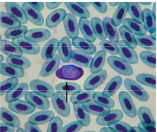
Microscopical examination of the starling’s blood smears revealed numerous roundish Leucocytozoon
Ozlem Ozmen - Ramazan Adanir - Mehmet Haligur - Bayram Ali Yukari 156
agents in its red blood cells. The nucleus of the parasite was generally round in shape and the nucleolus was prominent. However, the nuclei of the bird’s erythrocytes were pushed aside and were markedly deformed. Numerous L. dubreuili gametocytes were observed in the prepared blood smears (Figure 1). The parasites were located peripherally in the host’s cells and had evident dumbbell-shaped structure with marked thickening at both ends (Figure 2). The agents were identified as L.
dubreuili, according to their morphological appearance
and relevant literature. No clinical symptom was observed related to haemoparasites in the bird. Morphology of the L. dubreuili in this study was identical with the classical knowledge (10).
Leucocytozoon spp. is one of the most common
blood parasites in the wild birds. Leucocytozoon species are considered nonpathogenic, while only a few species of this parasite are considered pathogenic. If a bird is under stress or is immunocompromised, then the
Leucocytozoon can cause clinical problems (3,4). In this
study, numerous intracytoplasmic L. dubreuili gametocytes were observed in the blood smears of the bird. No pathological findings were observed in any organ of the bird, related to haemoparasites. Furthermore, at histopathological examination, there was no developmental stage of L. dubleuli, as well as no inflammatory reaction around the vessels in any organ.
Leucocytozoon spp. is transmitted from infected to
uninfected birds by a variety of biting flies. Simuliidae is a common fly throughout the Turkey. The prevalence of this blood parasite in wild birds is still unknown. Thus, we can say that, this is the first report of L. dubreuili identification in starlings.
References
1. Cample TW (1994): Identification of Common Blood
Parasites. 190-191. In: BW Ritchie, GJ Harrison, LR
Harrison, YM Saif (Eds.), Avian Medicine: Principles and Application, Wingers Publishing Inc., Florida.
2. Forrester DJ, Foster G., Morrison JL (2001):
Leucocytozoon toddi and Haemoproteus tinnunculi
(Protozoa: Haemosporina) in the Chimango Caracara (Milvago chimango) in Southern Chile. Mem. Inst. Oswaldo. Cruz. Rio de Janeiro. 96, 1023-1024.
3. Forrester DJ, Greiner EC (2008): Leucocytozoonosis. 54-107. In: CT Atkinson, NJ Thomas DB Hunter (Eds), Parasitic Diseases of Wild Birds. Wiley-Blackwell, Iowa. 4. Greiner EC, Ritchie BW (1994): Parasites. 1007-1029.
In: BW Ritchie, GJ Harrison LR Harrison (Eds.), Avian Medicine: Principles and Application, Wingers Publishing Inc., Florida.
5. Levine ND, Corliss JO, Cox FEG, Derroux G, Grain J, Honinberg BM, Leedale GF, Loeblich III AR, Lom J, Lynn D, Merinfeld EG, Page FC, Poljansky G, Sprague V, Vavra J, Wallace FG (1980): A newly revised
classification of the protozoa. J. Protozool, 27, 37-58.
6. Marzal A, Albayrak T (2012): Geographical variation of
haemosporidian parasites in Turkish populations of Kruper’s Nuthatch Sitta krueperi. J Ornithol. DOI
10.1007/s10336-012-0853-z.
7. Ozmen O, Haligur M, Adanir R (2009): Identification of
different protozoa species from a common buzzard (Buteo buteo). Turk J Vet Anim Sci. 33, 257-260.
8. Ozmen O, Haligur M, Yukari BA (2005): A study on the
presence of Leucocytozoonosis in wild bird of Burdur district. Turk J Vet Anim Sci. 29, 1273-1278.
9. Springer WT (1997): Other blood and tissue protozoa. 900-905. In: BW Calnek, HJ Barnes, HJ Beard, LR McDougald, YM Saif (Eds), Diseases of Poultry. Iowa State Press: Iowa.
10. Valkiunas G. (2005): Family Leucocytozoidae. 737-908. In: Avian Malaria Parasites and Other Haemosporidia. CRC Press, Florida.
Geliş tarihi: 03.08.2012 / Kabul tarihi: 13.11.2012
Address for correspondence:
Prof.Dr. Ozlem Ozmen
University of Mehmet Akif Ersoy,
Faculty of Veterinary Medicine, Department of Pathology, 15030, Istiklal Yerleskesi, Burdur-TURKEY
e-mail: [email protected]
Figure 1. Leucocytozoon dubreuili gametocytes (arrows) in blood smear, starling, Giemsa stain, Bar= 50µm.
Şekil 1. Kan frotisinde Leucocytozoon dubreuili gametositi, sığırcık, Giemsa, Bar= 50µm.
Figure 2. Higher mahnification of a Leucocytozoon dubreuili gametocyte (arrow) in blood smear, starling, Giemsa stain, immersion oil.
Şekil 2. Bir Leucocytozoon dubreuili gametositinin yakından görünümü, sığırcık, Giemsa, immersiyon yağı.