DAHİLİ BİLİMLER / MEDICAL SCIENCES Araştırma Makalesi / Research Article
Received: 18.02.2010 • Accepted: 22.03.2010 Corresponding author
Zeliha Kayaaltı
Institute of Forensic Medicine, Ankara University, Ankara, TURKEY Phone : +90 (312) 319 27 34
Fax : +90 (312) 319 20 77 E-mail Address : [email protected]
Aim: The aim of this study was to determine 4977 bp and 7436 bp mitochondrial DNA (mtDNA)
deletions and to investigate whether there is an association between the mtDNA deletions and myocardial infarction (MI) in cardiac muscle samples from autopsies.
Material and Method: In this study, a total number of 66 heart tissues from autopsies were
stud-ied, 33 of them were obtained from individuals with MI and the rest without MI. In order to deter-mine 4977 and 7436 bp mtDNA deletions, mtDNAs in heart muscles from controls and heart tis-sues from MI region were isolated and amplified with polymerase chain reaction (PCR) technique. The amplified products separated on 2% agarose gel electrophoresis.
Results: 4977 bp mtDNA deletion was observed in 97% of the heart tissues of the individuals
with MI. On the other hand, this ratio was only 39.4% for the control group (p<0.001). Similarly, the resultant deletion ratios for 7436 bp mtDNA were 97% for the heart tissues of subjects associ-ated with MI, and 12.1% for the control group (p<0.001). Furthermore, there was a statistically significant association between the average age of all individuals and both deletions mentioned (p<0.01). Even though a strong effect of aging on deletions was identified; stronger effect of MI was undeniable.
Conclusion: In this study, it is showed that mtDNA deletions may contribute to diagnosis of MI
in autopsies.
Key Words : Miyokardiyal infarktüs, mtDNA delesyonları, Otopsi
Amaç: Bu çalışmanın amacı, 4977 bp ve 7436 bp mitokondriyal DNA (mtDNA) delesyonlarını
belirlemek ve otopsi kalp kası örneklerindeki miyokardiyal infarktüs (MI) ve mtDNA delesyonları arasında ilişki olup olmadığını araştırmaktır.
Materyal ve Metod: Çalışmada 33 MI’lı ve 33 MI’sız kişiden alınan toplam 66 otopsi kalp dokusu
kullanıldı. 4977 ve 7436 bp mtDNA delesyonlarını belirlemek için, kontrollerin kalp kaslarından ve MI’lı kalp dokularından mtDNA izole edildi ve polimeraz zincir reaksiyonu (PCR) tekniği ile çoğaltıldı. Çoğaltılan ürünler %2’lik agaroz jel elektroforezinde ayrıştırıldı.
Bulgular: MI’lı kişilerin kalp doklarının %97’sinde 4977 bp mtDNA delesyonu gözlemlendi. Diğer
taraftan kontrol grubunda bu oran sadece %39.4’tü (p<0.001). 7436 bp mtDNA delesyon oranı da benzer şekildeydi; MI’lı kalp dokularında bu delesyonun oranı %97 iken, kontrol grubunda %12.1 bulundu (p<0.001). MI arasında istatistiksel olarak önemli ilişki saptandı (p<0.001). Ayrıca Bahse-dilen her iki delesyonla kişilerin ortalama yaşı arasında da istatistiksel olarak önemli ilişki belirlendi (p<0.01). Delesyonlarda yaşlanmanın güçlü bir etkisi olduğu görülse de, MI’nın delesyonlardaki etkisinin de kuvvetli olduğu yadsınamaz.
Sonuç: Bu çalışmada, mtDNA delesyonlarının otopsi örneklerinde MI’ın tanısına katkıda
bulunabileceği gösterilmiştir.
Anahtar Sözcükler: Myocardial infarction, mtDNA deletions, Autopsy
1 Institute of Forensic Medicine, Ankara University, Ankara, TURKEY 2 Ankara Oncology Hospital, Ankara, TURKEY
*Authors are grateful to Dr. Görkem Mergen for his valuable support
and contributions.
Detection of Mitochondrial DNA Deletions in Heart Tissue with
Acute Myocardial Infarction
*Akut Miyokardiyal İnfarktüslü Kalp Dokularında Mitokondriyal DNA Delesyonlarının Belirlenmesi
Zeliha Kayaaltı
1, Nergis Cantürk
1, Levent Şahiner
2, Miyase Odabaşı
1, Tülin Söylemezoğlu
1Human mitochondria contain their own genetic material in the form of mito-chondrial DNA (mtDNA), which is the only extrachromosomal DNA in human cells. mtDNA is a super coiled, double-stranded circular molecule of 16,569-bp in size that encodes 13 polypeptides (7 NADH, 1 Cyt-b, 3
COX and 2 ATPase) which constitute the respiratory enzyme complexes (1). The mitochondrial genome is more sus-ceptible to increased oxidative dam-age than nuclear DNA due to its lack of histone protection, limited repair capacity, and close proximity to the
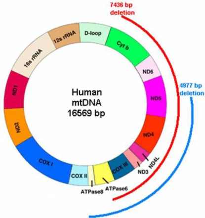
electron transport chain (2). Mito-chondria are not the only major site of energy production in the cell, but also the major intracellular source of reactive oxygen species (ROS) and free radicals due to electron leakage from the respiratory chain. The free radicals cause peroxidation of membrane lip-ids, accumulation of oxidized dysfunc-tional proteins, and increased mtDNA damage. Especially impaired functions of energy dependent tissues such as heart are affected by mitochondrial dysfunction (3). The increased gen-eration of reactive oxygen species is associated with mitochondrial damage and dysfunction in the failing heart, mtDNA defects may thus play an im-portant role in the development and progression of myocardial failure (4). The accumulation of mtDNA muta-tions and oxidative damage is poten-tial contributor to human diseases, such as age-related pathologies and cardiovascular disease (5). The most studied common mitochondrial DNA deletions are 4977 bp and 7436 bp mtDNA deletions (Figure 1). 4977 bp mtDNA deletion is detected at high frequencies in various tissues from aging humans as well as in ischemic myocardium (6). Also a significant accumulation of 4977 bp mtDNA has been demonstrated in the atrial tissue of patients with clinical atrial fibrillation (7). Some studies have documented the presence of mtDNA 4977 deletions in cardiac muscle from patients suffering from coronary ath-erosclerotic heart disease (8,9). Myocardial infarction is defined as
myo-cardial cell death due to prolonged ischemia. Cell death is categorized pathologically as either coagulation or contraction band necrosis, or both, which usually evolves through oncosis, but can result to a lesser degree from apoptosis. After the onset of myocardi-al ischemia, cell death is not immedi-ate but takes a short period of time to develop. It takes 6 h before myocardial necrosis can be identified by standard macroscopic or microscopic post-mortem examination (10). This may sometimes have considerable medi-colegal importance, especially when cardiac diseases have been considered to cause a traffic or other accident and
in some criminal circumstances (11). It is very important to reach the exact diagnosis and many authors work on this issue especially for early stages of myocardial infarction (12,13). The aim of the present study was to clarify the
relationship between mtDNA deletion and acute myocardial infarction (MI) in cardiac muscle samples from autopsies.
Material and Method
In this study, we examined 66 heart tissues from autopsies, 33 of them obtained from individuals who had died be-cause of MI and the rest from individ-uals died due to noncardiac cases (con-trol group), all in the range of 25-89 years of age. All samples were stored at -20 °C until the analysis. The samples were acquired from The Council of Forensic Medicine, Ministry of Justice, Ankara. All of the autopsy cases were from the same ethnic and geographical origin, living in the Central Anatolia region of Turkey. A small question-naire for gathering the demographic information was given to the family
or relative of each autopsy cases. Only Turkish subjects were included in the study. The study design was approved by Ankara University Faculty of Medi-cine Ethics Committee (approval No: 121-3212 in 2007). Autopsy heart tis-sues were stored at -20 ºC before the analyses and autopsies were performed in accordance with the principles of The Declaration of Helsinki.
DNA Extraction
Total DNAs were extracted from 25 mg he-art muscles from controls and hehe-art tis-sues from MI region by “Qiagen DNA Mini Kit” according to the method re-commended by the manufacturer. Determination of 4977 bp and 7436 bp mtDNA deletions
In order to determine 4977 and 7436 bp mtDNA deletions, the regions were amplified with polymerase chain re-action (PCR) technique using the F1-R1, F2-R2 and F3-R3 primer pairs as performed in the previous study (14).
Figure 1. Schematic presentation of mitochondrial wild type genome and common deletions (4977 bp and
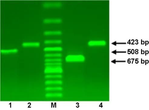
675 bp, 423 bp and 508 bp oligonuc-leotides were produced in the pre-sence of total mtDNA, the 4977 bp dmtDNA and 7436 dmtDNA, respec-tively (Table 1). The polymerase chain reaction products were separated on a 2% agarose gel electrophoresis, visuali-zed by ethidium bromide staining un-der an ultraviolet illuminator, scanned and photographed using Syngene Mo-nitoring System (Figure 2).
Amplification was carried out on a Tech-ne Tc 512 PCR System in a 50 µl re-action mixture containing 200 µM of dNTPs, 10 pmol each of forward (F) and reverse (R) primers, 1 U Hot Star Taq DNA polymerase (Qiagen),
1 X PCR buffer (Qiagen) and 100 ng DNA. The PCR cycling conditions consisted of an initial denaturation step at 94 ºC for 5 min, followed by 33 cycles of 94 ºC for 30 s, 55 ºC for 30 s, 72 ºC for 30 s, and final extensi-on step at 72 ºC for 10 min.
Statistical Analysis
Fisher’s exact test was used for categorical variables to compare the differences of mtDNA deletions in with and without MI groups. Differences of mtDNA de-letions among the age groups were as-sessed with one way anova technique. All statistical procedures were carried
out by the Scientific Package for Social Sciences (SPSS), version 16.0, Statis-tical analysis software. Values of p less than 0.05 were considered as statisti-cally significant.
Results
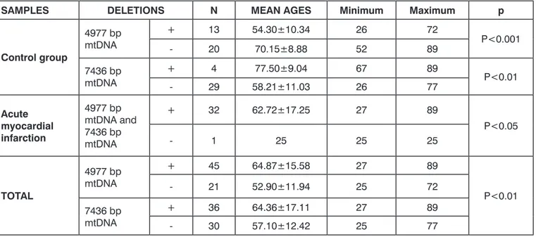
4977 bp mtDNA deletion was observed in 97% of the heart tissues of the in-dividuals who had died because of MI whereas this ratio was only 39.4% for the control group. Similarly the re-sultant deletion ratios for 7436 bp mtDNA were 97% for the heart tissu-es of the individuals who had died be-cause of MI, and 12.1% for the cont-rol group. There was a statistically sig-nificant difference between myocardial infarction and both deletions mentio-ned (p<0.001) (Table 2). Furthermore, a significant association was also found between the average age of control and patient group with 4977 bp and 7436 bp mtDNA deletions. It was determi-ned that the mean ages of the cont-rol group with 4977 bp and 7436 bp mtDNA deletions were 70.15±8.88 and 77.50±9.04 respectively. On the other hand, the mean ages of the in-dividuals without these deletions were 54.30±10.34 and 58.21±11.03 (p<0.001 and p<0.01, respectively). Also same statistical significant relati-on was found in the acute myocardial infarction patients. (Table 3)
Conclusion
It is well known that up to 200 different deletions in the mitochondrial geno-mes are detectable in postmitotic tis-sues. However, among these 200, the Table 1. Oligonucleotid primers used for PCR amplification of the mtDNA with 4977 bp and 7436 bp mtDNA deletions.
PRIMER
PAIRS PRIMER SEQUENCES
Size of the PCR product amplified from mtDNA with
the length mutation (bp) Aim of this PCR
F1-R1 F1: 5’-GGAGTAATCCAGGTCGGT-3’R1: 5’-AATGATGGCTAGGGTGACTT-3’ 675 Determination of thetotal mtDNA F2-R2 F2: 5’-GCCCGTATTTACCCTATAGC-3’R2: 5’-GGGGAAGCGAGGTTGACCTG-3’ 423 Determination of the4,977 bp dmtDNA F3-R3 F3: 5’-CTCTAGAGCCCACTGTAAAG-3’R3: 5’-GTTGAGGGTTGATTGCTGTAC-3’ 508 Determination of the7436 bp dmtDNA
Figure 2. Representative gel image of 4977 bp and 7436 bp mtDNA deletions. M=100 bp ladder;
Lane1= PCR products for 7436 bp mtDNA deletion (508 bp).
Lane2 and Lane4= PCR products for 4977 bp mtDNA deletion (423 bp). Lane3= PCR products for total mtDNA (675 bp).
most studied common mitochondrial DNA deletions are 4977 bp and 7436 bp mtDNA deletions. Most studies showed these mtDNA deletions were important contributors to ageing and degenerative diseases. The age-related increase of the common mtDNA dele-tion is not universal, but clearly tissue-specific and different organs or even different areas of the same organ accu-mulate these deletions at different ra-tes (15). Pathological mtDNA deleti-ons are associated with ultrastructu-rally abnormal mitochondria; mitoc-hondrial DNA defects reduce the spe-cific mitochondrial enzyme activity le-vels and change the mtDNA integrity (16). Consequently, the accumulation
of abnormal mtDNA contributes to the progression of mitochondrial dise-ases and heart failure (17).
The threshold for mutation and deletions in mtDNA causing symptoms vari-es between tissuvari-es, being lower in tho-se tissues that are post-mitotic, highly metabolically active and dependent on oxidative phosphorylation for energy production, such as the heart musc-le, brain, skeletal muscle etc. (18). The threshold level varies not only between tissues but also between different types of mutation. It has been demonstrated threshold level for mtDNA deletions is 60%. Most pathogenic mutations are heteroplasmic; if they were
homoplas-mic, they would often be lethal (19). In forensic medicine, sensitive
biochemi-cal markers for the post-mortem di-agnosis of acute myocardial infarction are needed. When the deleted mtDNA reaches the threshold level of 60-80%, the cell functions will be affected. Ho-wever, when this ratio is homoplasmic, it will be lethal for the cell. In order to evaluate the mtDNA deletions as the biomarker for the MI diagnosis, it might also be better to determine he-teroplasmic ratio quantitatively (20). In our study, mtDNA deletions
investiga-ted and it has been showed that the re-sult may contribute diagnosis of MI in autopsy specimens.
Table 3. The association between the mtDNA deletions and mean age of individuals in control and acute myocardial infarction groups.
SAMPLES DELETIONS N MEAN AGES Minimum Maximum p
Control group 4977 bp mtDNA + 13 54.30±10.34 26 72 P<0.001 - 20 70.15±8.88 52 89 7436 bp mtDNA + 4 77.50±9.04 67 89 P<0.01 - 29 58.21±11.03 26 77 Acute myocardial infarction 4977 bp mtDNA and 7436 bp mtDNA + 32 62.72±17.25 27 89 P<0.05 - 1 25 25 25 TOTAL 4977 bp mtDNA + 45 64.87±15.58 27 89 P<0.01 - 21 52.90±11.94 25 72 7436 bp mtDNA + 36 64.36±17.11 27 89 - 30 57.10±12.42 25 77
Table 2. The frequencies of occurence of 4977 bp and 7436 bp mtDNA deletions in acute myocardial infarction and control groups.
SAMPLES N Mean Ages±SE
DELETIONS
4977 bp mtDNA deletion 7436 bp mtDNA deletion
+ - +% + - +%
Acute myocardial
infarction 33 61.58±18.21 32 1 97 32 1 97
Control group 33 60.55±12.45 13 20 39.4 4 29 12.1
KAYNAKLAR
1. Christen MA. Mitochondrial Dysfunction in Diabetes Mellitus. Drug Development Rese-arch 1999; 46:67–79
2. Zastawny TH, Kruszewski M, Olinski R. Comparison of oxidative base damage in mitochondrial and nuclear DNA. Free Rad Biol Med 1998; 24:722-725
3. Bindoff L. Mitochondria and the heart. Eur Heart J. 2003; 24:221-4
4. Tsutsui H, Kinugawa S, Matsushima S. Mi-tochondrial oxidative stress and dysfunction in myocardial remodelling. Cardiovasc Res. 2009; 81:449-56
5. Botto N, Berti S, Manfredi S. Detection of mtDNA with 4977 bp deletion in blo-od cells and atherosclerotic lesions of pati-ents with coronary artery disease. Mutat Res. 2005; 570:81-88
6. Corral-Debrinski M, Shoffner JM, Lott MT, et al. Association of mitochondrial DNA da-mage with aging and coronary atherosclero-tic heart disease. Mutat Res. 1992; 275:169-80.
7. Lai LP, Tsai CC, Su MJ, et al. Atrial fibrillati-on is associated with accumulatifibrillati-on of aging-related common type mitochondrial DNA deletion mutation in human atrial tissue. Chest. 2003; 123:539-44
8. Mohamed SA, Hanke T, Erasmi AW, et al. Mitochondrial DNA deletions and the aging heart. Exp Gerontol. 2006; 41:508-17 9. Bogliolo M, Izzotti A, De Flora S, et al.
Detec-tion of the “4977 bp” mitochondrial DNA deletion in human atherosclerotic lesions Mutagenesis. 1999; 14:77-82
10. Alpert JS, Thygesen K, Antman E, et al. Myocardial infarction redefined--a consen-sus document of The Joint European Society of Cardiology/American College of Cardio-logy Committee for the redefinition of myo-cardial infarction. J Am Coll Cardiol. 2000; 36:959-69
11. Knight B. The pathology of sudden death. In: Knight B, eds. Forensic Pathology. 2nd ed. London: Arnold. 1996; 487-515 12. Martínez Díaz F, Rodríguez-Morlensín
M, Pérez-Cárceles MD, et al. Biochemical analysis and immunohistochemical determi-nation of cardiac troponin for the postmor-tem diagnosis of myocardial damage. Histol Histopathol. 2005; 20:475-81
13. Osuna E, Perez-Carceles MD, Alvarez MV, et al. Cardiac troponin I (cTn I) and the postmortem diagnosis of myocardial infarc-tion. Int J Legal Med. 1998;111:173-6
14. Kayaaltı Z, Söylemezoğlu T. Türk populasyo-nunda mitokondriyal DNA delesyonlarında yaşlanma ve sigara kullanımının etkisi. Tur-kiye Klinikleri J Med Sci. Accepted 15. Meissner C, Bruse P, Mohamed SA, et al. The
4977 bp deletion of mitochondrial DNA in human skeletal muscle, heart and different areas of the brain: a useful biomarker or more? Exp Gerontol. 2008; 43:645-52 16. Arbustini E, Diegoli M, Fasani R, et al.
Mi-tochondrial DNA mutations and mitoc-hondrial abnormalities in dilated cardiom-yopathy. Am J Pathol. 1998; 153:1501-10 17. Marín-García J, Goldenthal MJ, Moe GW.
Abnormal cardiac and skeletal muscle mitoc-hondrial function in pacing-induced cardi-ac failure. Cardiovasc Res. 2001; 52:103-10 18. Rossignol R, Rossignol R, Faustin B, Rocher
C, et al. Mitochondrial threshold effects. Bi-ochem. J. 2003; 370:751-762
19. DiMauro S, Tanji K, Bonilla E, et al. Mitoc-hondrial abnormalities in muscle and other aging cells: classification, causes, and effects. Muscle Nerve. 2002; 26:597-607
20. Rossignol R, Malgat M, Mazat JP, et al. Threshold effect and tissue specificity. Imp-lication for mitochondrial cytopathies. J Biol Chem. 1999; 274:33426-32