Introduction
Haploid S. cerevisiae cells possess two loci on chromosomes which play a role during sexual reproduction. These are MATa (mating type a) and MATα (mating type α). The cells possessing the same mating type (MATa - MATa or MATα- MATα) do not mate. The mating phenomenon may occur between the cells possessing the opposite alleles. As a result, the mating partners join, their cytoplasms fuse and then their nuclei fuse to form a diploid nucleus. This later process of nuclear fusion is known as kariogamy (1). Fusion of the yeast cells possessing the same mating type (identical)
requires a different approach such as cell fusion techniques. A number of physical, chemical, and biological techniques have been used to induce fusion between cells. The best known chemical technique employs polyethylene glycol (PEG). A physical method, namely electrofusion, has been developed that rivals other fusion methods in terms of efficiency. The electrofusion method, combining the use of dielectrophoresis alignment and reversible membrane breakdown for cell fusion, was first reported by Zimmermann et al. (2). This method eliminates most of the disadvandages of chemical and virus-induced fusion techniques (3). However, electrofusion devices are often
Electrofusion of Saccharomyces cerevisiae Auxotrophic Mutants of
Identical Mating Type Using a Laboratory-Made System
Gökhan CORAL*
Department of Biology, Faculty of Arts and Sciences, Mersin University, 33342 Çiftliköy-Mersin - TURKEY Ömer ÇOLAK
Department of Biology, Faculty of Arts and Sciences, Çukurova University, 01330 Balcal›-Adana - TURKEY M. Nisa ÜNALDI
Department of Biology, Faculty of Arts and Sciences, Mustafa Kemal University, 31040 Hatay - TURKEY Burhan ARIKAN
Department of Biology, Faculty of Arts and Sciences, Çukurova University, 01330 Balcal›-Adana - TURKEY
Received: 10.12.2001
Abstract: Yeast protoplasts from haploid auxotroph strains DC6 (MATα) and FY73 (MATα) were exposed to an inhomogeneous
alternating field (AC 200 V/cm). Due to the dielectrophoretic aggregation two or more cells with close membrane contact were formed between the two electrodes. The effects of different DC pulses were investigated to determine critical fusogenic pulse strength. In the results, the maximum fusant cells were obtained only when 8 kV field strengths were applied. These findings showed that the optimum fusogenic pulse strength for yeast protoplasts is 8 kV/cm under our experimental conditions. After fusion, the DNA contents of both hybrid and individual parental strains were compared. The amount of DNA in the hybrid was found to be about twofold higher than that in the individual parental strain.
Key Words: Electrofusion, Saccharomyces cerevisiae
‹dentik Eflleflme Tipine Sahip Saccharomyces cerevisiae Oksotrofik Mutantlar›n›n Laboratuvar Yap›m› Bir Sistem Kullan›larak Elektrofüzyonu
Özet: Bu çal›flmada DC6 (MATα) ve FY73 (MATα) haploid oksotrof maya sufllar›ndan elde edilen protoplastlar, homojen olmayan
bir alternatif alana (200 V/cm AC) maruz b›rak›ld›. Dielektroforetik agregasyona ba¤l› olarak, iki elektrot aras›nda kalan iki ya da daha fazla say›daki hücrede yak›n membran temas› oluflturuldu. Hücreleri füzyona u¤ratan optimum puls fliddetini belirlemek için farkl› DC pulslar›n›n etkisi araflt›r›ld›. Sonuçta, sadece 8 kV'luk fliddete sahip alan gerilimleri uyguland›¤›nda maksimum say›da füzant hücre elde edilebilmifltir. Bu bulgular, deney koflullar›nda maya protoplastlar› için optimum füzogenik puls fliddetinin 8 kV/cm oldu¤unu göstermifltir. Füzyondan sonra hibrit ve her bir parental suflun DNA miktarlar› k›yaslanm›flt›r. Hibritin DNA miktar›n›n, parental sufllar›n DNA miktarlar›ndan yaklafl›k 2 kat daha fazla oldu¤u bulunmufltur.
Anahtar Sözcükler: Elektrofüzyon, Saccharomyces cerevisiae
quite expensive ($10,000-$15,000) and not easily applicable to gene transfer by electroporation (4). Electrofusion is broadly applicable to a wide variety of cell and membrane types, including vesicular membranes (5). Most previous studies of electrofusion have focused on applications and the development of improved techniques. The use of electrofusion to study the mechanisms of cell fusion is relatively new. For example, electrofusion of human erythrocytes (6) and human erythrocyte ghosts (7) has been used as a model system to study the mechanisms of membrane fusion.
During recent years electrofusion methods have been used in many areas of biological research, including plant somatic hybridization (8), murine and human hybridoma and monoclonal antibody production (6). Most recently, electrofusion has been widely used in the cloning of several farm animals by transfer of nuclei of somatic cells to oocytes (9-11).
In the present study, our aim was to produce hybrids between Saccharomyces cerevisiae protoplasts of identical mating type with our laboratory-made electrofusion device. We also determined optimal fusion conditions with our device, such as dielectrophoresis time and critical fusogenic pulse strengths.
Materials and Methods
Strains:Saccharomyces cerevisiae DC6 (MATα, leu2, his4) and FY73 (MATα, ura3, his3) auxotroph mutant strains were obtained from UMIST, UK. These strains were used as fusion partners in the electrofusion experiments.
Media and culture conditions: M9 minimal medium supplemented with suitable amino acids was used for the selection of fusant cells. YPD medium containing 1% yeast extract, 2% polypeptone and glucose was used as a complete medium. Regeneration agar was made in 1.2 M sorbitol plus 5% YPD, 2% glycerol and 3% agar. Yeast cells were cultivated aerobically at 30 ºC.
Preparation of yeast protoplasts: Yeast cells were grown overnight at 30 ºC in 20 ml of YPD and then cells were collected by centrifugation at 2000 rpm for 5 min. Then 2.5 mg/ml Zymolase (SIGMA) was added, followed by incubation at 37 ºC for 60 min. Cells were centrifuged in a microfuge for 1 min and supernatants were discarded. Cells were resuspended in a solution of 1M
sorbitol, 0.1 M EDTA (pH 7.5). Finally protoplasts were examined under a microscope (1).
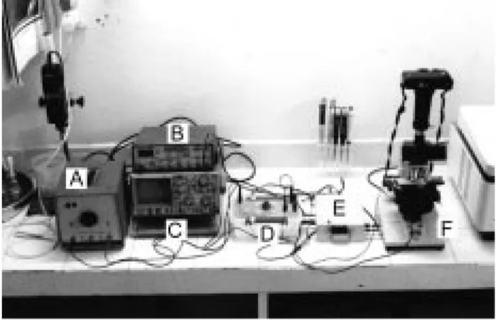
Electrofusion apparatus: A laboratory-made fusion system was used in all experiments. This system can also be used in electroporation at the same time. This system consists of a DC power supply (3000 V maximum), an AC power supply (20V, 2 MHz), an oscilloscope and a pulser (Figure 1). The pulser can generate exponential decay waveforms between 1 and 100 ms and it was equipped with both DC and AC power supplies (Figure 2). This includes a discharging output connected to the switching unit. The switching unit cuts AC voltage while DC pulses are applied. This unit was operated manually.
Electrofusion: Identical mating type S. cerevisiae DC6 and FY73 protoplasts were used in the electrofusion experiments. For the fusion experiments, the protoplasts were mixed in a 1:1 ratio and incubated in 10 mM Tris (pH 7.6), 10 mM CaCl2 and 1.2 M sorbitol. The
Figure 1. Electrofusion system. A) DC power supply, B) AC power supply, C) Oscilloscope, D) Pulser, E) Switching unit, and F) Light microscope. + -S1 C 3750 V S2 R2 470 K + -Output R1 30 MΩ Power supply input
Figure 2. Electronic scheme of the pulser, which produces exponential decay waveform pulses. S1 and S2: charging and discharging button respectively, C (capacitor): 3750 V, R1 (resistor): 30 MΩ, R2 (variable resistor): 470 K.
suspension density was about 107cells/ml. Then 10 to 20 µl of the protoplast suspension was pipetted into the gap between the two electrodes and the formation of pearl chains in the homogeneous alternating electrical field (200 V/cm, 2 MHz AC) was monitored under a microscope (12). The subsequent cell fusion was induced by electrical breakdown pulses (8 kV, 1 ms) applied with an interval of 5 s. After fusion of the cells and rounding off of the fusion products, the electrode gap was covered with regeneration medium and incubated at 30 ºC for 2 d. These cells were spread on M9 minimal medium supplemented with histidine (20 mg/ml w/vol.) and incubated at 30 ºC for 2 d. Finally hybrid cells were scored.
Determination of DNA quantity of parental and fusant cells: For determination of the DNA contents of the parental and fusant strains, cells were grown in YPD at 30 ºC for 3 d. Cells were harvested by centrifugation and washed twice. DNA extraction was carried out as described by Kaiser et al. (1). DNA quantities were determined by UV spectrophotometer as described by Maniatis et al. (13).
Cytological analysis: For cytological investigations, the mutant and fusant strains were inoculated into YPD medium. After 3 d, the shapes and dimensions of the cells were compared. The number of cell nuclei was also determined by acridine orange staining (fixing with 90% ethanol: staining for 2 min; observation under a microscope) (14).
Results and Discussion
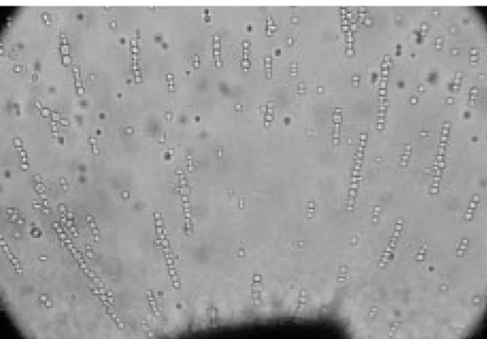
Optimization of dielectrophoretic parameters For the optimization of dielectrophoretic parameters appropriate for the laboratory-made electrofusion system, the most suitable field conditions for the best pearl chain formation of intact cells and protoplasts were tested. Dielectrophoresis applied for 1 min with 200 V/cm and 2 MHz AC frequency was found to be ideal for pearl chain formation of intact yeast cells (Figure 3). The same conditions were also investigated for yeast protoplasts; however, it was found that longer dielectrophoresis durations are required for pearl chain formation of yeast protoplasts. According to this, the best pearl chain formation for yeast protoplasts are 200 V/cm, 2 MHz AC frequency and 3 min dielectrophoresis (Figure 4). Complete alignment could take as little as a few seconds or as long as several minutes (7). It was reported that the best pearl chain formation for S. cerevisiae protoplasts was observed at an alternative field of 1 kV/ cm and 2 MHz frequency (12). The AC voltage value of our experimental settings was relatively lower than the settings in the literature, because our AC power supply can reach 200 V/cm maximally. Such relatively low alternative fields might extend the duration of dielectrophoretic alignment. In addition, electrofusion cannot be achieved under strict conditions. It was stressed that the frequency at which stable dielectrophoresis occurs varies from cell type to cell type and therefore must be detected empirically (5).
Figure 3. Pearl chain formation of intact yeast cells. Dielectrophoresis was applied for 1 min with 200 V/cm and 2 MHz AC frequency. (Magnitude 40X)
Figure 4. Pearl chain formation of yeast protoplasts. Dielectrophoresis was applied for 3 min with 200 V/cm, 2 MHz AC frequency. (Magnitude 40X)
The fusion solutions used in our experiments consisted of 10 mM Tris (pH 7.6), 10 mM CaCl2and 1.2 M sorbitol (15). For the stabilization of protoplasts, the use of 1-1.2 M sorbitol is essential (12). For this reason, sorbitol increased the density of the fusion medium used in the fusion experiments. However, the dielectrophoretic alignment of yeast protoplasts required more time than the intact yeast cells. We thought that this was the result of removal of the charged molecules from the cell walls during the protoplasting process (data not shown).
Optimization of pulse strength
After the dielectrophoretic optimization of yeast protoplasts, a set of experiments were carried out for determining the optimum fusogenic pulse strength. In the controlled fusion experiments, which used protoplasts from S. cerevisiae FY73 and DC6, pulse duration, AC field strength and frequency were kept constant at 1 ms, 200 V/cm and 2 MHz respectively and DC field strength was tested variably. Each test was carried out by using a fusion chamber with two parallel electrodes 1 mm apart. The tested electrical strengths were 3, 4, 5, 6, 7, 8 and 9 kV respectively. From the results of fusion experiments, maximum fusant cells were obtained only when 8 kV field strengths were applied (Figure 5). These findings showed that the critical fusogenic pulse strength for yeast protoplasts is 8 kV/cm under our experimental conditions. It was found that the optimum condition for obtaining hybrids of auxotrophic mutants of S. cerevisiae was 7 kV/cm (16). This value is 1 kV smaller than our critical voltage. This may be due to the difference in the fusion solution used during the electrofusion process and
the conductivity of the fusion solution would affect the pulse strength. The conductivity of these solutions is low and it was reported that this is an important factor for cell viability (17).
Electrofusion of haploid S. cerevisiae protoplasts of identical mating type
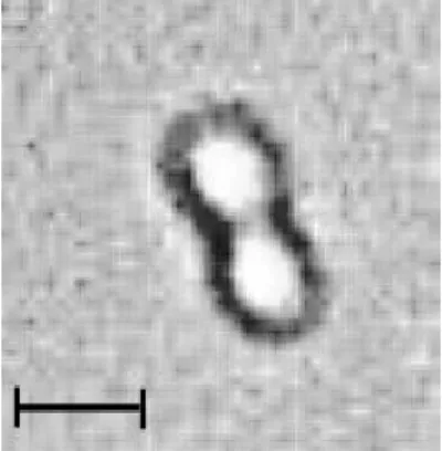
A mixture of protoplasts from two yeast strains DC6 and FY73 was exposed to an nonuniform alternating field of 200 V/cm strength and 2 MHz frequency. The inhomogeneity of the field in the electrode gap gave rise to dielectrophoresis, which led to the formation of pearl chains of cells. The number of cells within a pearl chain depended on the suspension density. The fusion of neighbouring cells within a pearl chain was brought about by two field pulses of 8 kV/cm intensity and 1 ms duration. The pulses were applied with an interval of about 5 s (Figure 6). From the experiments, it was concluded that the optimal field strength to produce hybrids is 8 kV/cm when exposed to two DC pulses for 1 ms with an interval of 5 s. It was reported that multiple pulse applications reduce cell viability, but, optimally, it was concluded that two pulse applications are efficient for yeast cells (12,18). The ideal interval between the two pulses was found to be 5 s.
A maximum of 28 hybrid cells were isolated from histidine supplemented minimal media according to their auxotrophic requirement. The morphological appearence of hybrids was similar to that of parental strains. The amount of DNA in the hybrid was found to be about twofold when compared with the individual haploid parental strain (Table). Acridine orange staining of both
0 5 10 15 20 25 30 3 4 5 6 7 8 9 Pulse Strength (kV)
Number of Fusants (cfu)
Figure 5. Effects of the field strength on the formation of fusant cells. Yeast protoplasts were exposed to a nonuniform alternating field of 200 V/cm strength and 2 MHz frequency. Dielectrophoresis was applied for 3 min and two pulses were applied with an interval of 5 s.
Figure 6. Fusion of two dielectrophoretically adhered protoplasts. Picture was taken 1 min after the application of two field pulses resulting in membrane breakdown (8 kV/cm, 1 ms, applied with an interval of 5 s). (Bar size is 5 µm)
hybrids and individual parental strains revealed that they possess only one nucleus. The increased genetic material is therefore situated in one nucleus. These findings confirmed that the fusant is a real diploid (2n). The complementing of this hybrid's defects suggests plasmogamy with subsequent karyogamy. After these results were obtained, no further genetic analyses were
needed, because our goal was only to find out whether hybrids form or not. If genetic complementation occurred between haploid auxotrophic mutants, this means that our lab-made system was successful; but further studies are needed to improve fusion yields. There may be irreproducibility in the fusion data owing to some experimental detail or a biological characteristic which is simply not obvious. Insufficient knowledge of the mechanism may also be a limitation. It was reported that, for unknown reasons, some cells or membrane systems may be resistant to electrofusion (7).
In conclusion, electrofusion is truly an interdisciplinary biophysical technique. Therefore, its successful use requires either an appreciation and familiarity with physics and electronics that is not common among biologists or an approach which duplicates the necessary setup, whether it be a laboratory-made system or a commercially available system.
Table. Comparison of DNA quantities between individual parental and fusant yeast strains.
Strains Average DNA quantity
absorbance (µg/ml) (OD260) DC6 2.8 140 FY73 2.6 130 Fusant 6.2 310 References
1. Kaiser, C., Michaelis, S., and Mitchell, A., Methods in yeast genetics, A Cold Spring Harbor Laboratory Course Manual. 1994. 2. Zimmermann, U., and Vienken, J., Electric field induced cell to cell
fusion. J. Membr. Biol. 67: 165-182, 1982.
3. Pilwat, G., Richter, H.P., and Zimmermann, U., Giant culture cells by electric-field induced fusion. FEBS Lett. 133: 169-172, 1981. 4. Potter, H., Application of electroporation in recombinant DNA
technology. Methods in Enzymol. 217: 461-477, 1993. 5. Y›lmazo¤lu, ‹., Einseitige und wechselseitige electrofusion
biologisher (humanplacenta) und küntstlicher vesikel (liposomen) charakterisierung der fusion producte. Dissertation zur des doktorgrades des Chemie der Universitat Hamburg, 1988. 6. Chang, D.C., Saunders, J.A., Chassy, B.M., and Sowers, A.E.,
Overview of electroporation and electrofusion. In: Guide to electroporation and electrofusion. Academic Press Inc. 1992. 7. Hui, S.W., and Stenger, D.A., Electrofusion of cells: Hybridoma
production by electrofusion and polyethylene glycol. Methods in Enzymol. 220: 212-228, 1993.
8. Sowers, A.E., Membrane electrofusion: A paradigm for study of membrane fusion mechanisms. Method Enzymol., 220: 196-211, 1993.
9. Kato, Y., Tani, T., Sotomaru, Y., Kurokawa, K., Kato, J., Doguchi, H., Yasue, H., and Tsunoda, Y., Eight calves cloned from somatic cells of a single adult, Science, 282: 2095-2098, 1998. 10. Hill, J.R., Winger, Q.A., Long, C.R., Looney, C.R., Thompson,
J.A., and Westhusin, M.E., Development rates of male bovine nuclear transfer embryos derived from adult and fetal cells. Biol. Reproduc. 62: 1135-1140, 2000.
11. Iwasaki, S., Campbell, K.H.S., Galli, C., Akiyama, K., and Iwasaki, S. Production of live calves derived from embryonic stem like cells aggregated with tetraploid embryos, Biol. Reproduc. 62: 470-495, 2000.
12. Halfmann, H.J., Emeis, C.C., and Zimmermann, U., Electro-fusion of haploid Saccharomyces yeast cells of identical mating type. Arch. Microbiol. 134: 1-4, 1983.
13. Manitais, T., Fritsch, E.F., and Sambrook, J., Molecular cloning: A laboratory manual. Cold Spring Harbor, New York, 1982. 14. Sipiczki, M., and Ferenczy, L., Protoplast fusion of
Schizosaccharomyces pombe auxotrophic mutants of identical mating-type, Mol. Gen. Genet. 151: 77-81, 1977.
15. Guthrie, E.C., and Fink, G.R., Guide to yeast genetics and molecular biology. Methods in Enzymol. 194: 809, 1991. 16. Noda, K., Togowa, Y., and Yamata, Y., Quantification of physical
and cyto-physiological conditions for the electrofusion of Saccharomyces cerevisiae. Agric. Biol. Chem. 54(8): 2023-2028, 1990.
17. Dower, W.J., Miller, J.F., and Ragsdale, C.W., High efficiency transformation of E. coli by high voltage electroporation. Nucl. Acid. Res. 16: 6127-6145, 1998.
18. Calvin, N.M., and Hanavalt, P.C., High efficiency transformation of bacterial cells by electroporation. J. Bacteriol. 170: 2796-2801, 1988.