The Gamma-Butyrolactone Model of Absence
Epilepsy: Acute and Chronic Effects in Wistar Rats
Absans Epilepside Gama-Butirolakton Modeli:
Wistar Sıçanlarda Akut ve Kronik Etkileri
Tuğba ERYİĞİT KARAMAHMUTOĞLU,
1Nihan ÇARÇAK,
2Melike ŞAHİNER,
3Özlem AKMAN,
4O. Carter SNEAD,
5Esat EŞKAZAN,
6Filiz ONAT
1Summary
Objectives: We studied the electroencephalographic (EEG) and behavioral changes of the chemical model of generalized absence epilepsy
induced by acute and chronic administration of gamma-butrolactone (GBL), a prodrug of gamma-hydroxybutyric acid.
Methods: Adult male Wistar rats under anesthesia were implanted with bilateral cortical recording electrodes. The rats were administered
30 intraperitoneal injections of GBL twice daily from Monday to Friday and EEG was recorded 20 min before and 40 min after GBL injections. In order to monitor spontaneous spike-and-wave discharges (SWDs), the baseline EEGs on the subsequent Monday mornings after the first, second and third weekends were recorded for 90 min.
Results: The intraperitoneal administration of GBL caused a rapid onset of bilaterally synchronous SWDs in the cortical EEG accompanied by
behavioral immobility, vacant-staring and vibrissal twitching. By repeated GBL injections, animals displayed spontaneous bilateral synchro-nous SWDs in the baseline EEG on the Monday morning session after the GBL-free weekend period (60 h after the Friday afternoon injection).
Conclusion: The present study reports the acute and chronic effects of the systemic administration of GBL. The chronic systemic application
of GBL may represent a model of epileptogenesis for absence epilepsy. Key words: Absence epilepsy; gamma-butyrolactone; wistar rats; EEG.
Özet
Amaç: Çalışmamızda, gama-hidroksibutiratın (GHB) ön ilacı olan gama-butirolakton (GBL) ile indüklenmiş kimyasal jeneralize absans epilepsi
modelinde GBL’nin akut ve kronik uygulanmasıyla oluşan EEG ve davranış değişiklikleri incelendi.
Gereç ve Yöntem: Çalışmada kullanılan yetişkin Wistar sıçanlara, anestezi altında stereotaksik cerrahi yöntemi uygulandı; hayvanlara iki
ta-raflı kortikal kayıt elektrotları implante edildi. Bir haftalık iyileşme sürecini takiben sabah ve akşam olmak üzere günde iki kez i.p. GBL enjek-siyonu uygulandı. Enjeksiyon uygulamasından önceki 20 dakika, sonrasında 40 dakika olmak üzere EEG kaydı alındı, enjeksiyon yapılmadan geçen haftasonlarını takiben Pazartesi sabahları enjeksiyon öncesi 40 dakika bazal EEG kaydı alındı.
Bulgular: Bu çalışmada sistemik GBL uygulamasının akut ve kronik etkileri tekrar ortaya kondu. Çalışmamızda yer alan hayvanlarda,
tekrarla-yan enjeksiyonlarla birlikte, enjeksiyon yapılmadan geçen haftasonlarını takiben Pazartesi sabahları enjeksiyon öncesi alınan bazal kayıtlarda spontan iki taraflı DDD’leri görüldü.
Sonuç: Kronik sistemik GBL uygulanması absans epilepsi için bir epileptogenez modeli ortaya koyabilir. GHB modeli antiepileptik ilaç
araştır-malarında, absans epilepsi modeli olarak kullanılabilir.
Anahtar sözcükler: Absans epilepsi; gama-butirolakton; wistar sıçanlar; EEG.
1
Department of Pharmacology and Clinical Pharmacology, Marmara University Faculty of Medicine, Istanbul;
2Department of Pharmacology, Istanbul University Faculty of Medicine, Istanbul;
3
Department of Physiology, Acıbadem University Faculty of Medicine, Istanbul, and Department of Physiology,
Istanbul Bilim University, Faculty of Medicine, Istanbul;
4
Department of Physiology, Istanbul Bilim University Faculty of Medicine, Istanbul;
5
Department of Pediatric Neurology, Hospital for Sick Childrens Hospital, and Department of Pediatrics,
University of Toronto Toronto, Canada;
6
Department of Pharmacology, Istanbul Bilim University Faculty of Medicine, Istanbul
© 2013 Türk Epilepsi ile Savaş Derneği
© 2013 Turkish Epilepsy Society
Submitted (Geliş) : April 25, 2013
Accepted (Kabul) : May 27, 2013
Correspondence (İletişim) : Filiz ONAT, MD
e-mail (e-posta) : [email protected] EXPERIMENTAL STUDY / DENEYSEL ÇALIŞMA
Introduction
A pharmacological modeling approach to absence epilepsy requires a reproducible and predictable method that has the EEG and behavioral characteristics of generalized typi-cal absence seizures in humans. The gamma-hydroxybutyr-ate (GHB) and gamma-butryrolactone (GBL) models meets these criteria.[1,2] The acute administration of GBL, which is a prodrug of GHB, has been shown to produce exactly the same EEG and behavioral effects of absence seizures as GHB [3] and GBL administration mimics typical absence epilepsy in humans.[4] GBL is preferred due to the consistency, pre-dictable dose response and rapidity of the onset of its ac-tion in rodents.[5-7] The systemic administration of a single dose of GBL produces absence seizures through the poten-tiation of the GHB-related neuromodulation and/or interac-tion with GABAergic inhibitory and excitatory neurotrans-mission in the cortico-thalamo-cortical circuit.
The objective of this study was to determine the EEG and behavioral changes induced by acute and chronic adminis-tration of GBL in male adult Wistar rats.
Materials and Methods
Animals
Adult (6-8 months old) Wistar male rats (n=20) weighing 270-300 g were used for all experiments. Animals were housed in a temperature controlled room (20±3 ºC) with a 12-h light–dark cycle in groups of four per cage and sepa-rated individually after the surgery with free access to com-mercial rat pellets and tap water. The experimental protocol was approved by the Animal Care and Use Committee of Marmara University (48.2011.mar).
Surgery
Animals were anaesthetized with ketamine (100 mg/kg, intraperitoneally) and xylazine (10 mg/kg, intraperitone-ally). Rats was placed in a stereotaxic instrument (Stoelting Model 51600, Stoelting Co., Illinois, USA) with the skull sur-face flat and bregma 0.0[8] and then the scalp was longitudi-nally incised for the implantation of stainless steel screws, for epidural recording and ground, placed bilaterally in the skull over frontal and parietal cortices for the record-ing of electroencephalogram (EEG). All of the electrodes were fixed to the skull with dental acrylic and in turn linked by insulated wires to a connector for EEG recordings. The animals were allowed to recover from surgery for 1 week
before any experimental protocols were started.
Experimental protocol
On the first day of the experiment, all animals were placed in Plexiglas recording cages. After an adaptation period, a baseline EEG was recorded for 20 min from as indicated in the experimental design. Electrical activity of the cortex was amplified (BioAmp ML 136) and recorded with a PowerLab 8S System running Chart v.5, (ADI Instruments, Oxfordshire, U.K.) before and after each GBL injection. GBL was pur-chased from Sigma Aldrich (St Louis, U.S.A) and prepared in saline for the intraperitoneal injection, a dose of 100 mg/kg of GBL was chosen based on the previous literature.[6,7] GBL has been shown to be readily hydrolyzed in vivo to its active congener, GHB.[9]
The rats were administered 30 intraperitoneal injections of GBL twice daily from Monday to Friday. The EEG was record-ed continuously 90 min after the first GBL injection to deter-mine the duration of the acute GBL effect. Then for the rest of the week, EEG was recorded 20 min before and 40 min after GBL injections to evaluate GBL-induced discharges with chronic administration of GBL throughout the 3 weeks. In order to monitor spontaneous SWDs, the baseline EEGs on the subsequent Monday mornings after the first, second and third weekends were recorded for 90 min.
A GBL-induced discharge was identified visually as such if its duration was ≥1 s with a complex of spike-and-wave (5-6 Hz) and amplitude at least three times the background amplitude of the EEG. The GBL-induced discharges were referred as SWDs captured on the EEG and the cumulative and mean duration of SWDs.
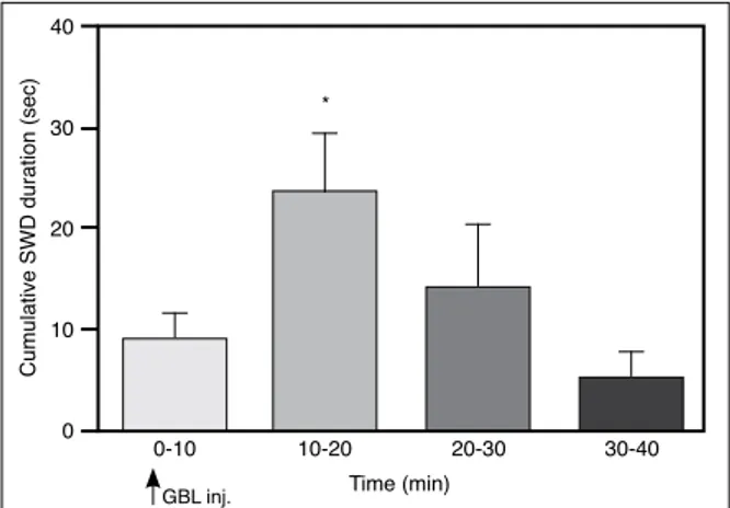
Figure 1. The cumulative duration of SWDs after the first GBL
injections over a 40 min period (*p<0.05).
0-10 10-20 Time (min) 20-30 30-40 * 0 20 10 30 40
Cumulative SWD duration (sec)
The effect of acute GBL administration
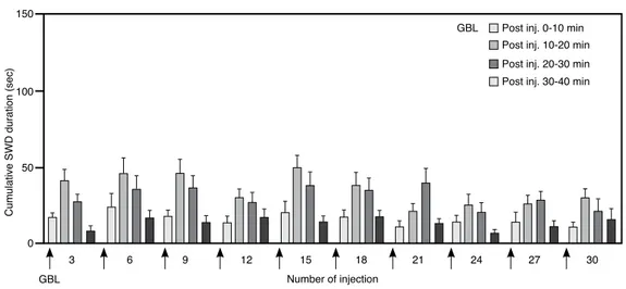
The intraperitoneal administration of GBL caused a rapid onset of bilaterally synchronous discharges in the cortical EEG accompanied by behavioral immobility, vacant-staring and vibrissal twitching. The SWD appeared 4.8±1.25 min after the first injection of GBL, reached maximum numbers and durations within 20 min (Figure 1). These complexes became regular, waxing and waning rhythmic SWDs with repeated chronic administration. The latency to onset of the first SWD complex was found to be delayed by repeated injections of GBL and reached 12.1±3.8 min after the 30th injection. The cumulative duration of SWDs by repeated chronic administration of GBL reached its maximum within 20 min as observed in the acute injections (Figure 2).
Statistical analysis
Data expressed as mean ± SEM were statistically evaluated by analysis of variance of repeated measures (ANOVA). A one-way ANOVA followed by the post hoc Bonferroni test was used to analyze cumulative duration of SWDs after the first GBL injection as well as the cumulative and mean dura-tion of spontaneous SWDs after the first, second and third weekends in the GBL group. The level of statistical signifi-cance was considered to be p<0.05.
Results
The baseline EEG recordings of all Wistar rats showed no ab-normal discharges.
Figure 4. The cumulative duration of spontaneous SWDs
throughout the 3-weeks experimental period, during the chronic GBL systemic administration (*p< 0.05).
1. week 2. week 3. week 0.0
1.0 0.5 1.5 2.0
Mean duration of SWD (sec)
Figure 3. The mean duration of spontaneous SWDs throughout
the 3-weeks experimental period, during the chronic GBL systemic administration.
1. week 2. week 3. week *
0 15
10
5
Cumulative SWD duration (sec)
Figure 2. The effect of repeated chronic administration of GBL. The changes of cumulative SWD duration by
repeated injections of GBL during the 40 min post injection periods.
Post inj. 0-10 min Post inj. 10-20 min Post inj. 20-30 min Post inj. 30-40 min
3 GBL Number of injection GBL 6 9 12 15 18 21 24 27 30 0 150 100 50
The effect of chronic GBL administration
By repeated intraperitoneal injections of GBL, animals dis-played spontaneous bilateral synchronous SWDs in the baseline EEG on the Monday morning session after the GBL-free weekend period (60 h after the Friday afternoon injection). The cumulative and mean duration of spontane-ous SWDs increased throughout the 3-weeks experimental period in the chronic administration (Figure 3 and 4). The statistical analysis revealed a significant increase in cumula-tive duration of spontaneous SWDs after the third weekend.
Discussion
The present study demonstrates that in animals received chronic administration of GBL, the GBL-induced discharges consisting of spike-and-wave complexes became more typical or mature form of SWD over the repeated intraperi-toneal injections of 100 mg/kg.
Additionally, the EEG characteristics of GBL-induced dis-charges with the acute administration of the dose of 100 mg/kg are in line with the previous observations.[6,10,11] The initial electrographic change in the baseline EEG recorded from the right and left frontoparietal cortices was a brief burst of spikes and thereafter quickly progressed to typi-cal SWDs of absence seizures as has been reported previ-ously by Snead.[6] These GBL-induced bilateral synchronous discharges in the acute administration show irregularity in rhythmicity, duration and amplitude. These electrographic discharges after the first injections were similar to the im-mature SWD paroxysms observed in postnatal 30 days old GAERS.[12] With repeated chronic administration of GBL, the SWD complexes appeared to mature with an increase in the power at the fundamental frequency and similar to that in adult GAERS or WAG/Rij rats.[13] It is accepted that the major disadvantage of the GHB and GBL models is that they represent an acute model of absence seizures rather than a chronic model of absence epilepsy such as the ge-netic models.[2] However, the maturation of GBL-induced SWDs over the time and spontaneous SWDs after the weekends in the present study indicated a fully synchro-nized paroxysmal activity, suggesting an augmentation in the cortico-thalamo-cortical network with repeated GBL administrations. The brief spontaneous bursts of bilaterally synchronous SWDs have been previously observed follow-ing the acute administration of GBL late as 72 h.[14] Indeed, the involvement of the cortico-thalamo-cortical circuitry has been well accepted in the pathophysiology of absence
epilepsy based on the studies in the genetic and chemical absence epilepsy models.[15-17] This synchronized paroxys-mal activity with the chronic administration of GBL sug-gests a change over time in the functional organization of absence seizure circuit that possibly occurs at the cellular and/or synaptic level in the thalamo-cortical circuitry. Tak-en together the chronic systemic application of GBL may represent an epileptogenesis model for absence epilepsy itself, deserving a particular attention in further studies.
Acknowledgement
This study was supported by Marmara University Re-search Council (BAPKO, D-300409-0116 and SAG-YLP-071211-0305).
References
1. Snead OC 3rd. Gamma hydroxybutyrate in the monkey. I. Elec-troencephalographic, behavioral, and pharmacokinetic stud-ies. Neurology 1978;28(7):636-42.
2. Snead OC 3rd. G-hydroxybutyrate and absence seizure activ-ity. In: Tunnicliff G, Cash CD, editors. Gamma-hydroxybutyrate: molecular, functional and clinical aspects. London: Taylor and Francis; 2002. p. 132-149.
3. Snead OC 3rd. Basic mechanisms of generalized absence sei-zures. Ann Neurol 1995;37(2):146-57.
4. Wong CG, Gibson KM, Snead OC 3rd. From the street to the brain: neurobiology of the recreational drug gamma-hydroxy-butyric acid. Trends Pharmacol Sci 2004;25(1):29-34.
5. Bearden LJ, Snead OC, Healey CT, Pegram GV. Antagonism of gamma-hydroxybutyric acid-induced frequency shifts in the cortical EEG of rats by dipropylacetate. Electroencephalogr Clin Neurophysiol 1980;49(1-2):181-3.
6. Snead OC 3rd. gamma-Hydroxybutyrate model of generalized absence seizures: further characterization and comparison with other absence models. Epilepsia 1988;29(4):361-8. 7. Snead OC 3rd. The gamma-hydroxybutyrate model of absence
seizures: correlation of regional brain levels of gamma-hy-droxybutyric acid and gamma-butyrolactone with spike wave discharges. Neuropharmacology 1991;30(2):161-7.
8. Paxinos G, Watson C. The rat brain in stereotaxic coordinates, 4th ed. San Diego: Academic Press; 1998.
9. Lettieri J, Fung HL. Improved pharmacological activity via pro-drug modification: comparative pharmacokinetics of sodium gamma-hydroxybutyrate and gamma-butyrolactone. Res Commun Chem Pathol Pharmacol 1978;22(1):107-18.
M, Micheletti G, et al. Effects of gamma-hydroxybutyrate and gamma-butyrolactone derivates on spontaneous general-ized non-convulsive seizures in the rat. Neuropharmacology 1988;27(7):683-9.
11. Depaulis A, Snead OC 3rd, Marescaux C, Vergnes M. Suppres-sive effects of intranigral injection of muscimol in three models of generalized non-convulsive epilepsy induced by chemical agents. Brain Res 1989;498(1):64-72.
12. Carçak N, Aker RG, Ozdemir O, Demiralp T, Onat FY. The rela-tionship between age-related development of spike-and-wave discharges and the resistance to amygdaloid kindling in rats with genetic absence epilepsy. Neurobiol Dis 2008;32(3):355-63.
13. Akman O, Demiralp T, Ates N, Onat FY. Electroencephalograph-ic differences between WAG/Rij and GAERS rat models of
ab-sence epilepsy. Epilepsy Res 2010;89(2-3):185-93.
14. Hu RQ, Cortez MA, Man HY, Roder J, Jia Z, Wang YT, Snead OC 3rd. Gamma-hydroxybutyric acid-induced absence seizures in GluR2 null mutant mice. Brain Res 2001;897(1-2):27-35. 15. Coenen AM, Van Luijtelaar EL. Genetic animal models for
ab-sence epilepsy: a review of the WAG/Rij strain of rats. Behav Genet 2003;33(6):635-55.
16. Cope DW, Di Giovanni G, Fyson SJ, Orbán G, Errington AC, Lorincz ML, et al. Enhanced tonic GABAA inhibition in typical absence epilepsy. Nat Med 2009;15(12):1392-8.
17. Velazquez JL, Huo JZ, Dominguez LG, Leshchenko Y, Snead OC 3rd. Typical versus atypical absence seizures: network mecha-nisms of the spread of paroxysms. Epilepsia 2007;48(8):1585-93.