tb't ■··'w ü'ií^ Чі*^ «f•Α ·-^o r * ^ '«Tw’s - 'Ç«^■Ч iV ■ -íc ^1·." ^ ..■ ;is¿! 'r^-> i. ‘^7 <*%■ " V·· ^ - i i -■- s | ^ ; - S 7 í Λ w··;·'^· . ·... $·; îè T i-rs :,.- m ı ^ I J . |< t « Ц P i» s A ·î* “·,,.·:· ··'· Ш. · » Ξ - I^"7jT U -w ¿ 'J-" · ^ '“ . ·Λ · « - . . 7 · , | 7 я V ! . » · · * « · ._J. ...^u·. ^ Α . »V , ÿ?·* .-Λ
A N A L Y SIS OF BRCAl A N D BRCA2 G EN ES IN TU R K ISH BREAST CANCER PA TIEN TS
A T H ESIS SU B M IT T ED TO THE D EPA R TM EN T OF M OLECULAR BIOLOGY A N D GENETICS A N D THE IN ST IT U T E OF EN G IN EER IN G A N D SCIENCE OF BILKENT U N IV E R SIT Y IN PARTIAL FULFILLMENT OF
THE R EQ UIREM ENTS FOR THE DEGREE OF D O C T O R OF PH ILO SO PH Y
BY
Bu çalışma TTGV172 no'lu proje
ve
TÜBİTAK Bütünleştirilmiş Doktora Programı
ile desteklenmiştir.
ilim ilim bilmektir
İlim kendin bilmektir
Sen kendini bilmezsin
Ya nice okumaktır
Okumaktan mana ne
Kişi hakkı bilmektir
Çün okudun bilmezsin
Ha bir kuru emektir
I certify that I have read this thesis and that in my opinion it is fully adequate, in scope and in quality, as a thesis for the degree of Doctor of Philosophy.
\
V A. V
Assoc. Prof. Tayfım Özçelik
I certify that I have read this thesis and that in my opinion it is fully adequate, in scope and in quality, as a thesis for the degree of Doctor of Philosophy.
y
Assist. Prof. Işık Yulug
I certify that I have read this thesis and that in my opinion it is fully adequate, in scope and in quality, as a thesis for the degree of Doctor of Philosophy.
J O
W F
I certify that I have read this thesis and that in my opinion it is fully adequate, in scope and in quality, as a thesis for the degree of Doctor of Philosophy.
f ^ · - 0 Ö- "
Prof. Dr. Ay Öğüş
I certify that I have read this thesis and that in my opinion it is fully adequate, in scope and in quality, as a thesis for the degree of Doctor of Philosophy.
. Ayşe Ayhan
Approved for the Institute of Engineering and Science
ABSTRACT
ANALYSIS OF BRCÁ1 AND BRCA2 GENES IN TURK ISH BREAST CANCER
PATIENTS
Hilâl Ö zd a ğ
Ph.D. in Molecular Biology and Genetics Supervisor: Assoc.Prof. Tayfiin özçelUc
June 2000,165 Pages
Breast cancer is the most frequent cancer type and the second cause of death among women. It is estimated that 10 to 15% o f breast cancer cases are hereditary. The majority o f hereditary breast cancers can be attributed to germ-line mutations in ^Jgeast CAncer susceptibility genes BRCAl and BRCA2.
In this study, germ-line BRCAl and/or BRCA2 gene mutations were screened in 50 Turkish breast and/or ovarian cancer patients divided into four groups of hereditary, familial, early onset, and male cancer by heteroduplex analysis and DNA sequencing. Two BRCA2 mutations, one novel (6880insG) and one previously reported (3034delAAAC), were found in the hereditary group. A novel BRCAl (1200insA) mutation was formd in the early onset group. All three mutations cause premature- termination codons. In addition, five BRCAl sequence variants have been identified in 23 patients. K654E (2080 A— >G), D693N (2196 G -> A ), P871L (2731 C— >T), and K1183R (3667 A—>G) result in a change of amino acids. 1013 T—^>C and 2201 C—>T are silent mutations. One patient in the early onset group was compound heterozygote for K654E and D693N. These results indicate that BRCAl and BRCA2 genes are
ÖZET
TÜRK M EM E K ANSERİ HASTALARINDA R R C 4İ VE BRCA2 GENLERİNİN
İNCELENM ESİ
Hilâl özdağ
Moleküler Biyoloji ve Genetik Doktorası Tez Yöneticisi: Doç. Dr. Tayfun özçelik
Haziran 2000,165 Sayfe
Meme kanseri kadmlar arasmda en sık görülen kanser tipidir ve ölüm nedenleri arasmda ikinci sırada yer alır. Meme kanseri olgularmın %10 ile 15’inin kalıtımsal olduğu tahmin edilmektedir. Kalıtımsal meme kanserlerinin büyük bir kısmı BRCAl ve BRCA2 Meme Kanseri yatkınlık genlerindeki eşey hücre mutasyonlanna atfedilebilir.
Bu çalışmada kalıtımsal, ailesel, erken yaş ve erkek meme kanseri gruplanna aynimış 50 Türk meme ve/veya över kanser olgusunda BRCAl ve /veya BRCA2 eşey hücre mutasyonlan heterodupleks analizi ve DNA dizilemesi ile taranmıştır. Kalıtımsal meme kanseri grubunda biri yeni (6880insG) biri önceden bildirilmiş (3034delAAAC) iki
BRCA2 mutasyonu saptanmıştır. Erken yaş grubunda yeni bir BRCAl mutasyonu (1200
insA bulunmuştur. Her üç mutasyonun da proteinde erken sonlanma kodonu oluşumuna neden olduğu tespit edilmiştir. Ayrıca 23 hastada beş adet BRCAl dizi varyantı tanımlanmıştır. Bunlardan K654E (2080 A ^ G ), D693N (2196 G->A), P871L (2731 C->T) ve K1183R (3667 A->G) tranzisyonlan aminoasit değişimine yol açmaktadır. 1013 T ^ C ve 2201 C-^'T ise sessiz mutasyonlardır. Erken yaş grubundaki bir hastanın K654E ve D693N için “compomıd”heterozigot olduğu tespit edilmiştir. Bütün bu sonuçlar BRCAl ve BRCA2 genlerinin Türk popülasyonundaki kalıtımsal meme kanserlerinin bir kısmmda rol aldığım ortaya koymaktadır.
TEŞEKKÜR
Yalnızca yazan kişi, jüri üyeleri ve belki konu ile ilgilenen birkaç araştmcı dışında kimsenin okumadığı her tezin ardmda uzun ve yorucu bir hikaye vardır. Bu uzun ve yorucu hikayenin başlamasında etkili olanlar, ilerlemesi ve sonlanmasmda desteği olanlar olay örgüsünün önemli birer parçasıdırlar.
Bu hikayenin başlaması iki kişi ile yakmdan ilgili. Hayatım boyunca beni olaylan sorgulamaya ve incelemeye iten ve böylece akılcılık çizgisini bilim yolunda bulmamda en önemli etken olan Sevgili .\nne ve Babam'a verdikleri tüm desteğin yamsıra özellikle bu nedenle teşekkür borçluyum.
Bu yolculuğa çıkmaya karar vermemi sağladığı için, üniversiteye başladığım ilk yıl, ilk gün, ilk saat karşıma çıkan ve öğrencilerine bilim felsefesini öğretmeye bütün bir hayatmı adamış olan değerli hocam Prof Dr. Ali Demirsoy'a ve aslında paslanmış çarklarm bile kişisel gayret ile çevrilebileceğini göstererek yolun henüz başındaki genç bir araştıncıya belki de en önemli teşviği vermiş olan değerli hocalarım Prof Dr. Reyhan öner ve Prof Dr. Cihan öner’e,
Bu uzun yolun başından itibaren, her çıkmaz yol levhasımn belirişinde yanımda bulduğum ve bilimsel perspektifi ile yolumu aydınlatan değerli hocam Prof Dr. Mehmet Öztürk'e,
Değerli tecrübelerini benimle paylaşan ve dostluklan ile bu yıllan zenginleştiren Doç. Dr. Uğur Yavuzer ve Dr. Cengiz Yakıcier'e; özellikle hakkı teslim etmekteki titizliği için destek ve yardımlanm esirgemeyen Doç. Dr. Işık Yuluğ’a,
Beraber gülüp ağlamanm, aynı evi, mutluluğu, hüznü ve dostluğu paylaşmanın güzelliğini yaşadığım Tuba Dinçer’e,
Uzun muhabbetlerini, her zor anda omuzunu, dertlerimizi ve mutluluklarımızı paylaştğı için inatçı, KÜÇÜK kardeşim Tolga Çağatay'a; sonsuz yardımseverliği, dosdoğru
dostluğu için Arzu Atalay'a; sakin ve soğukkanlı tavnyla her an yardıma hazır olan Hani Al’Otabi'ye,
İnamimaz keyifli, derin ve felsefi geyikleri, uzun yaz ve kış gecelerindeki lezzetli sofialar için sevgili kuzucuklarmı. Aslı Öztan (The Aquarius), Gülayşe İnce (Gülbahar Teyze), Bleda Bilican (Mösyö-Ahmet Can), N. Tolga Emre (Temre)'ye, (Toprak anamz sizleri seviyor)
On senelik kesintisiz dostluğun her anını doyasıya destek ve sevgileri ile paylaştığım Sibel Songür ve Yasemin Gülefe; uzun geceler ve telefon sohbetlerinde dostluğu için sevgili kuzenim Ayhn özgen'e.
Eşsiz dostluğu, güveni ve arkası kesilmez desteği ile hep yammda bulduğum Lütfiye Mesci'ye; hayatı aynntılarda aramanın keyfi, sımrsız güven ve desteği ile yanımda olduğu için Dr. Gürol Tunçman'a,
Farklı bilim dallarmda aynı uzun yolu katetmiş olan Doç. Dr. Ümit Özdağ, Dr. Hakan Özdağ ve Av. Savaş Özdağ (müstakbel Dr)'a, destek ve sevgileri ve herbiri nev’i şahsına münhasır ağabeylerim oldukları için,
en içten teşekkürlerimle.
TABLE OF CONTENTS SIGNATURE PAGE_ ABSTRACT______ ÖZET____________ TEŞEKKÜR TABLE OF CONTENTS LIST OF TABLES_____ LIST OF FIGURES ABBREVIATIONS 1 ii iii iv vii xiii xiv xvii / . INTRODUCTION 1.1. Cancer ____ 1.2. Oncogenes.
1. 3. Tumor suppressors “Gatekeepers and Caretakers’· 1. 4. Breast cancer____________________
1.4. 1. Etiology of breast cancer_____ 1. 4. 2 Genetic factors in Breast Cancer.
1.4.2.1. Somatic Mutations in Breast C ancer_ 1.4.2.2. Germline Mutations in Breast Cancer _ 1.4.2.2.1. Li-Fraumeni Syndrome {p53)____ 1.4.2.2.2. Lynch Type 11 Syndrome (MLHl) 1.4.2.2.3. Ataxia Teleangiectasia (A TM )___ 1.4.2.2.4. Cowden Disease {PTEN)_______ 1.4.2.2.5. Genetic Polymorphisms________
1.5. Hereditary Breast Cancer Genes_______
1.5.1 Breast Cancer susceptibility gene 1 {BRCAl)_ 1.5.1.1 Identification of BRCAl____________ 1.5.1.2. The Function of B R C A l___________ 1.5.1.3. Mutations in BRCA7 ______________ 1.5.2. Breast Cancer suscepdbility gene 2 (BRCA2)
1.5.2.1. Identification of BRCA2_____________ 1.5.2.2. The Function of BRCA2_____________ 1.5.2.3. Mutations in BRCA2_______________
1.5.3. Penetrance and Histoprognosis of BRCAl and BRCA2 genes
.10 _13 _17 _17 _19 _19 _19 .20 _20 _21 .23 -24 .24 .26 .30 .36 .36 .39 .40 46
1.5.4. Breast Cancer Susceptibility Genes and Risk Assesment. 1.5.5. Homozygous germ-line BRCAl mutations ___________
1.6. Mutation Screening
1.6.1. Protein Truncation T est__________________________________ 1.6.2. Denaturing Gradient Gel Electrophoresis (DGG E)____________ 1.6.3. Denaturing High Performance Liquid Chromatography (DHPLC) 1.6.4. Single Strand Confirmation Polymorphism (SSCP)____________ 1.6.5. Heteroduplex Analysis (HA)__________________ ___________ 1.6.6. Direct Sequencing______________________________________
2. MATERIALS AND METHODS
2.1. Materials _
2.1.1. Families.
2.1.1.1. Hereditary Breast Cancer Families, 2.1.1.2. Familial .Breast Cancer Families _ 2.1.2. Oligonucleotides__________________ 2.1.3. Chemicals and Reagents
2.1.4. PCR Materials________ 2.1.5. Sequencing Materials,
2.1.6. Standard Solutions and Buffers
2.2. Methods__________________
2.2.1. DNA isolation from peripheral blood. 2.2.2. Agarose Gel Electrophoresis______ 2.2.3. Polymerase Chain Reaction_______ 2.2.3.1. PCR for Heteroduplex analysis 2.2.3.2. PCR for DNA Sequencing___ 2.2.4. Protein Truncation T est__________
2.2.5. Cloning___________________
2.2.5.1. Ligation and Transformation. 2.2.5.2. L0W scale DNA Isolation
2.2.5.3. Restriction Endonuclease Digestion, 2.2.6. DNA Sequencing___________________
2.2.6.1. Cycle Sequencing.
2.2.7. Polyacrylamide Gel Electrophoresis (PAGE) 2.2.7.1 PAGE for Heteroduplex Analysis_____ 2.2.12. PAGE for PTT_____ ________ 2.2.1.2. Denaturing PAGE for DNA Sequencing
2.2.8. Capillaiy Electrophoresis_____________
3. RESULTS
3.1. DNA Isolation.
3.2. Polymerase chain reaction. 3.3. Protein Truncation Test 3.4. Heteroduplex analysis
3.3.1. BRCAl exon 2 _____ 3.3.2. BRCAl exon 5 _____ 3.3.3. BRCAl exon 11A_ 3.3.4. BRCAl exon 11B_ 3.3.5. BRCAl exon 11C_ 3.3.6. BRCAl exon 11D_ 3.3.7. BRCAl exon 11 E -47 51 .52 _53 _55 _56 _57 _58 59 62 ,62 _62 _62 _64 _65 _69 _70 _71 72 _76 _76 _77 _78 _78 _79 _81 _82 _82 _83 _84 _84 _84 _85 _85 _86 _86 -87 88 -88 -89 .91 .92 _92 _93 _94 _95 _96 _97 _98
3.3 3.3 3.J 3.3 3.3 3.3 3.3 3.3 3.3 3.3, 3.3, 3.3, 3.3 3.3 3.3 3.3 3.3 3.3 .8. 5/CC4/exon I I F ___ .%.BRCAl exon IIG___ .9.BRCA1 exon IIH___ 10. BRCAl exon 111__ .n.B R C A I exon I I J __ .12. BRCAl exon IIK __ .13. BRCAl exon 13___ .14. BRCAl exon 2 0 ___ . 1 5 . exon 2 4 ___ 16. BRCA2 exon 11a__ 17. BRCA2 exon 1 l b __ 18. BRCA2 exon lla-b 19. BRCA2 exon 11c__ 20. BRCA2 exon 1 I d __ .21. BRCA2 exon l i e __ .21. BRCA2 exon I l f __ ,22. BRCA2 exon l l g __ ,23. BRCA2 exon llg-26 3.4. C loning_____________________
3.4.1. Mini prep DNA Isolation________ 3.4.2. Restriction Endonuclease Analysis
3.4. DNA Sequence Analysis________
3.4.1. Hereditary Breast and/or Ovarian Cancer 3.4.2. Familial Breast Cancer_______________ 3.4.3. Early Onset Breast Cancer____________ 3.4.4. Male Breast Cancer_________________
4. DISCUSSION
4.1. Mutation Screening
4.2. Hereditary Breast Cancer.
4.3. Familial Breast Cancer__ 4.4. Early Onset Breast Cancer 4.5. Male Breast Cancer_____ 4.6. Conclusion_____________ _ 9 9 .100 -101 _102 .103 .104 .105 .106 .107 .108 .109 .110 -111 -112 -113 -114 -115 .116 120 .120 .121 123 .123 .126 .130 -134 REFERENCES 135 .138 .139 -142 .142 .145 .145 149
Table 1. Leading cause of death and their mortality rates in WHO member states____4
Table 2. TNM Staging_____________ 12
Table 4. BRCAl Primer sequences, expected fragments’ sizes and optimal [MgCb] _ 66 Table 5. BRCA2 Primer sequences, expected fragments’ sizes and optimal [MgCL] _ 67 Table 6. Hetcroduplex analysis results of BRCAl and BRCA2 genes in Turkish breast cancer patients_________________________________________________________ 117 Table 7. Mutations and polymorphisms identified in Turkish breast and breast-ovarian cancer patients.________________________________________________________ 137 Table 8. Phenotypes and mutations identified in Turkish Hereditary, and Familial breast and breast-ovarian cancer groups.__________________________________________ 140 Table 9. BRCAl and BRCA2 mutation frequencies in patient
groups______________ 147
LIST OF FIGURES
Figure 1. Caretaker-Gatekeeper Model (from Kinzler and Vogelstein, 1997). Figure 2. Main anatomic structures of breast, (from NCI database)________ Figure 3. Chromosomal location and exonic organisation of B R C A l______ Figure 4. Mutation type ratio in BRCAl (adapted from B ic ) ____________
_ 9 10 25 32 Figure 6. BRCAl mutation frequency in different populations in families with three or more cases of female breast and/or ovarian cancer (Adapted from Szabo and King, 1997)
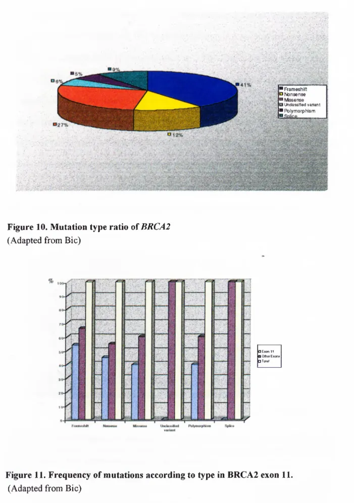
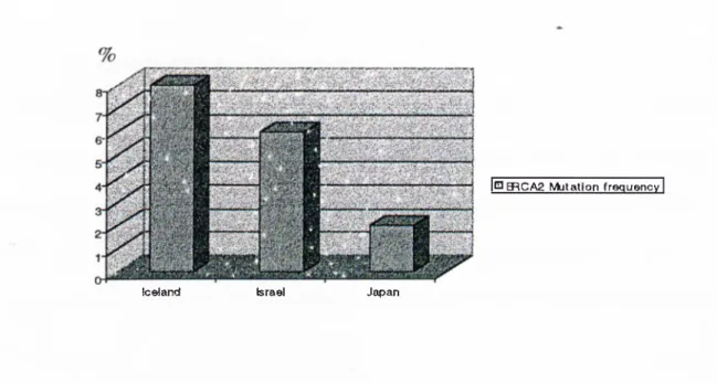
_____________________________________________________________________ 34 Figure 7. BRCAl mutation frequency in US, Hungarian and Icelandic families with male and female breast cancer._____________________________________________ 34 Figure 8. BRCAl mutation frequency in Italian, Israeli and Japanese population in breast and/or ovarian cancer patients not selected for family history. _____________ 35 Figure 9. Chromosomal location* and exonic organization of BRCA2**. _________ 37 Figure 10. Mutation type ratio of BRCA2____________________________________ 41 Figure 11. Frequency of mutations according to type in BRCA2 exon 1 1 ._________ 41 Figure 12. BRCA2 mutation frequency in English, Canadian, Finnish, French,
Hungarian, Icelandic, Swedish, Danish, and US populations in families with three or
more cases of female breast and/or ovarian cancer. 42
Figure 13. BRCA2 mutation frequency in US, Hungarian and Icelandic families with
male and female breast cancer cases. _______
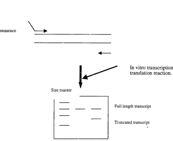
Figure 16. Schematic representation of protein truncation te s t._______ Figure 17. Comparison of SSCP, DGGE and HA in mutation detection. Figuréis. Pedigrees of hereditary breast cancer families.____________ Figure 19. Pedigrees of familial breast cancer families._____________ Figure 19. Analysis of extracted genomic DNA samples.
Figure 20. Analysis of PCR products.________________ Figure 21. PTT of BRCAl exon 11._________________ Figure 22. Heteroduplex analysis of BRCAl exon 2.
44 54 59 63 65 89 90 91 92
Figure 23. Heteroduplex analysis of BRCAl exon 5.__ Figure 24. Heteroduplex analysis of BRCAl exon 11 A. Figure 25. Heteroduplex analysis of BRCAl exon 1 IB. Figure 26. Heteroduplex analysis of BRCAl exon 11C. Figure 27. Heteroduplex analysis of BRCAl exon 1 ID. Figure 28. Heteroduplex analysis of BRCAl exon IIF. Figure 29. Heteroduplex analysis of BRCAl exon IIF _ Figure 30. Heteroduplex analysis of BRCAl exon IIG. Figure 31. Heteroduplex analysis of BRCAl exon 1IH. Figure 32. Heteroduplex analysis of BRCAl exon 11I._ Figure 33. Heteroduplex analysis of BRCAl exon 1IJ. _ Figure 34; Heteroduplex analysis of BRCAl exon IIK. Figure 35. Heteroduplex analysis of BRCAl exon 13._ Figure 36. Heteroduplex analysis BRCAl exon 2 0 .___ Figure 37. Heteroduplex analysis of BRCAl exon 24.___ Figure 38. Heteroduplex analysis of BRCA2 exon 11a.__ Figure 39. Heteroduplex analysis of BRCA2 exon 1 lb .__ Figure 40. Heteroduplex analysis of BRCA2 exon 1 la-b. Figure 41. Heteroduplex analysis of BRCA2 exon 1 I c __ Figure 42. Heteroduplex analysis of BRCA2 exon l i d __ Figure 43. Heteroduplex analysis of BRCA2 exon 1 l e __ Figure 44. Heteroduplex analysis of BRCA2 exon 1 I f __ Figure 45. Heteroduplex analysis of BRCA2 exon l l g . __ Figure 46. Heteroduplex analysis of BRCA2 exon 1 lg-26.
_ 93 _ 94 _ 95 _ 96 _ 97 _ 98 _ 99 100 101 102 103 104 105 106 107 108 109 110 111 112 113 114 115 116 Figure 47. Agarose gel electrophoresis of extracted mini prep DNA samples of 97/641 BRCAl exon IIB ._____________________________________________________ 121 Figure 48. Restriction endonuclease analysis of 97/641 mini prep DNA samples. 122 Figure 57. Secondary structure prediction of K654E substitution._______________ 144
ABBR EV IA TIO N S APS BIC bisacrylamide bp BRCAl BRCA2 cDNA cm dCTP del ddH20 ddNTP DNA dNTP EDTA g HA ins IPTG kb It M MCi min ml mM NCI ammonium persulfate
Breast cancer Information Core N,N, methylene bis-acrylamide base pair
BReast CAncer susceptibility gene 1 BReast CAncer susceptibility gene 2 complementary DNA
centimeter
cytosine deoxyribonucleoside triphosphate deletion deionized water dideoxynucleotide triphosphate deoxyribonucleic acid deoxynucleotide triphosphate ethylenediaminetetraacetic acid gram heteroduplex analysis insertion isopropyl "P-D thiogalactopyranoside kilobase liter Molar MiliCurie minute mililiter milimolar
nt nucleotide p32
phosphore 32
PCR polymerase chain reaction
pmol picomol
PTT protein truncation test
RNA dibonucleic acid
rpm revolution per minute
S35
sulphure 35
SDS sodium dodecyl sulphate
sec second
SSCP single strand conformation polymorphism
TBE Tris, Boric acid, EDTA
lEM ED N,N,N,N-tetramethyl-l,2 diaminoethane
U unit
uv ultraviolet
V volt
WHO World Health Organization
Xgal 5 Bromo-4 chloro-3 indolyl, P-D-galactopyranoside
|iCi microCurie
1. INTRO DUCTIO N
Cancer is one of the leading causes of death in human populations'. There have been many studies that aim to elucidate the mechanism of turnorigenesis, and many attempts to cure cancer in the 20 century. These efforts accumulate a huge amount of data about the nature and mechanism of tumorigenesis. Although the accumulated data is just the tip of the iceberg, it is now known that cancer appears as a result of the disruption of the genomic integrity. This disruption may arise in chromosomal or gene levels. In both ways there is either a gain (oncogene) or a loss (tumor supressor) of function of a particular set of genes that control the growth and the life time of a cell (Haber and Harlow, 1997). Thus the researches dealing with tumorigenesis are focused on either oncogenes or tumor supressor genes at several different scopes, all aiming to find out the underlying mechanisms of a cell’s life cycle.
Disruption of genomic integrity may occur in several different ways. It may develop sporadically, because of epidemiologic factors, lifestyle, or it may be hereditary. In either case, genomic integrity is disrupted. Recent experimental data has shown that some individuals are more susceptible to cancer because of their genomic heritage. Actually, more than a century ago, Paul Broca described four generations of breast
cancer in his wife’s family which underlined, probably for the first time, contribution of the heredity factor in tumorigenesis (Lynch etal. 1994).
Breast cancer is the most frequent cancer type and the second cause of death among women. Like many other cancer types, breast cancer may arise sporadically or hereditary factors may contribute to its development. It is estimated that 10 to 15% of breast cancer cases are hereditary (Feunteun 1999). In 1994 and 1995, as a result of an international collaboration of many research groups, two loci have been found to be linked to hereditary breast cancer (Miki et a/. 1994, Wooster et al. 1995). The subsequently identified genes were named as ^ e a s t C ancer susceptibility genes
BRCAl and BRCA2 are now considered to be responsible for approximately 60% of
hereditary breast cancer cases based on mutation analysis studies in many different populations (Szabo and King 1997). Functional studies, although not complete, show that these two genes are tumor suppressors that have roles in the maintenance of genomic integrity as caretakers (Vogelstein and Kinzler 1998).
Characterization of the mutations of a particular gene renders important information regarding the function of that gene. Besides, documentation of the mutation spectrum of a gene is one of the most reliable reflection of a population’s dynamics. The BRCAl and
BRCA2 mutation status of most Western populations has been studied during the last
five years. However, there is not enough data on these two structurally gigantic genes’ status in Eastern populations. Breast cancer is among the most common malignancies in Turkish women (Karaoguz and içli, 1993). The frequency and the types of germ-line mutations involved in Turkish breast/ovarian cancers are not well known. In order to
determine the contributions of BRCAl and BRCA2 mutations to the development of breast/ovarian cancer in the Turkish population, we screened pre-selected regions of these genes in four groups of patients composed of hereditary and familial cancer. As well as early onset and male breast cancer.
1.1. Cancer
Cancer is a common term used for all malignant tumors, and 2.3 million individuals die (Table 1) from cancer each year according to the 1998 estimates of WHO (The World Health Report, 1999). Although the ancient origins of this term is somewhat uncertain, it probably derives from the Latin for crab, “cancer”, presumably because a cancer “adheres to any part that it seizes upon in an obstinate manner like the crab”. Neoplasia means “new growth”. The term tumor was originally applied to the swelling caused by inflammation. Neoplasms also may induce swellings, and by long precedent the non- neoplastic usage of tumor has passed into limbo; thus, the term is now equated with neoplasm. Oncology (greek “oncos”= tumor) is the study of tumors or neoplasms (Robbins S. et al, 1984). Since the beginning of the century, researchers have studied cancer to understand the mechanisms of tumorigenesis. The original concept of negative regulators of tumor development can be traced to the earliest deseriptions of cancer as a genetic disease. The earliest known reference is to the German cell biologist Theodor Boveri, who proposed in 1902 that cancer might arise from the effects of both positive and negative regulators (Haber D. and Harlow E., 1997). It is known for many years that cancer arise as the result of genetic disruption which leads the cell to be immortalized
Table 1. Leading cause of death and their mortality rates in WHO member states
Mortality in WHO Member States
rank % o f total (000)
Botth s«*es ..;, ..'kjAk;...^
Ischaemic heart disease 1 13.7 7 375
Cerebrovascular disease 2 9.5 5 106
Acute lower respiratory infections 3 6.4 3 452
HIV/AIDS 4 4.2 2 285
Chronic obstructive pulmonary disease 5 4.2 2 249
Diarrhoeal diseases 6 4.1 2219
Perinatal conditions 7 4.0 2 155
Tuberculosis 8 2.8 1 498
Cancer of trachea/bronchus/lung 9 2.3 1 244
Road traffic accidents 10 2.2 1 171
.'.¡'v J..
Ischaemic heart disease 1 12.8 3 659
Cerebrovascular disease 2 8.2 2 340
Acutelowerrespiratory infections 3 6.1 1 753
Chronic obstructive pulmonary disease 4 4.3 1 240
fflV/AIDS 5 4.1 1 164
Diarrhoeal diseases 6 4.0 1 149
Perinatal conditions 7 3.9 1 121
Cancer of trachea/bronchus/lung 8 3.2 911
Tuberculosis 9 3.1 893
Road traffic accidents 10 3.0 855
S k / k k A 'i .
Ischaemic heart disease 1 14.6 3 717
Cerebrovascular disease 2 10.9 2 766
Acutelowerrespiratory infections 3 6.7 1 699
HIV/AIDS 4 4.4 1 121
Diarrhoeal diseases 5 4.2 1 070
Perinatal conditions 6 4.1 1 034
Chronic obstructive pulmonary disease 7 4.0 1 010
Tuberculosis 8 2.4 605
Malaria 9 2.1 538
Genetic disruption leading to tumorigenesis cause either a negative regulation - loss of function of tumor supressor genes, or a positive regulation - gain of function of oncogenes, in a cell’s life cycle. Tumorigenesis is a multistep process. Thus the genetic disruption is an accumulation of many genetic alterations. Some individuals are genetically susceptible to cancer, although most of the cases arise sporadically. This susceptibility comes from what is termed as “the first hit” . Knudson postulated that individuals with an autosomal dominant cancer susceptibility inherited a germ-line mutation in one of the alleles of a particular gene and that subsequent genetic alterations were required for tumor formation. Over the years Knudson’s hypothesis was refined to include a second hit at the same locus to inactivate the remaining normal allele. The past decade has seen numerous molecular confirmations of Knudson’s hypothesis (ex. APC,
R B ) and concrete demonstration of the multiple genetic events required for
tumorigenesis (ex. colon cancer) (Vogelstein and Kinzler, 1997).
1.2. O ncogenes
For many years it has been realised that damage to the DNA of a cell (mutation) is associated with changes that lead to cancer. It was initially believed that the genetic mutations responsible for cancer caused a deletion of essential regulatory genes restraining cell growth. Evidence for this has also come from cell fusion studies, which implied that malignant cells have lost functions that are dominantly expressed in normal cells, and are capable of suppressing malignancy. However, the discovery of retroviruses radically changed these ideas concerning the alterations that occur to genes in cancer; it
by genes that actively promote uncontrolled growth. The vast majority of non-viral oncogenes appear to be altered forms of cellular genes that encode proteins that participate in the pathway of cellular proliferation. The normal, intact cellular genes are known as proto-oncogenes. Cells receive signals to proliferate via growth factors that bind to receptors located on the outside surface of the cell. The signal is then transmitted into the cell, across the cytoplasm to the nucleus. There, the signal is converted into a growth response by means of transcriptional activation of the genes for proliferation. As a general rule, oncogenes are mutated forms of the cellular proto-oncogenes which encode the components of this signalling pathway. Direct comparisons of the DNA sequences of normal proto-oncogenes with their mutated oncogene derivatives have revealed the mechanisms by which proto-oncogenes become activated. Activation can occur either by changes to the coding sequence of the proto-oncogene so that a mutated protein -an oncoprotein- is produced with aberrant biochemical properties; or it can occur by mutations that deregulate the levels, and/or timing, of expression of the structurally unaltered proto-oncogene (Cooper GM, 1995).
The result of proto-oncogene activation is the gain of function in genes signalling proliferation to occur in cells which should not normally be proliferating. This inappropriate cell division will disrupt the normally ordered differentiation program of the cell. These activation processes can occur in a number of ways, which do not necessarily involve alteration of the structure of the proto-oncogene, but rather its forced expression at times in the development of the cell when proliferative genes should be silent and differentiative genes should be expressed (Burck KB et al, 1988).
Similarly, just as the overexpression of a normal gene product at the inappropriate time can lead to partial cell transformation, so the alteration of the coding sequence of a proto-oncogene can change the function of the protein product so that it relays its proliferative signals inappropriately. Such activation events can occur by deletions of portions of the coding sequence, by fusion of the sequence to other protein domains that alter the activity of the signalling domain, or by point mutations to the sequence at critical bases such that the amino acid sequence of the protein product is critically altered. The ras class of proto-oncogenes can become oncogenic by a change as small as a single base being changed in the sequence of the gene (Vile R., 1992)
1 .3 . Tum or suppressors “ Gatekeepers and Caretakers”
The molecular concepts of tumor suppressor genes stem from three lines of experimental evidence. The first is explained by Knudson’s model predicting the development of the childhood tumor retinoblastoma requires two rate-limiting genetic hits, subsequently shown to represent mutations of both alleles of a tumor suppressor gene. The hypothesis that children with inherited susceptibility to retinoblastoma harbour a germline hit, while sporadic cases have two somatic mutations, was confirmed with the cloning of the RB gene. A second essential observation was the fact that these genetic hits consisted of loss-of -function mutations. Loss of heterozygosity showed how loss of genetic material was the mechanism by which recessive mutations became manifest. A third experimental avenue was explored by Harris and co-workers in 1969, who found they could suppress the malignant phenotype by fusion of cancer cells with non-transformed
Many of the known tumor suppressor genes, including R B ,p53, W Tl, APC andp l6 , fulfill these basic early predictions-loss-of-function mutations, inactivation in both familial and sporadic tumors, and rescue of the tumor phenotype by the wild-type allele. For example, the classical concept that re-introduction of tumor suppressors into malignant cells inhibits their proliferation, is challenged by the new class of genes implicated in genomic integrity and DNA repair, whose loss leads to genetic damage that cannot be remedied simply by restoration of a wild-type allele.
The functions of known tumor suppressor genes are in a wide range of different cellular pathways. The current family of tumor suppressors includes DNA-binding transcription factors (p53 and W Tl), genes that may indirectly modulate transcriptional regulation
{RB, APC, possibly BRCAl), an inhibitor of kinases required for cell-cycle progression (pl6), a cell stmctural component (NF2), a novel phosphatase (P T E N M M A C l), a
potential mediator of mRNA processing (VHL), and genes implicated in signaling pathways including those of ras (NFl, TGF) Tumor suppressor genes involved in DNA repair and genomic stability (MSH2, M LH l, B R C A l and BRC A2) have also been described as caretakers.
Actually, recently Vogelstein and Kinzler divided most tumor suppressors broadly into two groups, named as “gatekeepers” and “caretakers” (Figure 1). Gatekeepers are genes that directly regulate tumor development by inhibiting its growth or by promoting its death. The functions of these genes are rate limiting for tumor growth, and as a result both the maternal and paternal copies of these genes must be inactivated for a tumor to
develop. The identity of gatekeepers varies with each tissue, such that inactivation of a given gene leads to specific forms of cancer predisposition. Because gatekeeping genes are rate limiting for tumor initiation, they tend to be frequently mutated in sporadic cancers through somatic mutation as well as in the germline of predisposed individuals.
In contrast, inactivation of caretakers does not directly promote growth of tumors. Rather, inactivation of caretakers leads to a genetic instability that only indirectly promotes growth by causing an increased mutation rate. Because numerous mutations are required for the full development of a cancer, inactivation of caretakers, with the consequent increase in genetic instability, can greatly accelerate the development of cancer (Vogelstein B. 1998). N O R M A L GATEKEEPER CARETAKER PATHWAY Mutation of a CARETAKER gene allele Mutation of 2“* CARETAKER gene allele leads to genetic instability Mutation of a GATEKEEPER gene allele Mutation of 2“* GATEKEEPER gene allele leads to tumor initiation N E 0 P L A S 1 A
Neoplasms constitute the most important lesions of the female breast. A great variety of tumors may occur in the female breast, made up as it is of a covering integument, adult fat, mesenchymal connective tissue, and epithelial structures (Robbins ei al, 1984). Breast cancer is the second leading cause of death among women of all ages according to the National Cancer Institute statistics. It becomes the first leading cause of death among women with age between 15-54.
1. 4. Breast cancer Rib ’ Muscle
p r
. ' v •Nipple -Areola-i / l/.i'V·' ' ... k k k ' ; , v-fakpFigure 2. M ain anatom ic structures o f breast, (from NCI database)
Each breast has 15 to 20 overlapping sections called lobes. Within each lobe are many smaller lobules, which end in dozens of tiny bulbs that can produce milk. The lobes.
lobules, and bulbs are all linked by thin tubes called ducts. These ducts lead to the nipple in the center of a dark area of skin called the areola. Fat fills the spaces around the lobules and ducts. There are no muscles in the breast, but muscles lie under each breast and cover the ribs. Each breast also contains blood vessels and vessels that carry colorless fluid called lymph. The lymph vessels lead to small bean-shaped organs called lymph nodes. Clusters of lymph nodes are found near the breast in the axilla (under the arm), above the collarbone, and in the chest. Lymph nodes are also found in many other parts of the body.
Breast carcinoma arises from the epithelium of the mammary gland, which includes the milk-producing lobules and the ducts that carry milk to the nipple. Malignant transformation of the stromal, vascular, or fatty components of the breast is not included in this definition and is extremely rare. The transition from normal to malignant breast epithelium has not been as well studied as the parallel changes in colonic epithelium; however, there is increasing evidence that the breast epithelium undergoes a transformation from normal to hyperplastic, followed by the appearance of atypia in association with the hyperplasia, ultimately becoming malignant. Malignant cells continue to evolve from noninvasive carcinoma, typified by in situ to invasive carcinoma, and ultimately to cells with metastatic potential (Kinzler and Vogelstein,
1997). Breast cancer is generally classified as invasive or non-invasive. Invasive cancer originates in the lobules and/or milk ducts, while while non-invasive or in situ cancers are confined to the lining of the lobules or ducts. The most important criteria for assessing future risk of invasive disease is to determine whether cancer cells have
according to growth rate. Well-differentiated tumors are termed as grade I; moderately differentiated tumors as grade II; and poorly differentiated tumors are termed as grade III. The most common type of breast cancer begins in the lining of the ducts and is called ductal carcinoma. Another type, called lobular carcinoma, arises in the lobules. When breast cancer spreads outside the breast, cancer cells are often found in the lymph nodes under the arm (axillary lymph nodes). If the cancer has reached these nodes, it may mean that cancer cells have spread to other parts of the body—other lymph nodes and other organs, such as the bones, liver, or lungs via the lymphatic system or the bloodstream.
The treatment and prognosis of a women with breast cancer are strongly influenced by the stage at the time of diagnosis. Multiple staging systems have been proposed, but the most commonly used system is the one adopted by both the American Joint Committee (AJC) and the International Union against Cancer (UICC). This staging system is a detailed TNM (tumor, nodes, metastasis) but can be summarized as in Table 2.
Table 2. TNM Staging
Stage 0 Carcinoma in situ
Stage I Tumor 2 cm, axillary nodes not involved
Stage II Tumor between 2 and 5 cm and/or involved but mobile axillary lymph nodes
Stage III Tumor larger than 5 cm and/or fixed axillary lymph nodes; includes
inflammatory breast cancer
There are several determinants of breast cancer occurrence such as reproductive history, diet and alcohol consumption, endogenous hormones and estrogen receptor expression, and exogenous hormones. There are many case-control studies analyzing the effect of reproductive factors on breast cancer. The possibility of a protective effect for mothers of twins versus mothers of single births (odds ratio 0.7) received support from a large nested case-control study conducted in Sweden (Murphy et al, 1997). This association may be correlated with either to the influence of hormonal characteristics of pregnancy or to genetic differences between women who give birth to twins and those who do not. Women appear to be at higher risk of developing breast cancer during or shortly after pregnancy, and the prognosis for such pregnancy associated breast cancers was observed to be poorer than for tumors unrelated to pregnancy (Bonnier,, et al, 1997). The possible influence of lactation on the risk of breast cancer is also under investigation. Some recent studies suggest that there is a protective association between breast feeding and breast cancer among both premenopausal and postmenopausal women (Enger et al,
1997).
Numerous studies have reported on the relationship between drinking alcohol and breast cancer, and the results have tended to indicate that alcohol consumption is associated with a slightly increased risk of breast cancer (Schatzkin, et al 1994)
Hormone replacement therapy is used in cardiovascular disease, in osteoporosis, and in the relief of menopausal symptoms. An analysis of studies on this topic showed that current hormone replacement therapy users were approximately 20% more likely to develop breast cancer than those who never used this therapy. The risk decreases in a nonuser 5 year after cessation of use (Coll. Group on Horm. Fact. In Breast Ca, 1997). These results suggest that estrogen acts late in the sequence of breast carcinogenesis and its tumor-promoting effects may be reversible (Alberg et al, 1998).
*Г) В <υ D H-3 Ö S, *0 u в î4> CÍ "елcö 2i PQ ГО e cd Рч Ч-»л вд с о 0 1ел О l·-! Рн с О Ή О е о <1> cd Рч 3 0> Рч h-:i3 3 JPо G В CQ 5 PQ »л с _о о о В о о > О Ü І- і О § Р-(U СА С О о DD « О OD -3 о о о л fi (U в о тз fi fi fi
ε
&D fi о fi fi fi u Ö D , f i fi > e {/} ■*■* *fi'M U O fO ? »Л *л *cd В o ‘Л m В Ό Р ч İd В Un Ό Я Рч 3 JG O G O t-< PQ ‘ЛON G Я о в в о й ÖÛ G 3 PQε
о 3 <υ hQε
о 3 <l> bQ 3 Λ о с 2 PQ VO ел »л о о 3 hQ РчI I
(N ГЛ о оε
рг 3 <ϋ ь-) Я Şİ r гл т г PQ РЗ с 2 ÇQ г -гл PQ (N В в ел 2 Рч с О *ο оε
в о P Í о § Рч b cd > О ’ SЦм РР §о X t <ύ О о G cd Рч ΓΝ ГЛ гл о гл г -<N X Έ(D О G 'Su« PQ ë •С о О •3 G W С ‘Со о T J с PQ с о υ сю ; ζ о гл(Ν РР н <tîо 00 § О с о çr\ 00гл »л УП в во о (В о G cd Рч ’ S ) тз о д S ' я <tî о 00 b cd >о
ІЛ гл fi fi Q 'ΐ 3 о σ1 ч- t 0 0 ο ο Ον Ον τ-Η 2І 3 ' ΰ СЛ С I—( ν-( <ύ ο fi fi и * f i fi o fi ϊ ζ o Ч-Ч P O fi • rHEvidence concerning reproductive history and exogenous estrogen use suggest that estrogen is involved in the etiology of breast cancer (Alberg et al 1998). Several studies, including a multi country study for the World Health Organization, suggest that oral contraceptives (used by 61 million women worldwide) are associated with a relative risk of 1/3 to 1/5 for breast cancer that will be diagnosed before the age of 40-45 (Hulka and Stark, 1995).
That hormones influence risk and prognosis of breast cancer has been known for decades. Breast cancer is rare in men, suggesting an influence of sex-steroid hormones. Women who have had bilateral oophorectomy early in life are at markedly reduced risk of subsequently developing breast cancer; the earlier oophorectomy is done, the greater the reduction. Early age at menarche (11 years or less) and late age at natural menopause (55 or older) are associated with increased risk (Hulka and Stark, 1995). Antiestrogens are employed clinically because of their ability to inhibit the growth of breast cancer cells. In addition, disease progression inevitably occurs, indicating a transition to hormone resistance even in the presence of the estrogen receptor (Eisen and Weber,
1998). The responsiveness of breast tissue to estrogen exposure would be expected to lead to differences in susceptibility to breast cancer. This is supported by the results of a case-control study which showed that the normal breast tissue of women with breast cancer was three times more likely than the breast tissue of the control patients to have estrogen receptor overexpression (Khan, et al, 1997).
1.4.2 G enetic factors in Breast Cancer
I.4.2.I. Som atic M utations in Breast Cancer
The study of sporadic breast cancers is important in order to understand the pathogenesis of breast cancer. Sporadic breast cancer which constitutes approximately 90% of all breast cancer cases, have fundamental molecular genetic differences. These somatic alterations are in growth factors and receptors, intracellular signalling molecules, regulators of cell cycle, adhesion molecules and proteases. In addition, LOH of many loci, such as chromosomes 17p, 17q, 16q, 13q, lip . Ip, 3p and 18q (Cleton-Jansen, 1994; Gudmundsson, 1993; Cropp,1993; Mathew, 1994; Dorion-Bonnet, 1995).
Members of the epidermal growth factor receptor (EGFR) family are frequently altered in sporadic breast cancer. These proto-oncogenes, EGFR, erbB-2 or HER-2, and erbB4, become oncogenic through gene amplification or overexpression resulting in the aberrations in signal transduction pathways and thus deregulation o f cellular proliferation (Bacus, 1994).
Disruption of the cell cycle checkpoints leads to uncontrolled growth and cancer. The tumor suppressor protein p53 which is known also as genome guardian, plays a central role in regulating progression through cell cycle. p53 Alterations in breast cancer are shown by analyzing the coding region for mutations or using antibody demonstrating aberrant localization or altered levels of the protein. p53 mutations have been detected in
15 to 45% of human breast cancer specimens in several studies (Deng ei al, 1994; Andersen et al, 1993; Saitoh et al, 1994)
Like p53, RB regulates cell cycle progression. Dephosphorylated RB inhibits cellular proliferation by halting cell cycle progression in G l. Structural rearrangements and inactivation of RB is detected in breast cancer (T’ang et al, 1988; Lee et al, 1988). In addition it is shown that estradiol decreases expression of RB (Gottardis et al, 1995). Another growth inhibitory protein TGF-(3 which is shown to be hormonally regulated is found to be decreased in breast cancer (Jeng et al, 1993). Cyclins which play important roles in different cell cycle checkpoints are studied in breast cancer. Cyclin D1 which regulates the Gl-S transition, is shown to be overexpressed in breast cancer (Musgrove
etal, 1991).
The programmed cell death, apoptosis, is an important physiological defense mechanism of cells to avoid cancer. The proto-oncogene, bcl-2, which functions to suppress apoptosis is found to be overexpressed in 30-45% of breast cancer cases (Johnston et al, 1994). Another proto-oncogene, c-myc, is also found to be overexpressed in breast cancer (Pavelic etal, 1992).
1.4.2.2. Gerinline Mutations in Breast Cancer
The search for reliable genetic markers for the susceptibility to develop breast cancer has proven to be difficult, since the disease itself is not due to a single gene mutation and even the predisposition can be based on different gene defects. There are several tumor suppressor genes associated with several syndromes that are thought to be involved in genetic predisposition to breast cancer.
I.4.2.2.I. Li-Fraumeni Syndrome (p53)
This rare dominantly inherited cancer syndrome is characterized by a predisposition to sarcomas, breast cancer, brain tumors, leukemia and adrenocortical carcinoma in children and young adults. Approximately 50% of the women with the Li-Fraumeni syndrome may develop breast cancer. Germ-line mutations found in the p53 tumor suppressor gene explains this disorder (Malkin et al, 1990). However, it has been shown that less then 1% of the women who develop breast cancer at a very young age carry a germ-line mutation in the p53 tumor suppressor gene (Borresen et al, 1992)
I.4.2.2.2. Lynch Type II Syndrome (M LH l)
The Lynch type II syndome is associated with a dominantly inherited susceptibility to a variety of tumors including breast, colon, uterine and ovarian cancers and melanomas. The risk of developing breast cancer is increased in individuals from families with the
Lynch type II syndrome, and the relative risk for breast cancer in first-degree relatives of index cases identified because of colon cancer has been estimated to be about 5% (Krainer, 1994).
L4.2.2.3. Ataxia Teleangiectasia (ATM)
It has been suggested that heterozygotes for the gene for ataxia teleangiectasia (A'l) may represent a large proportion of those at high risk for breast cancer, since the relative risk for breast cancer in A T heterozygotes is said to be between 7% and 15%, and the
heterozygote frequency is estimated to be 1.4%. However, a linkage to the locus in
familial breast cancer could not be found (Wooster, ei al, 1993).
I.4.2.2.4. Cowden Disease (PTEN)
Cowden disease (CD) is an autosomal dominant syndrome characterized by the development of hamartomas in multiple organ systems, and an increased risk of developing breast and thyroid cancers. The CD locus was mapped to chromosome 10q22-23 in 1996 (Nelen, ei al, 1997). Two groups independently identified a tumor suppressor gene (PTEN) at 10q23 using loss of heterozygosity analysis in glioblostoma multiforme (Steck ei al, 1997; Li el al, 1997). Mutations in this gene were found in primary gliomas and carcinomas of the breast and kidney as well as in related cell lines. Subsequently, mutations in P T E N have been identified in multiple CD kindreds identifying PTEN as the gene responsible for CD when mutated. So far no germ-line
mutations in breast cancer families have been identified. These data provide strong evidence that PTEN does not account for hereditary breast cancer susceptibility outside
of families that are affected by CD. Finally, somatic mutations in primary breast
cancer specimens have been identified only very rarely (Rhei el al, 1997, Teng, et al, 1997).
I.4.2.2.5. Genetic Polymorphisms
Environmental contaminants have potential to influence breast cancer risk. The most v/ell-defined environmental risk factors are radiation exposure and alcohol ingestion. Diet is clearly related to the increased incidence of breast cancer in developed countries, but its precise role is not yet established. Epidemiologic and écologie investigations must take into account the very complex etiology of breast cancer, and the knowledge that tumorigenesis can arise from different mechanisms. Genetic polymorphisms exist in genes that govern capacity to metabolize environmental contaminants. Higher risk may occur among persons whose enzymes either are more active in the production of procarcinogens or fail to detoxify carcinogenic intermediates formed from chemicals in the environment (Wolff and Weston, 1997).
Catechol-O-methyltransferase (COMT) participates in the metabolism of potentially harmful estrogen metabolites. In a nested case-control study, the low activity COMT allele was not strongly associated with breast cancer overall, but was associated with increased risk among postmenopausal women, particularly those with higher body mass
and breast cancer overall, but it was associated with decreased risk among postmenopausal women, and increased risk among premenopausal women in the study
(Thompson a/, 1998).
The results of a nested case-control study showed a strong association between putative high risk genotypes for three of the glutathione S-transferases (GiST^), which act to detoxify carcinogens. Women with all three hypothesized high risk genotypes were over three times more likely to develop breast cancer than women with all three putative low risk genotypes (Gharrier et al, 1999). Other studies, however, did not observe significant associations between breast cancer and the GSTMl positive or GSTM l null genotypes
(Ambrosone a/, 1999).
The CYP17 gene may influence the risk of breast cancer by influencing estradiol production. In a case-control study, little association was present between CYP17 polymorphisms and breast cancer overall but the presence of an A2 allele was associated with a 2.5-fold excess risk among women with advanced stage disease (Haiman et al,
1999).
Slow and rapid acetylator individuals exist in most human populations, and the mutations responsible for the slow-acetylator genotype have been determined. Slow acetylators, is at higher risk of developing breast cancer, and fast acetylators, have an increased risk of developing colon cancer (Rodrigo et al, 1999). Previous findings suggested that cigarette smoking was a risk factor for postmenopausal breast cancer
among women with the N-acetyltransferase 2 slow acetylation genotype (Zheng et al, 1999).
Besides all of these genetic factors that are supposed to be involved in genetic predisposition to breast cancer, it has been found that two genes are acting a major role in familial breast cancer.
1.5. H ereditary Breast Cancer Genes
Most genetic changes arise spontaneously (sporadic mutations) and are not inherited from either parent. Familial and hereditary are terms that refer to increased cancer risk within families; however, they are not synonymous. Familial cancer is defined as the simple clustering of disease within families. Hereditary cancer is a more specific term, referring to a subtype of familial disease with a pattern of distribution consistent with Mendelian inheritance of a susceptibility gene. Families in which the predisposition for malignant disorders appears to be restricted to breast cancer are known as “breast specific” families. There are also families with an increased risk for ovarian cancer along with breast cancer. Furthermore, there is evidence that these families show a higher incidence of prostate, colon and pancreas cancer (Krainer, 1994). Women are not all at equal risk for contracting breast cancer. The most important factor other than age that determines who will and who will not manifest breast cancer is the family history of breast and related cancers. Descriptive studies of pedigrees of breast cancer-prone families and epidemiologic studies of breast cancer in the general population are the
he described four generations of breast cancer in his wife’s family, an enormous number of breast cancer-prone families have been reported. Usually the pattern of breast cancer occurrences in these families is most consistent with an autosomal dominant mode of inheritance (Lynch, etal, 1994).
1.5.1 Breast Cancer susceptibility gene 1 (BRCAl)
1.5.1.1 Identification o f BRCAl
Description of families with multiple cases of breast cancer has led the researchers to hunt for a breast cancer susceptibility genes. Linkage analysis allows the localization of human disease genes in families with high incidence of the disease. This analysis relies on the identification of one or more markers in the genome that segregate with the disease. In 1990, Hall et al., made a genetic analysis on 23 extended families with 146 cases of breast cancer. These families share the epidemiological features that are characteristic of familial, versus sporadic, breast cancer: younger age at diagnosis, frequent bilateral disease, and more frequent occurrence of disease among men. The result of this study showed that familial breast cancer is linked to 17q21 (Hall, et al, 1990) . Several confirmations of this result were subsequently published. After the first confirmation by Narod and his coworkers in 1991 (Narod, et al. 1991) this breast- ovarian cancer locus has been formally labelled ''B R C A l” (Solomon and Ledbetter, 1991) . Then a third confirming report came from Breast Cancer Linkage Consortium for the locus on 17q21 (Easton, et al, 1993).
After the localization of BRCAl to 17q21, great efforts were made to identify the gene, its transcript, and protein. Miki and his coworkers developed a detailed map of transcripts for the 600 kb region of 17q21 between D17S1321 and D17S1325 (Figure 3). Sixty-five candidate expressed sequences within this region were identified. Three expressed sequences eventually were merged into a single transcription unit whose characteristics strongly suggest that it is BRCAl. Conceptual translation of the cDNA revealed a single, long open reading frame encoding a protein of 1863 amino acids. Homology searches revealed that the protein contains a zinc finger domain in its amino terminus. Probing of genomic DNA samples from different species with B R C A l sequences revealed strong hybridizations in tissues from humans, mice, rats, rabbits, sheep, and pigs. In the same study of Miki and co-workers an 11 bp deletion, a 1 bp insertion, a nonsense, a missense and a regulatory mutation that were segregating with breast cancer in families linked to 17q21 were identified (Miki Y. et al, 1994).
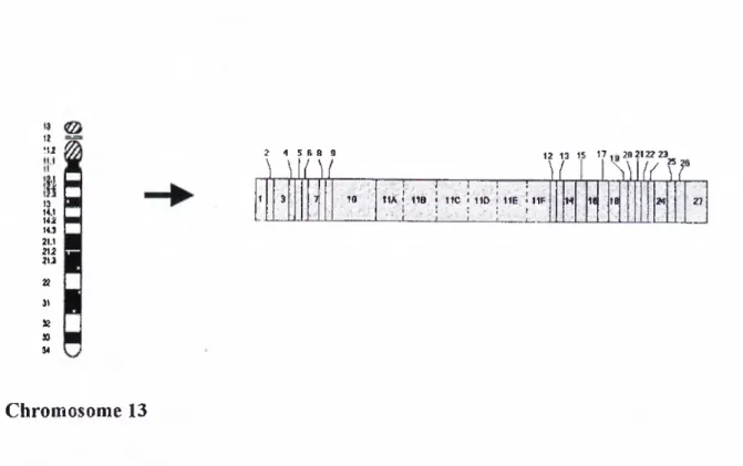
I) li Hi 11.1 21.1 2!i 21,3 n 23 H 2S 9 10 t|2 14 19 20 21 s ? ;|8 i j ■ ; ; ■ i l A : '· ■ · l i e '· ·'■ ;■■■ ; i : i u r , ; : IS · ;■ -i!S; ■ ■ ' Chromosome 17
Figure 3· Chromosomal location and exonic organisation of BRC Al * adapted from NCBI database
BRCA] appeared to encode a tumor suppressor protein. In order for B R C A l to be a
tumor suppressor gene it must bear the most important characteristic of a tumour suppressor. That is BRCAl protein should be present in normal breast and ovarian tissue and should be altered, reduced or absent in breast and ovarian tumors to fulfill the loss- of-function characteristic of a tumor suppressor gene. After the localization of BRCAl to 17q21 many research groups studied whether BRCAl has this characteristic or not, by loss of heterozygosity studies (Smith, ei al, 1992; Lindblom, el al, 1993; Cornelis, el al, 1993; Neuhausen, el al, 1994). Just six months before the identification of BRCAl it was shown that transfer of an intact human chromosome 17 led a breast cancer cell line (MDA-MB-231) to loose its capacity to induce tumors in nude mice. This result obtained from one clone (MDA-231/H17) which bears the long arm of chromosomel7, indicated that at least one gene mapping to this region could be a tumor suppressor, confirming the BRCAl identification and LOH studies (Negrini, elxtl, 1994). Another study by Rao and co-workers also provided evidence for the tumor suppressive effect of
BRCAl. In this report, it is stated that antisense RNA to BRCAl transforms mouse
fibroblasts (Rao, etal, 1996).
I.5.I.2. The Function of BRCAl
After the identification of BRCAl and definition of a zinc finger motif in its amino terminus in 1994, it is thought that the gene product is a tumor suppressor that may be a transcription factor (Miki, el al, 1994). Until 1996 there was no definite evidence about
the function of BRCAl protein. That year two successive reports stated that the carboxy terminal of the protein (nt 1528-1863) bears a transcriptional activation function (Chapman and Verma, 1996; Monteiro, et al, 1996).
A very exciting finding appeared in 1996 by Jensen et al, stating that BRCAl is actually a protein that is secreted, and exhibited properties of a granin. This result was relied on homology search and immunological studies. This was a very important finding because BRCAl would be the first secreted tumor suppressor protein (Jensen, et al, 1996). But four months later a report by Wilson et al discussed the specificity of the antibody used in this paper, and found out that the detected protein was actually EGFR but not BRCAl (Wilson e/a/, 1996).
Gudas and co-workers found that BRCAl mRNA and protein levels are regulated by the steroid hormones and progesterone (Gudas, et al, 1995). Later, another group has suggested iheX BRCAl expression is not directly responsive to estrogen but it is induced as a result of the mitogenic activity of estrogen in estrogen receptor positive cells (Marks, etal, 1997).
Experimental inhibition of BRCAl expression with antisense oligonucleotides produced accelerated growth of normal and malignant mammary cells, but had no effect on non mammary epithelial cells (Thompson, et al, 1995). It was also shown that retroviral transfer of the wild-type BRCAl gene inhibits in vitro growth of all breast and ovarian cancer cell lines tested (Holt, et al, 1996). An evidence on how BRCAl mediate growth
transactivated expression of cyclin-dependent kinase inhibitor p21 in a /?53-independent manner, and that B R C A l inhibits cell-cycle progression into the S-phase (Somasundaram et al, 1997). There is also a report on the induction of apoptosis by
BRCAl (Shao etal, 1996).
Little is known about the role of BRCAl in human development. But there are several studies on the role of BRCAl in mice development. These results suggest that BRCAl is crucial in the embriyonic development and that BRCAl homozygous mutants were not able to survive (Liu et al, 1996; Lane et al, 1995, Gowen et al, 1996). It is also found
ihniBRCAl is expressed in rapidly proliferating cell types undergoing differentiation. In
the mammary gland, B R C A l expression is induced during puberty, pregnancy, and following treatment of ovariectomized animals with estrogen and progesterone (Marquis
et al, 1995). The role of BRCAl in human development remains unclear. There is one
report on a human B R C A l gene knockout. The reported case was a women who had breast cancer diagnosed at age 32 (Boyd et al, 1995). This data implies that the role of
BRCAl in human development is not as crucial as in mice development or B R C A l
mutations’ penetrance are rather wide.
It was in 1994 just before the publication of BRCAl identification in a review Vogelstein and Kinzler stated some predictions on the possible functions of BRCAl protein. There they suggested that BRCAl gene product was part of a repair or replication complex that only indirectly affects the growth of breast cancer cells in a similar way to the mismatch repair genes (Kinzler and Vogelstein, 1994). Their predictions were actually confirmed by several different studies. First report has come from Scully and his coworkers noting
that BRCAl is associated with RadSl, a protein that functions in DNA double stranded break repair, in mitotic and meiotic cells (Scully et al, 1997). In a following report, Scully and his coworkers stated that BRCAl is a component of the RNA polymerase II holoenzyme, linking BRCAl with the transcription process (Scully ei al, 1997). The finding of BRCT module (BRCAl C-terminus) has emphasized the potential importance of the BR.CA1 C-terrninal region for BRCAl-mediated breast cancer suppression, as this domain showed similarities with the C-terminal regions of a p53-binding protein (53BP1), the yeast RAD9 protein involved in DNA repair.
Another evidence confirming and supporting the function of BRCAl in DNA repair and transcription process came from Gowen and co-workers stating that mouse embryonic stem cells deficient in BRCAl are defective in the ability to carry out transcription- coupled repair of oxidative DNA damage, and are hypersensitive to ionizing radiation and hydrogen peroxide (Gowen ei al, 1997). A more precise evidence that links BRCAl to transcription process noted that BRCAl protein is linked to the RNA polymerase II holoenzyme complex via RNA helicase A (Anderson ei al, 1998). The most recent evidence that supports the role of BRCAl protein in DNA repair shows that BRCAl interacts in vitro and in vivo with hRadSO, which forms a complex with hM rell and p95/nibrin. Upon irradiation, BRCAl was detected in discrete foci in the nucleus, which colocalize with hRadSO. Formation of irradiation-induced foci positive for BRCAl, hRadSO, hMrel 1, or p95 dramatically reduced in HCC/1937 breast cancer cells carrying a homozygous mutation in BRCAl but was restored by transfection of wild-type BRCAl (Zhongeia/, 1999).
The important function of BRCAl defined in DNA repair puts it in a crucial role as a caretaker gene. This finding lead to the studies that search its relation with the genome guardian, p53. It was shown that BRCAl physically associates with p53 and stimulates its transcriptional activity. BRCAl and p53 cooperatively induce apoptosis of cancer cells indicating that they may coordinately regulate gene expression in their role as tumor suppressors (Zhang ei al, 1998; Ouchi et al, 1998). It was also found that breast tumors with mutant alleles of BRCAl show high frequency of p53 mutations (Glebov, 1994). This finding is attributed to its role in DNA repair implying a mutator phenotype to BRCAl associated breast tumors. Although this finding is supported by a study demonstrating that disruption of BRCAl causes genetic instability and triggers further alterations, including the inactivation of p53, that lead to tumour formation (Xu et al, 1999). Another study shows that the correlation between BRCAl and p53 is not because of the attributed role to BRC Al as a mutator, but to multiple mechanisms (Crook et al, 1998). Another finding suggested that BRCAl and BRC A l respond to DNA damaging agents, regulated by a p53-sensitive component (Andres etal, 1998).
It is found that in sporadic breast cancer cells 5’ aberrant méthylation of BRCAl CpG island leads to decrease in BRCAl mRNA suggesting a mechanism of BRCAl repression in sporadic breast cancer (Rice et al, 1998).
I.5.I.3. M utations in BRC Al
Demonstration of germ-line mutations that disrupt the function of BRCAl protein was cmcial to establish primary involvement of this gene in hereditary breast cancer. The