The relationship between positron emission
tomography‑computed tomography
imaging and histopathological features of
thyroid incidentalomas detected during
follow‑up for primary malignancy
ABSTRACT
Aim of the Study: While the rate of thyroid incidentaloma detected on positron emission tomography (PET) was reported as 4%, the malignancy rate was 14%–50%. We evaluated the thyroid nodules which were detected by PET‑computerized tomography (CT) in cancer patients and analyzed the pathological results of those thyroid nodules diagnosed by fine needle aspiration biopsy (FNAB) and their correlation with the maximum standardized uptake (SUVmax) value and PET imaging features.
Materials and Methods: FNAB were performed for 40 thyroid incidentalomas. We analyzed the relationship between the histopathological findings and radiological features by Pearson’s correlations and Chi‑square‑Fisher’s exact tests to evaluate the factors associated with SUVmax.
Results: The median SUVmax values were 5.4 for thyroid nodules. Totally, 14 malignancies were detected by FNAB (35%).The sensitivity and specificity of SUVmax value for diagnosis of malignancy were 87.5% and 52%, respectively. Positive and negative predictive values were 36.8% and 92.8%. The most common malignant and benign pathologies were classic variant papillary carcinoma and benign colloidal nodule. The median SUVmax was the higher in colon cancer thyroid metastasis and oncocytic neoplasia (SUVmax 14.5 and 13.6, respectively). Histopathological type was not related with nodule size but positively associated with categorical SUVmax (r = 0.318, P = 0.04) and negatively correlated with both the density of the thyroid nodule in PET‑CT (r = −0.0042, P = 0.01) and density of nodule in ultrasound (USG) (r = −0.305, P = 0.05). Margin of the thyroid nodule in USG (P = 0.007) and internal component of the nodule in PET (P = 0.03) were found to be important factors to differentiate benign or malignant lesion. Conclusion: If the thyroid nodule is detected with flouro‑2‑deoxy‑D‑glucose uptake, to differentiate benign nodule from malignant, cytological examination is noteworthy to diagnose the more aggressive type of thyroid nodule and also thyroid metastasis from primary cancer.
KEY WORDS: Biopsy, malignancy, positron emission tomography‑computerized tomography, thyroid incidentaloma
INTRODUCTION
Thyroid nodules have been reported in 5.3% of women and 0.8% of men.[1] With improvements
to radiological imaging modalities, incidental thyroid nodules are now found more frequently. Incidental thyroid nodules have been reported in 50%–60% of autopsy series without a palpable thyroid nodule clinically.[2] Thyroid
incidentaloma is an asymptomatic, unsuspected thyroid lesion that is diagnosed by an imaging study unrelated to the thyroid gland.[3] These
lesions are detected by ultrasonography (USG), c o m p u t e r i z e d t o m o g r a p h y ( C T ) o r flouro-2-deoxy-D-glucose (FDG) positron emission tomography (PET).[3] The detection rates of thyroid
incidentaloma by radiological modalities are 67% with USG, 16% with CT or MRI, and 2%–3% with
Bala Basak Oven, Zeynep Gamze Kilicoglu1, Ahmet Bilici2, Mehmet Tarik Tatoglu3, Sule Canberk4, Metin Tilki5, Fugen Aker Vardar4 Departments of Medical Oncology, 1Radiology, 4Pathology, 5General Surgery, Haydarpasa Numune Education and Research Hospital, 2Department of Medical Oncology, Medipol University, 3Department of Nuclear Medicine, Medeniyet University, Istanbul, Turkey For correspondence: Dr. Bala Basak Oven Ustaalioglu, Selimiye Mah, Sair Nesimi Sok, Kardesler Apt No. 1, Daire 4,34668, Uskudar, Istanbul, Turkey. E‑mail: basakoven@ yahoo.com
Access this article online Website: www.cancerjournal.net DOI: 10.4103/jcrt.JCRT_889_16 Quick Response Code:
This is an open access journal, and articles are distributed under the terms of the Creative Commons Attribution‑NonCommercial‑ShareAlike 4.0 License, which allows others to remix, tweak, and build upon the work non‑commercially, as long as appropriate credit is given and the new creations are licensed under the identical terms.
For reprints contact: [email protected]
Cite this article as: Oven BB, Kilicoglu ZG, Bilici A, Tatoglu MT, Canberk S, Tilki M, et al. The relationship between positron
emission tomography-computed tomography imaging and histopathological features of thyroid incidentalomas detected during follow-up for primary malignancy. J Can Res Ther 2019;15:589-95.
PET.[3] The risk of cancer in the thyroid nodules detected by
USG is 1.5%–10%.[4]
PET is a rapidly developing imaging modality which is used for diagnosis and tumor staging, to assess treatment response and to perform follow-up in oncology; it can also detect thyroid nodules incidentally.[5] In contrast to USG or CT, PET provides
not only the anatomical location of nodules, but it also provides information on metabolism and biological activity.[6] FDG is
trapped by metabolically active tissue so that tumor tissue may be seen as a hypermetabolic focus on PET imaging.[3]
Metabolic information, together with anatomical information detected by PET, means that detected thyroid incidentalomas are more likely to be malignant than thyroid incidentalomas detected by USG or CT.[6] While the normal thyroid has no FDG
activity on PET, abnormal FDG uptake in the thyroid gland may be benign or malignant. The risk of malignancy can be predicted by radiological features. Hypoechogenicity, irregular margins, larger size, and punctate calcifications detected by USG increase the suspicion of malignancy.[3,7] The sensitivity of
these features to detect malignancy has been reported to be 94%.[8] On the other hand, focal or unilateral FDG uptake on
PET imaging is more likely to be associated with malignancies than diffuse uptake.[3] Diffuse uptake is generally associated
with benign diseases such as thyroiditis. It is difficult to differentiate benign from malignant thyroid incidentalomas based on maximum standardized uptake (SUVmax) values only.[4]
The malignancy rate of thyroid incidentaloma detected by CT has been reported as 3.9%–11.3%.[9] The prevalence
of malignancy of thyroid incidentalomas detected by PET ranges from 14% to 47%.[6] Metastatic involvement of the
thyroid gland from a primary tumor was reported to be 1.2% in 162 patients with a known malignancy.[10] The American
Thyroid Association recommends fine needle aspiration biopsy (FNAB) for thyroid nodules >1 cm in diameter or nodules <1 cm in diameter with abnormal USG features.[11]
The aim of our study was to evaluate thyroid nodules which were detected by PET-CT in patients with malignancy unrelated to thyroid cancer. We analyzed the pathological results of those thyroid nodules diagnosed by FNAB and their correlation with the SUVmax value and PET imaging features.
MATERIALS AND METHODS
The data of 1840 patients with primary malignancy were evaluated retrospectively. A total of 40 cancer patients with thyroid incidentaloma who had been followed up at two Medical Oncology Centers in Turkey between 2009 and 2014 were included in this study. All patients underwent FNAB to histopathological confirmation. Thyroid incidentalomas were detected by PET-CT, which were performed for diagnosis, staging, and treatment response assessments for a primary malignancy unrelated to the thyroid. Eligibility was limited to patients with a primary malignancy and patients who
had incidental thyroid nodules detected by FDG activity on PET-CT during diagnosis or treatment of a primary malignancy. Thyroid USG was performed; then, all patients underwent FNAB guided by thyroid USG. Patients with a known history of thyroid malignancy, patients who had insufficient primary disease information or thyroid nodule biopsy results, and patients who did not agree to undergo thyroid biopsy were excluded from the data analysis. All pathological slides of primary malignancies and thyroid nodules were re-evaluated to confirm the histopathological subtypes and the diagnosis of metastasis from known malignancies by an experienced cytopathologist at both centers.
Thyroid USG imagings were reported according to internal echogenicity, density, margins, and presence of calcifications. The presence of solidity, hypoechogenicity, irregular margins, microlobulation, and microcalcification was accepted as USG abnormalities.[7] PET-CT imagings were also evaluated
according to thyroid nodule density, internal component, and SUVmax values. The age of the patients at diagnosis, primary tumor, histopathological type, tumor stage, thyroid nodule size, thyroid nodule size, and USG and PET-CT findings of thyroid nodules including data on the margins, internal components, density, presence of calcification, cervical lymphadenopathy, and history of cervical radiotherapy were retrospectively obtained from patient charts after written informed consent was obtained from patients or their relatives.
Statistical analysis
After SUVmax was obtained from the PET-CT images, it was categorized as 0, 2.2–2.9, 3–4.9, 5–6.9, 7–9.9, or >10. Histopathological diagnosis was also divided into benign or malignant. The clinicopathological factors of the patients were compared using the Chi-square and Fisher’s exact tests according to the categorical data of the histopathological diagnosis. We also evaluated any factors associated with the SUVmax categories using the Chi-square and Fisher’s exact tests. We analyzed the relationship between the histopathological findings and radiological features by Pearson’s correlations. All P values are two-sided, and P < 0.05 was considered statistically significant. All data were analyzed using SPSS 16.0 (SPSS Inc., Chicago, IL, USA) software.
RESULTS
In total, 40 cancer patients with a median age of 58 years (range: 36–84 years) were retrospectively analyzed. Most of the patients were female (n = 26, 65%). The primary malignancies were breast (37.5%), lung (20%), colon (17.5%), pancreatic (7.5%), renal cell carcinoma (5%), ovarian (5%), gastric cancer (2.5%), melanoma (2.5%), and sarcoma (2.5%) in order of frequency. Sixteen (40%) thyroid nodules were right sided and 18 nodules (45%) were left sided, whereas five presented with bilateral (12.5%) and one (2.5%) with an isthmus location. Only four patients had a history of previous
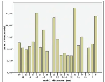
cervical radiotherapy, and seven patients had a history of thyroid disease such as goiter or hyper- or hypo-thyroidism. Median thyroid nodule diameter was 18 mm (range: 8–59 mm). Thyroid USG revealed solid nodules in 50% of patients; in addition, 18 nodules were hypoechoic (45%), 11 nodules (27.5%) were microlobular, 15 (37.5%) had irregular margins, and three nodules (7.5%) had microcalcifications. Cervical lymphadenopathies were detected in four patients (10%) by USG or PET-CT. PET-CT detected 31 (77.5%) focal areas of thyroid uptake; 25 (62.5%) nodules detected by PET were hypoechoic, 27 (67.5%) were solid, and 30% had irregular margins. The results of the frequency of clinical and radiological features are shown in Table 1. The median SUVmax values were 5.4 (range: 0–17.8) and 8.5 (range: 0–23.9) for thyroid nodules and primary cancer, respectively [Figure 1].
In total, 14 malignancies (35%) were detected by FNAB in thyroid nodules. The sensitivity and specificity of the PET-CT SUVmax value for the diagnosis of malignancy were 87.5% and 52%, respectively. On the other hand, the positive and negative predictive values were 36.8% and 92.8%, respectively. While the most common malignant pathology was classic variant papillary carcinoma (25%), others included papillary carcinoma oncocytic variants (2.5%), papillary microcarcinoma (2.5%), and thyroid metastasis from colon carcinoma (2.5%). Benign colloidal nodules were the most frequent benign pathology (n = 18, 45%). The remaining benign pathologies were lymphocytic thyroiditis (10%), atypia with unknown significance (7.5%), and oncocytic neoplasia (2.5%). The median SUVmax was higher in thyroid metastasis from colon cancer and oncocytic neoplasia (median SUVmax 14.5 and 13.6, respectively) and lower in benign colloid nodules (median SUVmax 4) [Table 2]. Figure 2 shows the benign and malignant thyroid nodules detected by PET/CT.
Thyroid nodule USG margins (P = 0.007) and thyroid nodule PET internal components (P = 0.03) were found to be important factors to differentiate thyroid nodules as benign or malignant. Nodules with irregular margins on USG and hypoechoic nodules on PET-CT were more malignant histopathologically than hyperechoic nodules with regular margins. Although all malignant nodules had one of the USG abnormalities, this was not statistically significant (P = 0.07). Table 3 shows the relationship between the pathological, clinical, and radiological features of the detected thyroid nodules.
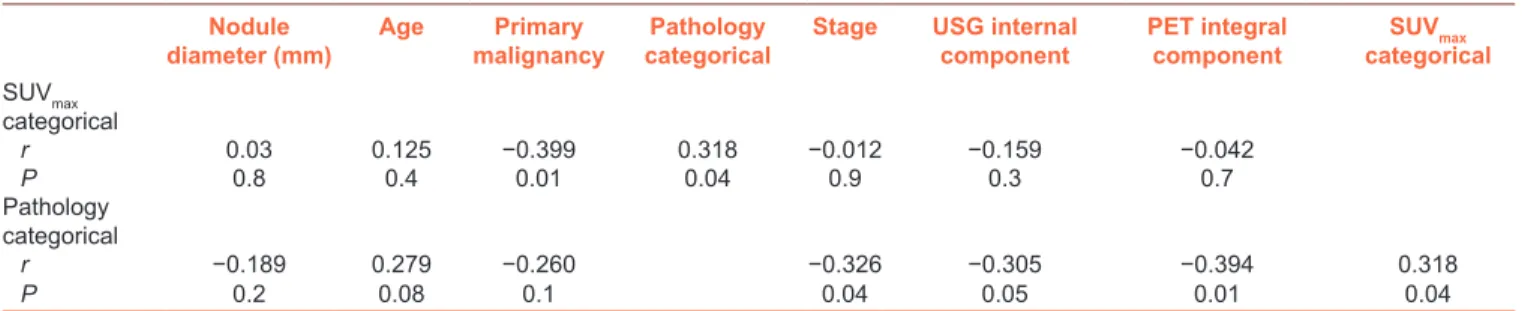
We could not find any correlation between nodule size and histopathological findings or SUVmax categorical values by the Pearson’s correlation analysis. However, histopathologically benign or malignant nodules were positively associated with the SUVmax categories (r = 0.318, P = 0.04) and negatively correlated with the density of the thyroid nodule by PET-CT (r = −0.394, P = 0.01), the density of nodule by USG (r = −0.305, P = 0.05), and the stage of the primary tumor (r = −0.326, P = 0.04). Furthermore, SUVmax categorical
values were negatively associated with primary malignancy Contd...
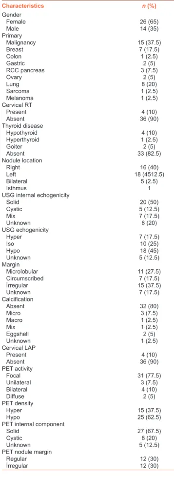
Table 1: The characteristics of the study group
Characteristics n (%) Gender Female 26 (65) Male 14 (35) Primary Malignancy 15 (37.5) Breast 7 (17.5) Colon 1 (2.5) Gastric 2 (5) RCC pancreas 3 (7.5) Ovary 2 (5) Lung 8 (20) Sarcoma 1 (2.5) Melanoma 1 (2.5) Cervical RT Present 4 (10) Absent 36 (90) Thyroid disease Hypothyroid 4 (10) Hyperthyroid 1 (2.5) Goiter 2 (5) Absent 33 (82.5) Nodule location Right 16 (40) Left 18 (4512.5) Bilateral 5 (2.5) Isthmus 1
USG internal echogenicity
Solid 20 (50) Cystic 5 (12.5) Mix 7 (17.5) Unknown 8 (20) USG echogenicity Hyper 7 (17.5) Iso 10 (25) Hypo 18 (45) Unknown 5 (12.5) Margin Microlobular 11 (27.5) Circumscribed 7 (17.5) İrregular 15 (37.5) Unknown 7 (17.5) Calcification Absent 32 (80) Micro 3 (7.5) Macro 1 (2.5) Mix 1 (2.5) Eggshell 2 (5) Unknown 1 (2.5) Cervical LAP Present 4 (10) Absent 36 (90) PET activity Focal 31 (77.5) Unilateral 3 (7.5) Bilateral 4 (10) Diffuse 2 (5) PET density Hyper 15 (37.5) Hypo 25 (62.5)
PET internal component
Solid 27 (67.5)
Cystic 8 (20)
Unknown 5 (12.5)
PET nodule margin
Regular 12 (30)
(r = −0.399, P = 0.01). Table 4 shows the results of the Pearson’s correlation analysis.
DISCUSSION
The frequency of thyroid nodules detected incidentally in PET-CT, which was performed for primary malignancy unrelated to thyroid cancer, has increased with widespread use of PET imaging in cancer patients. In the literature, the incidence of thyroid incidentaloma detected by PET is reported to be 2%–3%.[6,12] In addition, the rate of malignancy
has been reported as 14%–50% by histopathology in thyroid incidentaloma, which was determined by PET imaging.[13] In
a Korean study, 3% of thyroid incidentalomas were detected by PET imaging, which was performed for cancer screening in healthy subjects, but only two nodules (20%) were malignant.[14] All patients in our study population had a
primary malignancy. The retrospective evaluation of PET scans from 1330 patients detected thyroid incidentalomas in 2.2% of them. Fifteen patients underwent surgery, and a malignancy rate of 26.7% was reported.[14] Another large
series included PET imaging data on 4.252 patients and revealed 102 (2.3%) thyroid incidentalomas; malignancy was detected in seven nodules.[15] We retrospectively analyzed the
PET-CT data of 1.840 patients with malignancy and revealed 40 thyroid nodules with focal or diffuse FDG uptake (2.1%). FNAB is the main diagnostic modality to evaluate incidental thyroid nodules.[3] The rate of tissue biopsy for thyroid
incidentalomas detected by PET has been reported to occur in 1%–83% of patients in the literature.[6] The most
common malignancy detected by FNAB was papillary thyroid carcinoma, in the range of 81.3%–83%.[16] All of our patients
underwent FNAB to identify primary thyroid malignancy or metastasis from the primary tumor. Our results show that 14 malignant (35%) and 26 benign nodules were diagnosed, similar to rates in the literature. The most common malignant and benign lesions were classic papillary carcinoma (71.4%) and benign colloidal nodule (69.2%), respectively. Two (14.2%)
thyroid nodules were diagnosed as papillary carcinoma oncocytic variants.
Clinically detectable thyroid nodules have a 5% of malignancy risk.[17] A higher SUV
max of thyroid nodule on PET has been
associated with a greater risk of malignancy.[3] However,
there is no definite SUVmax value that differentiates benign from malignant.[18] If PET associated thyroid incidentaloma
is diagnosed as malignant, it is usually a more aggressive histological type and has a worse prognosis.[14] Increased FDG
uptake may be associated with a more aggressive histological type,[6] but there is controversy about the positive correlation
between SUVmax values and malignancy potential.[6] The FDG
uptake pattern of thyroid nodules can predict the malignant potential of thyroid nodules. Unilateral and focal FDG uptake is more likely to be associated with malignancy.[6] Focal uptake
of FDG has been associated with a 34.8% risk of malignancy.[19]
Choi et al. showed that 17 out of 44 thyroid nodules with focal FDG uptake on PET were diagnosed histologically as malignant.[4] Although we detected 31 nodules with focal FDG
uptake, this was not related to malignancy. The prevalence of thyroid focal lesions on PET was 4% (70/1763) in the Choi et al.’s study.[4] Diagnostic FNAB was performed on 29 of 70 patients,
and 17 nodules were found to be malignant by histopathology. In the present study, the SUVmax of malignant lesions was higher than that of benign lesions (6.7 vs. 10.7, P < 0.05). Diffuse FDG uptake could indicate normal variation, chronic thyroiditis, or Graves’ disease.[19] In the literature, 47.4%
of 133 cases of thyroid diffuse uptake were diagnosed as autoimmune thyroiditis or hypothyroidism.[20] Although 22.5%
of our nodules showed diffuse FDG uptake, 65% of the FNAB results were benign, and this was not found to be an important factor to differentiate benign from malignant lesions. There is no consensus on the role of SUVmax to differentiate benign from malignant thyroid nodules.[19] A SUV
max cutoff
value 9.1 has been reported as being sensitive (81.6%) and specific (100%) to diagnose malignant nodules[21] Hales
Figure 1: The histogram of standardized uptake value maximum and
thyroid nodule size
Table 1: Contd... Characteristics n (%) Diffuse 2 (5) Unknown 14 (35) Pathology Benign 26 (65) Malignant 14 (35)
SUVmax nodule
0 5 (10) 2.2-2.9 9 (12.5) 3-4.9 10 (22.5) 5-6.9 4 (25) 7-9.9 8 (10) >10 4 (20) Thyroid surgery Present 13 (32.5) Absent 27 (67.5)
RT=Radiotherapy, LAP=Lymphadenopathy, FNAB=Fine needle aspiration biopsy, Ca=Cancer, RCC=Renal cell carcinoma, SUVmax=Maximum standardized uptake value, PET=Positron emission tomography, USG=Ultrasonography
et al. reported that the sensitivity and specificity of PET to predict malignancy of thyroid nodule are 57% and 50%, with
positive and negative predictive values of 50% and 57%, respectively.[14] However, these authors evaluated only eight
FDG-positive patients. In one meta-analysis, it was shown that the sensitivity and specificity of PET for the detection of cancer in thyroid nodules were 89% and 55%, respectively.[22]
The best cutoff SUVmax value for differentiation was 2.05. In another study, it was reported that every unit increase in SUVmax is associated with poor survival.[23] While we found that
the sensitivity and specificity of PET-CT to identify malignancy were 87.5% and 52%, the positive and negative predictive values were 36.8% and 92.8%, respectively. The low positive predictive value of our results indicates that FDG uptake alone is not enough to predict malignancy. Therefore, the features of imaging combined with USG findings and FNAB may be more valuable.
Kim et al. reported that greater tumor and cervical lymph node metastasis detected by PET were associated with positive FDG uptake in thyroid cancer patients compared to FDG-negative patients. They reported 71.6% FDG uptake of thyroid cancer.[24]
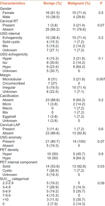
Table 3: The comparison of characteristic in respect to pathological features
Characteristics Benign (%) Malignant (%) P
Gender Female 16 (61.5) 10 (71.4) 0.5 Male 10 (38.5) 4 (29.6) Cervical RT Present 1 (3.8) 3 (21.4) 0.07 Absent 25 (95.2) 11 (78.6) USG internal Echogenicity 10 (38.4) 10 (71.4) 0.2 Solid cystic 4 (15.3) 1 (7.2) Mix 5 (19.2) 2 (14.2) Unknown 7 (27.1) 1 (7.2) USG echogenicity Hyper 4 (15.3) 3 (21.6) 0.1 Iso 8 (30.6) 2 (14.2) Hypo 9 (23.4) 9 (64.2) Unknown 5 (30.7) 0 Margin Microlobular 8 (31) 3 (21.6) 0.007 Circumscribed 7 (27) 0 İrregular 5 (19.5) 10 (71.4) Unknown 6 (22.5) 1 (7.1) Calcification Absent 23 (88.6) 9 (64.2) 0.2 Micro 1 (3.8) 2 (14.2) Macro 0 1 (7.2) Mix 0 1 (7.2) Eggshell 1 (3.8) 1 (7.2) Unknown 1 (3.8) 0 Cervical LAP Present 3 (11.4) 1 (7.2) 0.6 Absent 23 (88.6) 13 (92.8) USG anomaly Present 21 (80.7) 14 (100) 0.07 Absent 5 (19.3) 0 PET density Hyper 10 (50) 5 (45.7) 0.8 Hypo 10 (50) 9 (64.3)
PET internal component
Solid 14 (53.8) 13 (92.8) 0.03
Cystic 7 (26.9) 1 (7.2)
Unknown 5 (19.3) 0
SUVmax categorical
2.2-2.9 5 (19.2) 0 0.09 3-4.9 7 (26.9) 2 (14.3) 5-6.9 5 (19.2) 5 (35.7) 7-9.9 4 (15.3) 0 >10 3 (11.5) 5 (35.7) 0 2 (7.9) 2 (14.3)
RT=Radiotherapy, LAP=Lymphadenopathy, SUVmax=Maximum standardized uptake value, PET=Positron emission tomography, USG=Ultrasonography
Figure 2: The positron emission tomography-computerized tomography
imaging of patients with thyroid metastasis of colon cancer
Table 2: The pathological findings and maximum standardized uptake value of the thyroid nodules
Benign pathology Malignant pathology n Median SUVmax
Oncocytic neoplasia 1 (2.5) 13.6
Lymphocytic thyroiditis 4 (10) 7.1
Atypia with unknown significance 3 (7.5) 3.1
Benign colloidal nodule 18 (45) 4
Classical papillary carcinoma 10 (25) 5.9
Papillary microcarcinoma 1 (2.5) 6
Papillary carcinoma oncocytic variant 2 (5) 5.9
Colon carcinoma metastasis 1 (2.5) 14.5
Only 10% of our patients had FDG-positive cervical lymph nodes by imaging. Neither cervical lymph nodes nor a previous history of cervical radiotherapy was found to be related to malignancy.
Kao et al. analyzed 942 PET scans retrospectively and reported a 7% incidence of thyroid incidentaloma;[25] 21 of the nodules
were FDG-avid incidentalomas, while 45 were not FDG avid. Only six patients underwent FNAB, and malignancies were identified in three of them (two papillary, one medullary thyroid cancer). The positive predictive value of FDG-avid thyroid incidentaloma for underlying malignancy was 50%. These authors did not find a statistically significant relationship between the SUVmax value and malignancy potential. However, not all of their patients had malignancy. Mitchell et al.[26] reported that 9 of 15 thyroid carcinomas
were FDG positive (sensitivity of 60%). We found only four thyroid incidentalomas without FDG uptake on PET-CT, and 90% of thyroid incidentalomas had FDG uptake with a median SUVmax of 5.4. According to our histopathological findings, the median SUVmax was 4.5 and 6.1 for benign and malignant lesions, respectively (P = 0.09). In another study, Brindle et al. reported that 30 thyroid incidentalomas were further investigated by biopsy (37%), and seven malignant nodules (23%) were diagnosed.[27] The median SUV
max were
5.4 and 9.9 for benign and malignant lesions, respectively. The most frequent primary malignancies associated with PET imaging were lung, colorectal, lymphoma, and esophageal cancer in order of frequency. These authors did not provide a convincing SUVmax to differentiate between benign and malignant lesions. The primary malignancies in our study were breast, lung, colon, and other cancers in order of frequency. PET-CT is mostly used in our oncology center for the staging of breast and lung cancer.
There is no exact factor predicting malignancy for thyroid incidentalomas. Thyroid nodule size has not been shown to predict malignancy,[3] but the FDG uptake pattern is related
to malignancy potential.[6] We analyzed radiological factors
to differentiate benign and malignant lesions and found that thyroid USG irregular margins and PET-CT solid components were associated with malignancy, but not the FDG uptake pattern. Although malignant lesions had a tendency toward a higher SUVmax than benign lesions, this was not statistically
significant (P = 0.09). SUVmax categorical values were positively related with pathological findings in the correlation analysis. Furthermore, benign or malignant lesions were negatively correlated with the stage of the primary disease, USG and PET lesion solidity. There was no relationship between tumor nodule diameter and radiological features.
The risk of malignancy among thyroid incidentalomas detected by PET is 47%,[6] so tissue diagnosis is required, especially
to differentiate aggressive forms of thyroid carcinoma or primary cancer thyroid metastases. All of our patients had a known malignancy, so decisions regarding thyroid nodules depended on the stage of the primary malignancy. Although thyroid cancer has an indolent nature, thyroid metastases and oncocytic type papillary carcinoma have aggressive features. Although the thyroid is a rare site for tumor metastasis,[19] the rate of metastasis has been reported to be
3.9%–24.2%.[19] Renal cell, breast, and lung cancer are the
most common primary tumors which can metastasize to the thyroid, whereas colorectal cancer metastases are rarely seen and are rather associated with lung and liver metastases.[28]
Choi et al. reported that one metastasis from esophageal carcinoma and 16 papillary carcinomas were detected by biopsy of incidental thyroid nodules.[4] Similarly, only one
of our 14 (7.1%) malignant thyroid nodules was metastatic from colon cancer. Because of this solitary thyroid metastasis without extra-thyroid metastases, this patient underwent total thyroidectomy with curative intent and was followed without disease for 12 months.
CONCLUSION
PET is a rapidly developing imaging modality. Although incidental thyroid uptake on PET is commonly related to benign histopathology, malignancy is detected one-third of these lesions by biopsy. FDG-avid malignant thyroid nodules such as oncocytic variants or metastases from the primary tumor tend to be a more undifferentiated and more aggressive histopathological type. Recognition of the pattern of FDG uptake and SUVmax combined with USG findings can guide appropriate treatment.
Financial support and sponsorship
Nil.
Table 4: The Pearson’s correlation analysis for the results of ultrasonography and positron emission tomography
Nodule
diameter (mm) Age malignancyPrimary categoricalPathology Stage USG internal component PET integral component categoricalSUVmax
SUVmax categorical r 0.03 0.125 −0.399 0.318 −0.012 −0.159 −0.042 P 0.8 0.4 0.01 0.04 0.9 0.3 0.7 Pathology categorical r −0.189 0.279 −0.260 −0.326 −0.305 −0.394 0.318 P 0.2 0.08 0.1 0.04 0.05 0.01 0.04
Conflicts of interest
There are no conflicts of interest.
REFERENCES
1. Tunbridge WM, Evered DC, Hall R, Appleton D, Brewis M, Clark F, et al. The spectrum of thyroid disease in a community: The Whickham survey. Clin Endocrinol (Oxf) 1977;7:481-93.
2. Furmanchuk AW, Roussak N, Ruchti C. Occult thyroid carcinomas in the region of Minsk, Belarus. An autopsy study of 215 patients. Histopathology 1993;23:319-25.
3. Jin J, McHenry CR. Thyroid incidentaloma. Best Pract Res Clin Endocrinol Metab 2012;26:83-96.
4. Choi JY, Lee KS, Kim HJ, Shim YM, Kwon OJ, Park K, et al. Focal thyroid lesions incidentally identified by integrated 18F-FDG PET/CT: Clinical significance and improved characterization. J Nucl Med 2006;47:609-15.
5. Hall NC, Kloos RT. PET imaging in differentiated thyroid cancer: Where does it fit and how do we use it? Arq Bras Endocrinol Metabol 2007;51:793-805.
6. Katz SC, Shaha A. PET-associated incidental neoplasms of the thyroid. J Am Coll Surg 2008;207:259-64.
7. Yoon JH, Cho A, Lee HS, Kim EK, Moon HJ, Kwak JY, et al. Thyroid incidentalomas detected on 18F-fluorodeoxyglucose-positron emission tomography/computed tomography: Thyroid imaging reporting and data system (TIRADS) in the diagnosis and management of patients. Surgery 2015;158:1314-22.
8. Kim EK, Park CS, Chung WY, Oh KK, Kim DI, Lee JT, et al. New sonographic criteria for recommending fine-needle aspiration biopsy of nonpalpable solid nodules of the thyroid. AJR Am J Roentgenol 2002;178:687-91.
9. Shetty SK, Maher MM, Hahn PF, Halpern EF, Aquino SL. Significance of incidental thyroid lesions detected on CT: Correlation among CT, sonography, and pathology. AJR Am J Roentgenol 2006;187:1349-56.
10. Are C, Hsu JF, Schoder H, Shah JP, Larson SM, Shaha AR, et al. FDG-PET detected thyroid incidentalomas: Need for further investigation? Ann Surg Oncol 2007;14:239-47.
11. American Thyroid Association (ATA) Guidelines Taskforce on Thyroid Nodules and Differentiated Thyroid Cancer, Cooper DS, Doherty GM, Haugen BR, Kloos RT, Lee SL, et al. Revised American Thyroid Association management guidelines for patients with thyroid nodules and differentiated thyroid cancer. Thyroid 2009;19:1167-214. 12. Are C, Hsu JF, Ghossein RA, Schoder H, Shah JP, Shaha AR, et al.
Histological aggressiveness of fluorodeoxyglucose positron-emission tomogram (FDG-PET)-detected incidental thyroid carcinomas. Ann Surg Oncol 2007;14:3210-5.
13. Van den Bruel A, Maes A, De Potter T, Mortelmans L, Drijkoningen M, Van Damme B, et al. Clinical relevance of thyroid fluorodeoxyglucose-whole body positron emission tomography incidentaloma. J Clin Endocrinol Metab 2002;87:1517-20.
14. Kang KW, Kim SK, Kang HS, Lee ES, Sim JS, Lee IG, et al. Prevalence
and risk of cancer of focal thyroid incidentaloma identified by 18F-fluorodeoxyglucose positron emission tomography for metastasis evaluation and cancer screening in healthy subjects. J Clin Endocrinol Metab 2003;88:4100-4.
15. Cohen MS, Arslan N, Dehdashti F, Doherty GM, Lairmore TC, Brunt LM, et al. Risk of malignancy in thyroid incidentalomas identified by fluorodeoxyglucose-positron emission tomography. Surgery 2001;130:941-6.
16. Soelberg KK, Bonnema SJ, Brix TH, Hegedüs L. Risk of malignancy in thyroid incidentalomas detected by 18F-fluorodeoxyglucose positron emission tomography: A systematic review. Thyroid 2012;22:918-25. 17. Hegedüs L. Clinical practice. The thyroid nodule. N Engl J Med
2004;351:1764-71.
18. Bertagna F, Giubbini R. F18-FDG-PET/CT thyroid incidentalomas and their benign or malignant nature: A critical and debated issue. Ann Nucl Med 2011;25:151-2.
19. Agrawal K, Weaver J, Ngu R, Krishnamurthy Mohan H. Clinical significance of patterns of incidental thyroid uptake at (18)F-FDG PET/CT. Clin Radiol 2015;70:536-43.
20. Karantanis D, Bogsrud TV, Wiseman GA, Mullan BP, Subramaniam RM, Nathan MA, et al. Clinical significance of diffusely increased 18F-FDG uptake in the thyroid gland. J Nucl Med 2007;48:896-901.
21. Agrawal K, Weaver J, Ul-Hassan F, Jeannon JP, Simo R, Carroll P, et al. Incidental Focal Thyroid Uptake on 18F-FDG PET Study d Large Retrospective Single Centre Experience in the United Kingdom. In: 27th Annual Congress of the European Association of Nuclear Medicine, 18e22 October, Gothenburg, Sweden; 2014. Abstract No. OP 693; 2014.
22. Wang N, Zhai H, Lu Y. Is fluorine-18 fluorodeoxyglucose positron emission tomography useful for the thyroid nodules with indeterminate fine needle aspiration biopsy? A meta-analysis of the literature. J Otolaryngol Head Neck Surg 2013;42:38.
23. Pryma DA, Schöder H, Gönen M, Robbins RJ, Larson SM, Yeung HW, et al. Diagnostic accuracy and prognostic value of 18F-FDG PET in hürthle cell thyroid cancer patients. J Nucl Med 2006;47:1260-6. 24. Kim BS, Kim SJ, Kim IJ, Pak K, Kim K. Factors associated with positive
F-18 flurodeoxyglucose positron emission tomography before thyroidectomy in patients with papillary thyroid carcinoma. Thyroid 2012;22:725-9.
25. Kao YH, Lim SS, Ong SC, Padhy AK. Thyroid incidentalomas on fluorine-18-fluorodeoxyglucose positron emission tomography-computed tomography: Incidence, malignancy risk, and comparison of standardized uptake values. Can Assoc Radiol J 2012;63:289-93.
26. Mitchell JC, Grant F, Evenson AR, Parker JA, Hasselgren PO, Parangi S, et al. Preoperative evaluation of thyroid nodules with 18FDG-PET/CT. Surgery 2005;138:1166-74.
27. Brindle R, Mullan D, Yap BK, Gandhi A. Thyroid incidentalomas d i s c o v e re d o n p o s i t r o n e m i s s i o n t o m o g r a p h y C T scanning – Malignancy rate and significance of standardised uptake values. Eur J Surg Oncol 2014;40:1528-32.
28. Wychulis AR, Beahrs OH, Woolner LB. Metastasis of carcinoma to the thyroid gland. Ann Surg 1964;160:169-77.