The effects of focused ultrasound pulsation of nucleus accumbens in
opioid-dependent rats
Erdem Deveci a, Alperen Kılıça*, Onur Yılmaz a, Aynur Nabi a, Arif Sanlı Ergün b, Ayhan Bozkurt c, Ayşe Kurtulmuşa, Ahmet Öztürk a, Mukaddes Eşrefoğlud, MehmetŞerif Aydıne, EbruŞahan aand İsmet Kırpınara
a
Department of Psychiatry, Bezmialem Foundation University Medical Faculty,İstanbul, Turkey;bFaculty of Engineering, TOBB University of Economics & Technology, Ankara, Turkey;cFaculty of Engineering and Natural Sciences, Sabancı University, İstanbul, Turkey;dDepartment of Histology, Bezmialem Foundation University Medical Faculty,İstanbul, Turkey;eRegenerative and Restorative Medicine Research Center, İstanbul Medipol University, İstanbul, Turkey
ABSTRACT
Background: Deep Brain Stimulation (DBS) is the only modality proven to be effective on selective stimulation of the deep brain structures. It was previously reported that, by using DBS, stimulation of nucleus accumbens (NA), a region that plays a pivotal role in the pathogenesis of substance addiction, is effective for the treatment of substance addiction. Objective: The purpose of the current study was to observe how the morphine-conditioned place preference changed in rats by stimulating NA with a non-invasive method, focused ultrasound (US) and to detect whether there would be any tissue damage caused by US waves. Methods: We used low-intensity focused ultrasound (LIFU), a noninvasive modality, in a place conditioning model to stimulate NA in rats.
Results: At the initial stage of our study, we used morphine to induce place preference. As expected, morphine administration caused significant place preference. After the place preference was obtained by morphine, we divided the rats into two groups. One group received LIFU waves to NA and the other group received only sham, that is, no stimulation with US waves. Rats in both groups were continued to receive morphine. Then, we investigated whether LIFU and sham will reduce morphine-induced place preference or not. We observed that morphine-induced place preference had an ongoing raise in the sham group while no raise was detected in the ultrasound group. Although LIFU prevented the rats from the raise, it did not cause a significant reduction of morphine preference.
Conclusion: We state that there is a need for future studies to investigate the effects of low-intensity focused ultrasound as an alternative treatment modality in addiction.
ARTICLE HISTORY Received 28 February 2019 Accepted 11 June 2019 KEYWORDS
Morphine dependence; low intensity focused ultrasound; nucleus accumbens; conditioned place preference; deep brain stimulation
Introduction
Opioid addiction, which is a type of substance depen-dence, has been defined by American Society of Addic-tion Medicine as“being extremely preoccupied in mind with opioid intake despite sufficient level of analgesia is obtained, and loss of control on opioid use” [1]. Meso-corticolimbic dopamine reward pathway, which con-sists of Ventral Tegmental Area (VTA), Nucleus Accumbens (NA) and Ventral Pallidum (VP), plays a critical role in substance dependence [2,3].
Neuromodulation is defined by The International Neuromodulation Society as “changing nerve activity by giving electrical stimulation or chemical substances to target areas in the body,” which is performed for normalizing and modulating nerve function. Vagal stimulation, Transcranial Magnetic Stimulation (TMS), Transcranial Electrical Stimulation (tES), Deep Brain Stimulation (DBS), High Intensity Focused
Ultrasound (HIFU) and Low Intensity Focused Ultra-sound (LIFU) can be listed in main neuromodulation methods.
Due to its deep location in the brain, NA cannot be specifically stimulated by noninvasive techniques such as TMS. For this reason, the DBS technique seems to be suitable for NA stimulation. Literature review on these studies revealed that DBS wasfirst applied to NA on 2007 for the treatment of a patient with treatment-resistant panic disorder and had no effect on panic dis-order, however, comorbid alcohol dependence was reported to be remitted [4]. High-frequency stimulation of NA with DBS was shown to be effective for lowering Conditioned Place Preference (CPP) test scores stimu-lated by morphine in rats [5–7]. But DBS is an invasive process that involves surgically placing an electrode deep in the brain. It has risks and complications regard-ing surgery, stimulation and stimulation pathway [8].
© 2019 The Author(s). Published by Informa UK Limited, trading as Taylor & Francis Group
This is an Open Access article distributed under the terms of the Creative Commons Attribution License (http://creativecommons.org/licenses/by/4.0/), which permits unrestricted use, distribution, and reproduction in any medium, provided the original work is properly cited.
CONTACT Onur Yılmaz [email protected] Department of Psychiatry, Bezmialem Foundation University Medical Faculty, Adnan Menderes Boulevard (Vatan Road) Fatih,İstanbul 34093, Turkey
*Present address:İstanbul Medipol University Hospital, İstanbul, Turkey
Supplemental data for this article can be accessed athttps://doi.org/10.1080/24750573.2019.1631942. 2019, VOL. 29, NO. 4, 748–759
In recent years, ultrasound energy focused on deep brain structures at low energy levels has been shown to stimulate or inhibit small brain areas without causing neuronal damage [9–11]. There has been a remarkable increase in studies concerning neuromodulator features of LIFU waves. A study has shown that, short term ultrasound (US) stimulations on peripheral nerves acti-vate and long term stimulations inhibit amplitude and speed of the action potential [12]. US is an energy type that has a waveform, which is considered to gener-ate a neuromodulator effect due to mechanic vibration [13]. Low intensity US neuromodulation, spreading as short pulses, was suggested to reduce energy storage on tissues for a specific time, and therefore may cause action potentials with mechanical effect by altering neuronal transmission [2,14].
The safety and efficacy of LIFU waves have been demonstrated in many studies performed with animals and the results of thefirst study on humans have been reported in 2014. In the relevant study performed with ten volunteers, the effect of focused ultrasound on two-point discrimination has been investigated and it was shown to increase somatosensory discrimination capacity in humans [15].
Although DBS has a very good spatial localization capacity, its usage is limited since it is an invasive method. TMS cannot reach deep tissues since it has a superficial effect and it cannot create a target-specific effect since its area of influence is relatively wide. Thus, an ideal neuromodulation method is expected to reach deep structures, to be specific for its target and to be non-invasive. There is no existing method that fully meets these criteria. Animal studies performed so far revealed that focused ultrasound can specifically stimulate and inhibit deep brain tissues without causing harmful effects outside of target point. This method is considered to become a significant step in the treatment of psychia-tric and neurological disorders with neuromodulation since it is noninvasive and does not require surgery and anaesthesia. Thus, focused ultrasound is argued to be an alternative method in the treatment of psychiatric and neurological disorders such as mood disorders, schizophrenia, depression, anxiety disorders, epilepsy, traumatic brain injuries and Parkinson’s disease in the future.
Our study has been designed on a dependency ani-mal model, one of the models that are considered to reflect human psychiatric disorders best. We suggest that focused ultrasound may open a new field in the treatment of opioid and other substance dependencies in humans and we decided to use rats since those dependencies of rats have similar pathophysiology with humans. In trials concerning substance depen-dency, various experimental animal models were estab-lished based on substance seeking behaviour and withdrawal symptoms of experimental animals. One of these models, CPP, is known to be a valid model
for assessment of reinforcing properties of addictive substances, and also scanning for abuse [16]. CPP test is a learning method that provides cooperation between reward and specific brain areas [17]. With the CPP test, it has been determined that experimental animals spend more time in areas which are peered with morphine, ethanol, encephalin, amphetamine and similar drugs and substances compared to the areas which are peered with physiological saline [18]
In the current study, our purpose was to determine the effects of stimulation of NA with a non-invasive method, focused ultrasound, on morphine-dependent place preference with a conditioned place preference model in rats. Our hypothesis in this CPP experimental model study was that LIFU focused on NA would reduce morphine dependency, without causing any tis-sue damage, via significantly effecting the time spent by rats in morphine-peered section.
Material and method
Selection of animals and sheltering environment
Our study has been conducted in Bezmialem Foun-dation University Hospital Psychiatry Clinics. Approval of the local ethics committee was taken as required (approval date: 29.01.2015; approval number: 2015/44). 26 adult Sprague-Dawley male rats, weighing between 300–490 g, were used. The details of the exper-iment environment are presented in the online sup-plementfile 1 (suppl 1).
CPP testing apparatus consisted of two Plexiglas boxes in square and rectangular shape and equal size, separated by a sliding (guillotine) door that can be lifted. In these two chambers of the same size, there were different clues (stimulants) such as the shape of thefloor and the colour of the walls that the animals can discriminate (suppl 2).
Steps of experimental addiction model and surgical procedure
Building an addiction model
In numerous previous studies, intraperitoneal mor-phine injection method was used for building addiction models in rats [19,20]. Likely in the current study, mor-phine hydrochloride dissolved in normal saline was administered via intraperitoneal (i.p.) route in equal volumes (0.5–1 ml).
Among the 26 rats included in the study, 18 were randomized to morphine group and the resting 8 were randomized to physiologic saline group. CPP test con-sisted of a three-stage and 14-day calendar. These stages were, before conditioning (pre-conditioning), con-ditioning and after concon-ditioning (post-concon-ditioning). Pre-conditioning stage lasted for 3 days and it was
applied to determine the place preference of animals. Each animal from all groups was placed every day in the central area with sliding guillotine door that allows transport, for about 900 s. On the third day, time spent by rats in each compartment was recorded with the camera (Table 1). As seen onTable 2, rats spent more time in the black section. This finding is quite likely based upon the need to hide from potential threats. For this reason, a method suitable for the biased design was selected during conditioning. Black section of CPP apparatus was matched with normal saline, while the white section was matched with morphine.
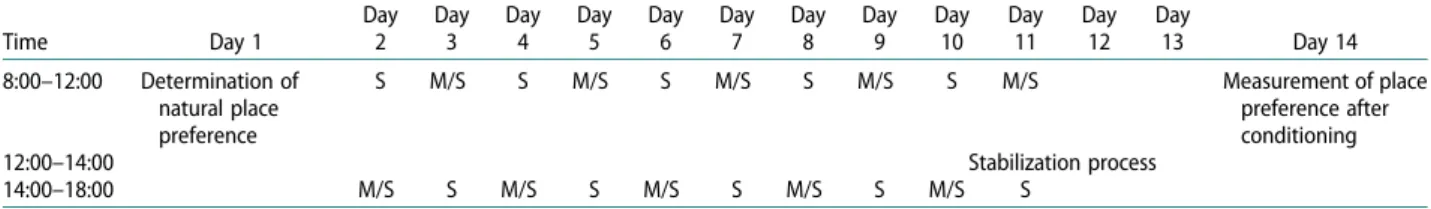
On thefirst day of the conditioning stage (4th day), normal saline was injected to rats in both groups and they were immediately placed to the black compart-ment matched with normal saline from 8:00 to 12:00.
After morphine injection to morphine group and normal saline injection to saline group was per-formed, rats were placed to white compartment matched with morphine from 14:00 to 18:00. On the second day, procedure was performed in reverse order, that is, after performing morphine injection to morphine group and normal saline injection to physiologic saline group, rats were placed to white
compartment matched with morphine from 8:00 to 12:00 and then normal saline was injected to rats in both groups, and they were placed in black compart-ment matched with normal saline from 14:00 to 18:00 (Table 1). This procedure continued until day 13. On the last 5 days of conditioning stage (days 9–13), ani-mals from all groups were put on stabilization appar-atus for 10 min in the period between two injections (12:00–14:00) and they were accustomed to waiting (Table 1). Morphine group received normal saline and morphine hydrochloride injections via i.p. route in 6-hour intervals throughout conditioning stage. Control group received only normal saline injections via i.p. route twice a day throughout the conditioning stage. Doses in preliminary likely studies were deter-mined between 5–50 mg/kg [21,22]. In the current study, throughout conditioning session, 10 mg/kg morphine hydrochloride was administered for 7 days via i.p. route, then it was stabilized at 20 mg/kg dose once a day. Guillotine doors were closed throughout conditioning stage.
Placing the magnets
On day 14, in order to determine the size of the brain area (NA) of LIFU wave administration, data outlined inFigure 1were used. Roughly, a volume sized 1.3 mm length in X axis, 2 mm width in Y axis and 2.1 mm depth in Z axis was determined to be the optimum geo-metrical volume for NA.
Two symmetrical points at 1.85 mm distance from bregma to nasal side, in 7 mm depth from bregma level and at 1.55 mm lateral from middle line at both right Table 1.Conditioned place preference study progress.
Time Day 1 Day 2 Day 3 Day 4 Day 5 Day 6 Day 7 Day 8 Day 9 Day 10 Day 11 Day 12 Day 13 Day 14 8:00–12:00 Determination of natural place preference S M/S S M/S S M/S S M/S S M/S Measurement of place preference after conditioning 12:00–14:00 Stabilization process 14:00–18:00 M/S S M/S S M/S S M/S S M/S S
S: Saline administration to both groups.
M/S: Morphine administration to morphine group and saline administration to saline group.
Table 2. Time spent by rats in all groups within white and black compartments in preliminary test stage.
Time spent in white compartment
Time spent in black
compartment Valuep Rats
(n = 26)
37,2 (0–188,8) s 862,8 (711,2–900) s <.001* Notes: Time spent in white and black compartments was stated as median
(minimum– maximum) values.
*Symbolizes statistically significant difference. n: Number of animals; s: seconds.
Figure 1.Stereotaxic imaging of nucleus accumbens in coronal sections of rat brain (taken with permission from The Rat Brain in
and left sides, were determined as middle focus points of sonication (for right and left NA) (Figure 2, suppl 3).
Transducers, designed with dimensions compatibly matching to the rat skull, produced ultrasound waves. In order to determine the focus of waves before soni-cation, a separate study was performed with a bullet hydrophone and a setting of three-dimensional
scanning device for measuring the US wave dynamics from behind the rat skull. The data obtained from three-dimensional scanning is presented in Figures 3 and 4. We determined the permeability of rat skull and focused US wave dynamics by scanning the rat skull spatially with a computerized XYZ scanner and by driving the transducer focused to computerized Figure 2.View of rat scalp; ultrasound focus points are shown in red shapes.
Figure 3.Transverse bundle shapes of the 10 channel, 2.4 MHz central frequency transducer at different depths. In these measurements, component phases are arranged as to obtain a focusing angle of 11 degrees. HIFU driver was drived at 2800 kHz with 10% amplitude. While it effects symmetrically, only the signal focused on left side is demonstrated.
Figure 4.Alteration of the transvers bundle shape with regard to depth after rat scale is put in front of the transducer. The centre of transducer array is set to be placed in 1.8 mm front of bregma point. Thus, ultrasound wave bundles are directed on nucleus accumbens. HIFU driver’s amplitude was used as 20%.
signal source and measuring the intensity of US with hydrophone and oscilloscope (Figures 3and4, suppl 4). In order to place the ultrasound probe, magnets were attached to all rats’ calvaria. One day after this step, the stage was started for installing a setting that will enable the transducer to be attached on the scalp of animals. Firstly, rats were anaesthetized with xyla-zine (10 mg/kg) and ketamine (80 mg/kg) i.p., and then the skin of the scalp was removed. Transducer was placed on the middle line, 1.70 mm away from Bregma on the nasal side, projection of installed mag-nets was marked on the scalp and magmag-nets were attached to these areas. Two circular magnets with 4 mm diameter and 2 mm thickness were used for keep-ing the transducerfixed on rats’ head throughout soni-cation period (SP) and two neodymium magnets with the same size were installed on transducer (suppl 5). Transducer was removed from the head at the end of each sonication. This process was also applied to rats in sham group, although they did not receive soni-cation (sham procedure). After the surgical procedure, the animals were left to healing for one day on day 15. Sonication
On day sixteen, 18 rats in the study group were ran-domly divided into two, as US group to receive ultra-sound stimulation and sham group to receive fake ultrasound stimulation. Both groups received normal saline injections in black compartment and morphine hydrochloride injections in white compartment for 30 min via i.p. route in 6-hour intervals twice a day for 10 days. In the meantime, 8 rats which were in physiologic saline group received only normal saline injections via i.p. route in 6-h intervals twice a day, and they were placed in black and white compart-ments for 30 min as in conditioning stage (Table 3). Before morphine administrations, rats in US group and sham group received isoflurane inhaler anaesthetic (1.5%) once a day for 10 days and taken into and fas-tened in the brain stimulation setting (suppl 6,7). Afterwards, ultrasound gel was administered to US group and LIFU stimulation was given for 10 min. Fake ultrasound stimulation was performed on sham group. Ten minutes after the stimulation, morphine was administered to rats and they were placed in white compartment for 30 min.
Reassessment of addiction level
Post-conditioning period started one day after last injections. Sliding guillotine doors were raised and
free access to all apparatus was given to rats for 900 s. Passing time of rats through settings were recorded with cameras. Assessment of CPP was calcu-lated with the time spent in white compartment matched with morphine. On the last day of the exper-iment, morphine-related CPP durations of US and sham groups were assessed.
Statistical analyses
In the statistical assessment of study data, non-para-metric Kruskal–Wallis test was used for comparison of more than two groups, and Dunn tests were used for subgroup analyses. For comparison of pairs, di ffer-ent groups were compared with non-parametric Mann–Whitney U test, and comparison of intra-group changes was performed by non-parametric Wil-coxon test. The value of .05 was assumed as the level of statistical significance in all tests.
Histologic study
Brain tissues of experiment groups werefixed for 72 h, in 10% neutral-buffered formalin (NBF). Then, they have been dehydrated through increasing series of alco-hol (70%, 90%, 96% and 100%) and pellucided with xylene. Following this, they have been kept in 60°C paraffin overnight and embedded in paraffin. These paraffin blocks were cut in 5-μm thickness and placed on microscope slide. For microscopic study, they have been stained with hematoxylin–eosin. Stained sections have been observed with the light microscope (Nikon, Eclipse i5, Japan).
Immunostaining
Sections have been kept in the incubator overnight at 37°C, then, placed in, firstly pure, subsequently 96% alcohol for 10 min. Endogenous enzyme blockage was performed through being kept in 3% hydrogen peroxide prepared with methanol. After being washed firstly with tap water, then with distillated water, they were put in citrate buffer and subjected to 200 W microwave for 10 min. Sections were cooled in room temperature, then kept in blocking solution for 10 min. They were kept overnight in rabbit policlonal anti-JNK1, anti-caspase 3 and anti-MAP2 primary antibodies at +4°C. Then they were washed three times, each for 5 min with PBS, followed by marking with Alexa Fluor 488 conjugated secondary antibodies. Table 3.Conditioned place preference study progress/ sonication period.
Time Day 16 Day 17 Day 18 Day 19 Day 20 Day 21 Day 22 Day 23 Day 24 Day 25 Day 26
08:00–12:00 S M/S S M/S S M/S S M/S S M/S Last place preference assessment
14:00–18:00 M/S S M/S S M/S S M/S S M/S S
S: Saline administration to both groups.
Afterwards, they were closed, using closing matter which contains DAPI (Fluoroshield with DAPI) and viewed on confocal microscope.
A summary of the process throughout the study is presented as aflowchart onTable 4.
Results
After the CPP protocol, three animals from US group and one animal from physiologic saline group died during anaesthesia.
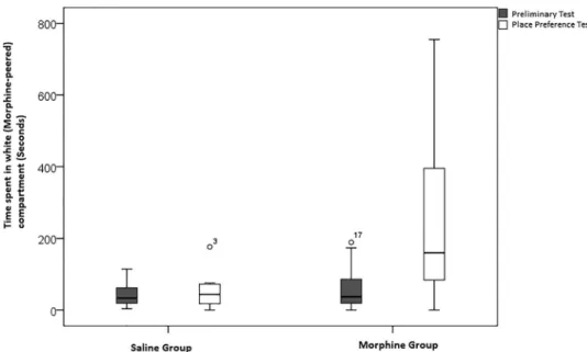
For CPP, time spent in seconds (s) by rats in pre-liminary test stage within the white compartment and the black compartment is presented in Figure 5, and the time spent by physiologic saline and morphine groups within the white compartment during prelimi-nary test and place preference test is presented in Figure 6. In Figure 7, LIFU was shown to affect the place preference of rats which have developed CPP to
morphine and are continuing to morphine
conditioning.
Results of statistical analyses
Upon comparing the time in seconds spent by all rats in the white and black compartments in preliminary test stage, time spent in the black compartment was sig-nificantly higher than time spent in the white compart-ment (Table 2). Thus, the experiment procedure was prepared to suit biased design.
In preliminary test stage, no significant difference was determined upon comparing time spent in the white compartment by rats in saline [33,4 (3,4−114,4) s] and morphine [37,2 (0–188,8) s] groups (p > .05). In place preference test stage, upon comparing time spent by groups in the white compartment, time spent by morphine group [159,7 (0,0–754,9) s] was signifi-cantly higher than the time spent by saline group [43,7 (0,3–176,3) s] (p < .05). Furthermore, time spent by Table 4.Flowchart of methodology throughout the study.
Time
Day 8:00–12:00 12:00–14:00 14:00–18:00
1 Comparing the time spent by rats in white and black compartment, demonstrating the natural place preference 2 3 4 S NPP M/S 5 M/S S 6 S M/S 7 M/S S 8 S M/S 9 M/S Stabilization Process S 10 S M/S 11 M/S S 12 S M/S 13 M/S S
14 Measurement of place preference after conditioning,
anaesthetizing rats with xylazine and ketamine, removing the skin of the scalp, attaching magnets to rats’ calvaria 15 Leaving animals for healing after surgical procedure
16a Dividing the rats into two groups: sonication group (9 for LIFU and 9 for sham) and physiologic saline group
16b S NPP M/S 17 M/S S 18 S M/S 19 M/S S 20 S M/S 21 M/S S 22 S M/S 23 M/S S 24 S M/S 25 M/S S
26 Raising guillotine doors, giving free access to all apparatus to rats, measuring the eventual place preference 27 Decapitation of rats, histologic study and immunostaining
Notes: While random division of the sample and then sonication process both started on day 16, we marked the relevant day as 16a and 16b respectively. S: Saline administration to both groups.
M/S: Morphine administration to morphine group and saline administration to saline group. NPP: No procedure performed (waiting time).
LIFU: Low Intensity Focused Ultrasound.
Figure 5.Time spent by rats in preliminary test stage within white compartment and black compartment.
morphine group within the white compartment in place preference test [159,7 (0,0–754,9) s] was significantly higher than the time spent within the white
compartment in preliminary test [37,2 (0–188,8) s] (p < .01). Upon comparing the place preference test time and the preliminary test time of both groups with regard to time spent in the white compartment, time difference of morphine group [93,7 (−86,0–733,1) s] was signifi-cantly higher than time difference of saline group [1,9 ± (−45,2–148,0) s] (p < .01) (Tables 5and6).
Figure 6.Time spent in white compartment by physiologic saline and morphine groups during preliminary test and place prefer-ence test.
Figure 7.LIFU effect on the place preference of rats.
Table 5. Comparison of saline and morphine groups with regard to time spent in white compartment and time difference. Saline (n = 8) Morphine (n = 18) Valuep Preliminary Test 33,4 (3,4 −114,4) s 37,2 (0188,8) s– .470 Place Preference Test 43,7 (0.3–
176,3) s
159,7 (0.0– 754,9) s
.013* Place Preference Test–
Preliminary Test Time Difference 1,9 ± (-45,2– 148,0) s 93,7 (−86.0– 733,1) s .009**
Notes: Time spent in white compartment was stated as median (mini-mum– maximum) values.
*p < .05 **p < .01.
n: Number of animals; s: seconds.
Table 6. Comparison of saline and morphine groups with regard to time spent in white compartment in preliminary and place preference tests.
Preliminary test Place preference test p Value Saline (n = 8) 33,4 (3,4–114,4) s 43,7 (0.3–176,3) s .779 Morphine (n = 18) 37,2 (0–188,8) s 159,7 (0.0–754,9) s .001* Notes: Time spent in white compartment was stated as median
(mini-mum– maximum) values *Symbolizes p < .01
Statistical analysis has shown a serious increase in the white compartment preference of rats in sham group who are continuing morphine administration.
Expected decrease could not be shown for the white compartment preference of rats in US group, however, no significant increase was observed in the white com-partment preference either.
US group [165,8 (35,5–754,9) s] spent significantly more time in the white compartment compared to the saline group [43,7 (0,0–176,3) s] in place preference test stage (p < .05). There was no significant difference between time spent by sham [158,6 (0,0–687,4) s] and US [165,8 (35,5–754,9) s] groups within the white com-partment in place preference test (p > .05).
In post-stimulation measurements, sham group [719,0 (41,1–828,6) s] spent significantly more time in the white compartment compared to saline group [96,3 (0,0–348,7) s] (p < .05) and no significant differ-ence was determined between US group [450,6 (12,0– 839,5) s] and saline group [96,3 (0,0–348,7) s] and between US group [450,6 (12,0–839,5) s] and sham group [719,0 (41,1–828,6) s] with regard to time spent in the white compartment.
Time spent by saline group in the white compart-ment after stimulation showed no significant differ-ence compared to place preferdiffer-ence test (pre-stimulation) stage. Time spent by sham group in the white compartment was significantly different from place preference test stage (p < .05). In addition, time spent by US group in the white compartment showed no significant difference from place preference test stage. With regard to time spent in the white compart-ment, upon comparing the differences of post-stimu-lation time and place preference test time; no significant difference was observed between time difference of saline group and time difference of sham group, despite the p value of .064. Similarly, no significant difference was observed between time difference of saline group and time difference of US group and between time difference of US group and time difference of sham group.
Results of histological examination
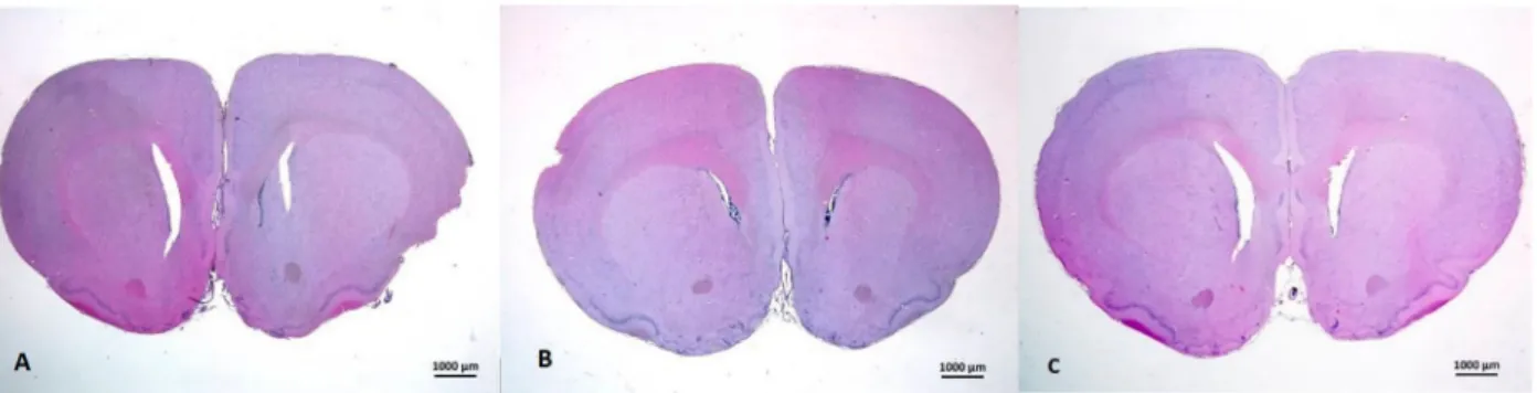
In all groups, histopathologic screening with hematox-ylin–eosin showed the area where NA was located (Figure 8). NA areas were examined in terms of Figure 8.Coronal section of nucleus accumbens of rats in saline (a), sham (b) and US (C) groups respectively. Hematoxylin–Eosin, ×1 lens.
Figure 9.Sections of brain tissues of Saline (A), Sham (B) and US (C) groups respectively. Nucleus accumbens around anterior commissure are seen. Hematoxylin–Eosine, ×20 lens.
impairment of tissue integration, necrosis, apoptosis, infiltration of cells and loss of myelinisation. In saline group, NA area was seen to have normal morphology. Likewise, sham and US groups were seen to have nor-mal morphology and protected tissue integrity. US group did not demonstrate tissue damage like cell infiltration, impairment of tissue integration, necrosis
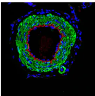
or loss of myelinisation. In light microscopic study, US group and saline group showed no difference (Figure 9). Immunostaining study with anti-MAP2 did not show difference between groups (Figure 10). This reveals that US has no increasing or reducing effect on microtubules, which are a component of the cytoskeleton of neurons or glial cells in rats’ brain tis-sue sections of NA region.
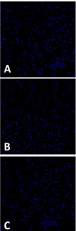
After anti-JNK1 immunostaining, no positive cells were found in blood vessels or neuronal tissue in NA region (Figures 11 and 12). While these antibodies are indicators of apoptosis, this finding exhibits that there were no apoptotic cells in NA region of any group. Thus, US stimulation does not seem to have apoptotic effect on NA area.
Brain sections were stained with Fluoro- Jade C, which is used to determine the neuronal damage. There were no damaged neurons found in NA area, among any group. Thus, US stimulation does not seem to have any neuronal damage effect.
Discussion
The purpose of the current study was to observe mor-phine-conditioned place preference in rats by stimulat-ing NA with a non-invasive method, focused ultrasound and to detect whether there would be any tissue damage caused by US waves. No significant tis-sue damage caused by LIFU waves was detected. Place preference scores of sham group increased sig-nificantly, while scores of LIFU group did not and this result may be associated with LIFU stimulation. Although LIFU stimulation seemed to stop the increase of morphine-induced place preference scores, it was Figure 10.Sections of brain tissues of Saline (A), Sham (B) and
US (C) groups respectively. Anti-MAP2 immunostaining is per-formed to nucleus accumbens region. Microtubules stained with anti-MAP2 are seen in green colour. Nuclei are stained with DAPI (blue). Concerning anti-MAP2 immuostaining, there is not any variation between groups in terms of green fluorescent luminescence intensity (Correspondingly, US stimu-lation is concluded to have no harmful effect on cytoskeleton). Anti-Map2 immünostaining, DAPI, ×40 oil-immersion lens.
Figure 11.Anti-JNK1 immünostaining. For positive control of Anti-JNK1 antibody, blood vessel is stained. Nuclei stained with DAPI (blue), anti-JNK1 positive cells on blood vessel (green) and internal elastic membrane located in intima layer of blood vessel (red autofluorescent luminescence) are seen, x40 oil-immersion lens.
not found to cause a significant decrease in those scores.
US performs a mechanical pressure when passing through one side of the tissue to the other side [23]. The underlying source of ultrasonic neuromodulation is still not clear, despite being the subject of the original study of Harvey [24]. Biological effects of US are pri-marily thermal at high intensity [25]. For US wave form, possible cellular mechanisms have been defined as cavitation, thermogenic effects and mechanic stimu-lation [26]. Unlike HIFU, effects of LIFU waves are not
thought to be thermogenic. Investigators have suggested that neuromodulation provided with short pulses in LIFU may alter the neural conduction and may cause potential changes via mechanic path rather than thermal path [14]. There are studies showing that focused ultrasound waves can change action potentials in hippocampal sections of rats [27], can quench stimulated potentials of rat hippocampus [7], and that stimulation and subsequent reduction in action potential is obtained with short (0.5 milliseconds) focused ultrasound blasts in sciatic nerve preparation of frogs [28]. There are hypotheses stating that, US induced neuromodulation occurs through mechanical stress of lipid layer [29].
No changes were observed in the structure of neural membranes even with chronic stimulation of LIFU waves [9]. Low frequency and low energy LIFU waves are much lower than the damage-causing threshold value. Tissue may not be damaged at all even with high frequency and high energy [30,31]. The possible increase in temperature with LIFU is not in a level to pose negative effects on the neuronal activity [32]. For this reason, LIFU waves may alter the neuronal activity probably without causing tissue injuries.
In a study, the activity of the primary somatosen-sory cortices of humans was shown to be modulated with LIFU waves [15]. Focused ultrasound applied on thalamus has been shown to reduce the time of voluntary movement and reflex response to pinching in anaesthetized rats [33]. Focused ultrasound may also help the growth of new nerves. In a study, micro bubble-enhanced focused ultrasound was shown to increase hippocampal neurogenesis in the mice [34]. Therefore, LIFU waves may be suggested for use in neurodegenerative disorders, particularly Alzheimer’s. There are studies showing that focused ultrasound may even affect nerve cell growth and morphology [35].
Morphine-conditioned place preference scores were shown in the first stage of the current study, in line with the findings of DBS studies. However, on the second stage, positive effects of LIFU waves on morphine dependency could not be shown signifi-cantly as performed in DBS studies and
morphine-conditioned place preference scores have not
decreased. On the other hand, it can be stated that there is no significant increase in morphine-seeking behaviour of US group rats which have developed morphine-conditioned place preference and are conti-nuing morphine conditioning. Meanwhile, morphine-seeking behaviour was seriously increased in the sham group which continued receiving morphine. While there is a significant difference in morphine-seeking behaviour upon comparison of US group with saline group, this difference became insignificant after ultra-sound stimulation.
Figure 12.Anti-JNK1 immünostaining. Saline (A), Sham (B) and US (C) groups respectively are stained with anti- JNK1 antibody. None of the sections of NA region within all groups included positive cells. Nuclei are stained with DAPI (blue), ×40 oil-immersion lens.
The reason for inability to show the positive effects of LIFU waves on morphine dependency as in DBS studies may be that LIFU stimulation dur-ation was relatively short. Besides, in the study, stimulation was started immediately after condition-ing procedure, however, dependency learncondition-ing behav-iour may have been developed in conditioning stage. Although morphine-seeking behaviour may be atte-nuated by LIFU, stimulation for such a short time may have failed in changing the already learned behaviour since then.
In our study, NA focused LIFU waves were esti-mated to form a stimulant effect through mechanosen-sitive channels by activating voltage-gated ion channels mechanically and with stress. By this way, LIFU stimu-lation was thought to effect as a partial agonist and cause a partial dopaminergic increase in NA, and therefore it was considered to prevent the increase in morphine-seeking behaviour of rats. However we did not aim to show the mechanism affecting the present neurotransmission, which might be a subject of further studies.
To the best of our knowledge, this is the first study that examines LIFU stimulation effects on dependency behaviour. Moreover, while animals have received ultrasound stimulation under anaes-thesia in all previous studies, only inhaler isoflurane was used in this study and rats were taken in cus-tom-made stabilizing devices we have ordered. Stereotactic magnet-attachment procedure accord-ing to the point of ultrasound probe that takes Bregma point as reference was also performed firstly in this study.
One animal from physiologic saline group and 3 animals from US group died during anaesthesia pro-cedures. These deaths have lowered the number of rats in both groups (7 animals in physiologic saline group, 6 animals in US group) which may have dimin-ished statistical power. Increasing the number of ani-mals in further studies performed with a similar methodology may contribute to the significance of results.
Prolonged anaesthesia is a potential risk for death of at least a part of experimental animals. Thus, future studies should consider selecting anaesthetics with low mortality rates.
Current study showed that dependency behaviour of sham group is increased significantly, but no sig-nificant increase was shown in US group. In the future, more evident results may be obtained by plan-ning treatment modalities with longer stimulation. In this case, adjusting morphine doses may be beneficial since morphine tolerance may be developed with the longer conditioning period that is required, or the effect of LIFU on morphine dependence may be studied on different addiction methods. An
exper-imental model which excludes the effect of
conditioned learning, like self-administration, may be helpful to detect the exact role of LIFU on mor-phine dependence. However, this may require a remarkably high study funding.
Conclusions
The purpose of this study was to observe the change in morphine-dependent place preference by the stimu-lation of NA with a non-invasive method, focused ultrasound, in a CPP model with rats which are
mor-phine-conditioned and continuing
morphine-conditioning.
Although morphine-dependent place preference scores of US group were not decreased in the study, no significant increase was shown in morphine-seek-ing behaviour compared to pre-stimulation period, and this result may be associated with LIFU stimulation.
The limitations of this study include limited fund-ing resultfund-ing in the inability to show with microdialy-sis method whether the present neurotransmission is also a significant change in neuronal activation or not, and the fact that rat deaths lowered the number of animals required for strong statistical significance. Considering the number of deaths, increasing the number of animals in further studies performed with a similar methodology may contribute to the significance of results. Another remarkable limitation is that we used i.p. route for morphine injections. While morphine has a marked hepatic first-pass metabolism, this application route may have altered the exact effects of morphine. Further, we possibly sonicated the whole NA, both shell and core parts,
which may well effect the interpretations of
outcomes.
We think that the innovation of this study will pro-vide new approaches for LIFU studies in both estab-lishing longer treatment modalities and its use for treatment purposes. In the future, more significant results may be obtained with different dependency methods and by planning treatment modalities with longer stimulation.
We consider that novel studies are required on LIFU stimulation as an alternative method in the treatment of morphine and other opioid dependencies, which have become a serious medical and social issue.
Disclosure statement
No potential conflict of interest was reported by the authors.
Funding
This work was supported by the Bezmiâlem Vakif Üniversi-tesi [grant number 9.2015/20]; Türkiye Bilimsel ve Teknolo-jik Araştirma Kurumu [grant number 214S249].