Purpose: The presence of a pronounced tumor lympho-cytic infiltrate (TLI) is deemed to reflect the presence of an immunoinflammatory response against the tumor and may thus have prognostic significance. We investigated the prognostic value of TLI detected in pathological specimens collected following neoadjuvant chemotherapy (NACT) in patients with breast cancer.
Methods: 100 consecutive patients with breast cancer (mean age 47.8±11.4 years) who were scheduled to under-go anthracycline- and/or taxane-containing NACT were enrolled. Specimens collected after NACT were scored with the 4-point Klintrup scoring criteria for the pres-ence of TLI.
Results: 60 patients had low-grade TLI and 40 high-grade
TLI. Comparison of the patient population according to low-grade vs high-grade TLI revealed statistically signifi-cant difference both in terms of disease-free survival (DFS) (log rank=4.28, p<0.05) and overall survival (OS) (log rank=3.96, p<0.05), with high-grade TLI patients showing a better prognosis. Multivariate Cox regression analysis identified postoperative tumor size and low-grade TLI as the two main independent adverse prognostic factors. Conclusion: High-grade TLI may interfer with tumor growth and can represent a favorable prognostic factor in women with breast cancer undergoing NACT.
Key words: breast cancer, neoadjuvant chemotherapy, prognosis, tumor lymphocytic infiltrates
Summary
Introduction
Prognostic impact of tumor lymphocytic infiltrates in patients
with breast cancer undergoing neoadjuvant chemotherapy
Nilufer Avci1, Adem Deligonul2, Sahsine Tolunay3, Erdem Cubukcu1, Omer Fatih Olmez4,
Ozgur Altmisdortoglu5, Ozgur Tanriverdi6, Asude Aksoy7, Ender Kurt2, Turkkan Evrensel2
1Department of Medical Oncology, Ali Osman Sonmez Oncology Hospital, Bursa; 2Department of Medical Oncology, UludagUniversity Faculty of Medicine, Bursa; 3Department of Pathology, Uludag University Faculty of Medicine, Bursa; 4Department of Medical Oncology, Medipol University School of Medicine,Istanbul; 5Department of Radiation Oncology, Balikesir State Hospital, Balikesir; 6Department of Medical Oncology, Mugla Sitki Kocman University, Mugla; 7Department of Medical Oncology, Firat University, Elazig, Turkey
Correspondence to: Nilufer Avci, MD. Department of Medical Oncology, Ali Osman Sonmez Oncology Hospital, Alaadin Str 2, Osmangazi, Bursa, Turkey. Tel: +90 5052530618, Fax: +90 2242238213, E-mail: [email protected], [email protected]
Received: 16/02/2015; Accepted: 14/03/2015
Breast cancer continues to represent the most common cancer in women worldwide, comprising 23% of all malignancies [1]. According to the Amer-ican Cancer Society, approximately 1.3 million women are diagnosed with breast cancer annual-ly around the world and approximateannual-ly 465,000 cases will die from this neoplasm [2]. NACT is de-fined by the administration of chemotherapy be-fore locoregional treatment (with surgery and/or irradiation) [3]. NACT followed by cytoreduction has currently become a part of standard care for patients with locally advanced breast cancer [4].
There is now convincing evidence that NACT can offer several advantages, including downstaging of large tumors, providing information on tumor response to a specific chemotherapeutic agent, and improving clinical outcomes (presumably through early clearance of systemic micrometa-states) [5-7]. Despite being associated with sig-nificant clinical benefits, breast cancer patients undergoing NACT continue to have a wide range of clinical outcomes [6,7]. In this context, the identification of clinically applicable prognostic features that may allow an improved prognostic E-mail: [email protected]
stratification is eagerly awaited.
The presence of a pronounced TLI in patho-logical specimens has also repeatedly associated with favorable clinical outcomes in a variety of solid malignancies [8-11]. Because the type and density of immune cells in and around the tumor reflects the presence of an immunoinflammatory response against the neoplasm [12], TLI could be a significant determinant of tumor progression. In order to provide an objective assessment of TLI, Klintrup and coworkers [13] have proposed a simplified method for structured scoring of the inflammatory reaction at the tumor invasive edge. The Klintrup classification includes all white cell types and results in a binary score of low-grade or high-grade TLI [13].
The prognostic value of TLI in patients with breast cancer remains controversial. Mohammed et al. [14] have previously shown that a high lym-phocytic infiltrate was associated with improved survival, independent of clinicopathological char-acteristics including ER status, in primary oper-able ductal invasive breast cancer. However, a recent meta-analysis involving a total of 66 inde-pendent studies (totalling 34,086 patients) provid-ed no definite evidence of an association between tumor inflammatory cell infiltrates and clinical outcomes in primary operable breast cancer [15]. Inconsistencies between studies have been attrib-uted to lack of methodological validation for de-terming the presence of TLI, small sample sizes, and patient heterogeneity [15]. Another potential source of confounding is the use of chemotherapy, which may have a significant impact on antican-cer immune responses [16].
In order to shed more light on the clinical significance of TLI in breast cancer patients, we designed the current study to examine the prog-nostic value of TLI (as assessed using the Klintrup score) in pathological specimens collected follow-ing NACT in a sample of patients with breast can-cer.
Methods
Study participantsThe current investigation was designed as a ret-rospective, observational study of 100 consecutive pa-tients with breast cancer (mean age 47.8 ± 11.4 years) who were scheduled to undergo NACT before attempt-ing cytoreductive surgery at the Department of Oncol-ogy of the Uludag University Medical Center, Bursa, Turkey. Enrollment took place between September 2006 and August 2011. All patients were of Turkish
descent. The clinicopathological characteristics of the study participants were collected from pathological reports and medical charts. Lesion staging was per-formed according to the sixth edition of the American Joint Committee on Cancer (AJCC) staging manual for breast cancer. All participants received anthracycline- and/or taxane-containing NACT before surgery. The study protocol conformed to the tenets of the Declara-tion of Helsinki and was approved by the local ethics committee.
All patients gave oral informed consent. Assessment of TLI
The presence of TLI in tumor specimens collect-ed after NACT was assesscollect-ed using the Klintrup scoring system [13]. Briefly, specimens were scored according to a 4-point scale. Scores were based on appearances at the deepest area of tumor invasion. A score of 0 indi-cated no increase in inflammatory cells at the deepest point of the tumor’s invasive margin; a score of 1 de-noted a mild and patchy increase in inflammatory cells; a score of 2 indicated a prominent inflammatory reac-tion forming a band at the invasive margin with some evidence of destruction of cancer cell islands; finally, a score of 3 denoted a florid cup-like inflammatory infil-trate at the invasive edge with frequent destruction of cancer cell islands. For the purpose of analysis, these scores were then divided into two subgroups, i.e. low-grade TLI (scores 0 and 1) and high-low-grade TLI (scores 2 and 3) [13].
Study endpoints
Disease-free survival (DFS) was defined as time from surgery to the date of first relapse, second pri-mary malignancy, or death resulting from any cause (whichever occurred first). Overall survival (OS) was defined as time from surgery to the date of death re-sulting from any cause. Patients who were alive (for OS) and disease-free (for DFS) were censored at the date of the last follow-up.
Statistics
The data were checked for normality using the Kolmogorov-Smirnov test for continuous variables. Variables were expressed as means ± standard devi-ation, medians (lower quartile-upper quartile), or as counts (percentages) if categorical. Correlations were tested using the Spearman’s correlation coefficient. The association of each risk factor with DFS and OS was as-sessed by multivariate Cox proportional hazard regres-sion analysis. The multivariate Cox model included all the characteristics listed in Table 1. The appropriate-ness of the proportional hazards assumption was ver-ified using graphical methods and tested as described previously [17]. The assumption of linearity for the Cox models was examined through visual inspection, and no violation was found. Hazard ratios (HRs) and their
95% confidence intervals (CIs) were calculated with the estimated regression coefficients and their standard er-rors in the Cox models. DFS and OS curves were plot-ted using the Kaplan-Meier method, and the differences were compared using the log-rank test. All calculations were performed using SPSS software (version 17.0, SPSS Inc., Chicago, IL, USA). All tests were two-sided, and p<0.05 was considered as statistically significant.
Results
The general characteristics of patients with breast cancer undergoing NACT are summarized in Table 1. Of the 100 study participants, 90 had invasive ductal carcinoma, 6 invasive lobular cinoma, and the remaining 4 other forms of car-cinoma. The primary tumor status was T1 in 16 patients, T2 in 64 patients, T3 in 12 patients, and T4 in 8 patients. The lymph node status was as follows: N0 in 63 patients, N1 in 35 patients, N2 in 1 patient and N3 in 1 patient. The mean num-ber of NACT cycles was 5.7±1.0. The patients were followed up for a mean of 38.2±11.4 months.
TLI and clinical outcomes
The distribution of the Klintrup scores in the 100 breast cancer specimens collected after NACT was as follows: 7 patients had a score of 0, 53 had a score of 1, 15 had a score of 2, and 25 had a score of 3 (Table 1). Therefore, there were 60 pa-tients with low-grade TLI and 40 with high-grade TLI. We then examined the associations between the extent of TLI and the general characteristics of the study participants (Table 2). Interesting-Table 1. General patient, disease and chemotherapy
characteristics (patients, N=100)
Characteristics N (%)
Age (years) , mean±SD 47.8 ± 11.4 Postmenopausal status (yes/no) 55/45 Preoperative primary tumor stage
T1 16 (16)
T2 64 (64)
T3 12 (12)
T4 8 (8)
Preoperative axillary lymph node stage
N0 63 (63)
N1 35 (35)
N2 1 (1)
N3 1 (1)
Number of nodal metastases
No metastases 77 (77) 1-3 18 (18) 4-9 1 (1) ≥ 10 1 (1) Unknown 3 (3) Clinical TNM stage 1A 13 (13) 1B 0 (0) 2A 48 (48) 2B 29 (29) 3A 5 (5) 3B 4 (4) 3C 1 (1) Histology
Invasive ductal carcinoma 90 (90) Invasive lobular carcinoma 6 (6)
Mucinous carcinoma 2 (2) Tubulolobular carcinoma 1 (1) Unknown 1 (1) Neoadjuvant chemotherapy Anthracycline combinations 40 (40) Taxane/anthracycline combinations 48 (48) Unknown 12 (12)
Number of chemotherapy cycles,
mean±SD 5.7 ± 1.0
Postoperative tumor size (mm) median
(range) 18 (5-30)
Postoperative primary tumor stage
T1 62 (62) T2 28 (28) T3 10 (10) T4 0 (0) Characteristics N (%) Pathological TNM stage 0 13 (13) 1A 33 (33) 1B 1 (1) 2A 33 (33) 2B 17 (17) 3A 2 (2) 3B 0 (0) 3C 1 (1)
TLI (Klintrup score)
0 7 (7)
1 53 (53)
2 15 (15)
3 25 (25)
Data are expressed as counts, mean±standard deviation, or medi-an (rmedi-ange), as appropriate
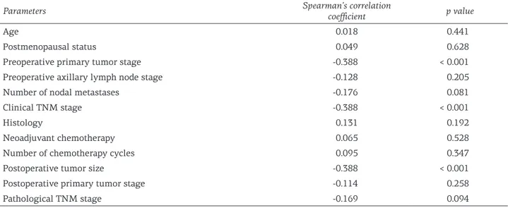
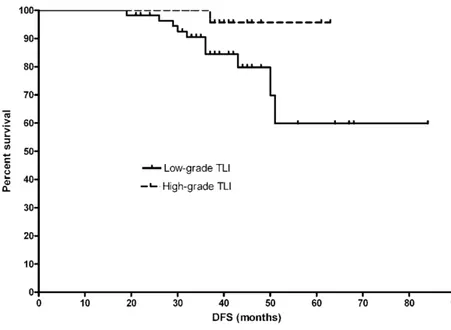
ly, we found significant associations of TLI with preoperative primary tumor stage, clinical TNM stage, and postoperative tumor size. These results suggested that higher TLI scores were associated with less severe preoperative primary tumor and clinical TNM stage as well as a lower postoper-ative tumor size. In the entire study cohort, the mean DFS was 36.5 ± 12.1 months, whereas the mean OS was 37.1±11.3 months (p=0.63). Cate-gorization of the patient population according to low-grade vs high-grade TLI revealed statistical-ly significant difference both in terms of DFS (log rank = 4.28, p<0.05, Figure 1) and OS (log rank = 3.96, p<0.04, Figure 2), with patients with high-grade TLI showing better prognosis. After
allow-ance for potential confounders, the results of mul-tivariate Cox regression analysis indicated that postoperative tumor size (DFS, HR=4.2, 95% CI = 1.1−10.4, p<0.05; OS, HR=4.4, 95% CI = 1.3−11.6, p<0.05) and low-grade TLI (DFS, HR=1.2 , 95% CI = 1.1−2.4, p<0.05; OS, HR=1.5, 95% CI = 1.1−3.6, p<0.05) were the two main independent adverse prognostic factors in our patients undergoing NACT (Table 3).
Discussion
This prospective investigation was designed to assess the association between the presence of TLI and prognosis in a series of breast cancer Table 2. Correlation analysis of TLI scores (Klintrup scoring system) with the general characteristics of breast cancer patients undergoing neoadjuvant chemotherapy (N = 100)
Parameters Spearman’s correlation coefficient p value
Age 0.018 0.441
Postmenopausal status 0.049 0.628
Preoperative primary tumor stage -0.388 < 0.001
Preoperative axillary lymph node stage -0.128 0.205
Number of nodal metastases -0.176 0.081
Clinical TNM stage -0.388 < 0.001
Histology 0.131 0.192
Neoadjuvant chemotherapy 0.065 0.528
Number of chemotherapy cycles 0.095 0.347
Postoperative tumor size -0.388 < 0.001
Postoperative primary tumor stage -0.114 0.258
Pathological TNM stage -0.169 0.094
Table 3. Results of multivariate regression analysis for disease-free survival and overall survival
Parameters Disease-free survival, HR (95% CI), p value HR (95% CI), p valueOverall survival, Age HR=1.1 (0.9−1.7), 0.85 HR=1.2 (0.9−1.5), 0.74 Postmenopausal status HR=1.5 (0.8−1.9), 0.56 HR=1.4 (0.7−1.8), 0.67 Preoperative primary tumor stage HR=2.7 (0.9−2.8), 0.78 HR=2.4, (0.9−2.3), 0.45 Preoperative axillary lymph node stage HR=1.8 (0.8−1.9), 0.49 HR=1.5 (0.7−1.5), 0.67 Number of nodal metastases HR=5.9 (0.8−6.4), 0.24 HR=5.5 (0.9−5.8), 0.18 Clinical TNM stage HR=2.3 (0.9−2.5) 0.70 HR=2.4 (0.8−2.6), 0.73 Histology HR=1.3 (0.4−2.5), 0.87 HR=1.4 (0.5−2.4), 0.90 Neoadjuvant chemotherapy HR=2.5 (0.8−2.8), 0.35 HR=2.6 (0.7−2.5), 0.24 Number of chemotherapy cycles HR=1.3 (0.4−1.5), 0.59 HR=1.5 (0.6−1.8), 0.68 Postoperative tumor size HR=4.2 (1.1−10.4), <0.05 HR=4.4 (1.3−11.6), <0.05 Postoperative primary tumor stage HR=1.1 (0.5−1.2), 0.56 HR=1.2 (0.7−1.3), 0.67 Pathological TNM stage HR=2.4 (0.9−2.9), 0.81 HR=2.3 (0.8−2.7), 0.74 Low-grade TLI HR=1.2 (1.1−2.4), p<0.05 HR=1.5 (1.1−3.6), <0.05
patients undergoing NACT. Because the clinical outcomes of this patient group continue to be het-erogenous [5-7], there is an urgent need of bio-marker tools that may improve their prognostic stratification. This study revealed three principal findings. First, significant associations of TLI with preoperative primary tumor stage, clinical TNM
stage, and postoperative tumor size were found. Second, patients with high-grade TLI after NACT showed a better prognosis both in terms of DFS and OS. Third, TLI was an independent prognos-tic factor in our patients undergoing NACT. Taken together, these results indicate that the assess-ment of TLI using the Klintrup criteria should be Figure 1. Kaplan-Meier plots for disease-free survival in breast cancer patients undergoing NACT according to the presence of low-grade vs high-grade TLI (p<0.05).
Figure 2. Kaplan-Meier plots for overall survival in breast cancer patients undergoing NACT according to the presence of low-grade vs high-grade TLI (p<0.04).
routinely included in the pathological reporting of breast cancer specimens obtained after NACT. Notably, the Klintrup method used in the present study allowed a structured assessment of all white cell types at the primary tumor and can be applied to routine hematoxylin and eosin specimens [13].
The association between TLI and prognosis in patients with breast cancer is still a matter of debate. Although TLI has been related to better clinical outcomes in some studies, other series have identified a significant association between inflammatory inflitrates and reduced survival [15]. The apparent controversy clearly highlights the need for further research on this topic. Our study supports the notion that the presence of TLI after NACT is a favorable prognostic factor in breast cancer patients. This association can be explained by the fact that tumor cells can be cleared by host innate and adaptive immuno-inflammatory cells, a process known as tumor immunosurveillance [18]. During tumor-specific adaptive immune re-sponses, some inflammatory cells infiltrating the tumor (e.g., cytotoxic T lymphocytes) can induce the production of tumor-associated antigens and the cytokine interferon-gamma (IFN-γ) [19]. No-tably, IFN-γ can modulate prognosis in patients with malignancies by influencing cell cycle ar-rest, apoptosis, differentiation, angiogenesis, and macrophage activity [20]. These observations may explain the favorable prognostic impact of TLI as observed in our report. In particular, we pos-tulate that an increased inflammatory response may have stimulated the clearance of rapidly di-viding tumor cells. All of the patients enrolled in
this study underwent NACT. In a previous study, Denkert et al. [16] reported a strong association between lymphocytic infiltrate and chemotherapy response in a large set of more than 1,000 sam-ples enrolled from two prospective, randomized clinical breast cancer trials. The authors speculat-ed that chemotherapy could trigger an immune response directed against the tumor cells, which can be particularly strong in the subset of patients in whom a sensitization of the immune system against some tumor antigens is present before the onset of chemotherapy [16]. Further studies are needed to shed more light on this possibility.
Our findings should be interpreted within the context of some limitations. First, this investi-gation was conducted in Turkish individuals, so results cannot be simply extrapolated to popula-tions with different ethnic backgrounds. Second, our study should be considered as an exploratory analysis and independent replication is needed to extend and confirm our results. Moreover, breast cancer patients were treated on an individual ba-sis according to each patient’s disease character-istics based on clinical trial data and influenced by the personal experience of the surgeon [7]. Finally, due to financial constraints, IFN-γ and molecular markers of white blood cell recruitment and infil-tration were not measured in this study.
Notwithstanding the study caveats, our re-sults suggest that the assessment of TLI provides independent prognostic information in breast can-cer patients undergoing NACT and should there-fore be considered for future inclusion in routine pathological reporting of this patient population.
References
1. Youlden DR, Cramb SM, Dunn NA, Muller JM, Pyke CM, Baade PD. The descriptive epidemiology of fe-male breast cancer: an international comparison of screening, incidence, survival and mortality. Cancer Epidemiol 2012;36:237-248.
2. DeSantis C, Siegel R, Bandi P, Jemal A. Breast cancer statistics, 2011. CA Cancer J Clin 2011:61:409-418. 3. Gampenrieder SP, Rinnerthaler G, Greil R. Neoadjuvant
chemotherapy and targeted therapy in breast cancer: past, present, and future. J Oncol 2013;2013:732047. 4. Connolly RM, Stearns V. Current approaches for
neo-adjuvant chemotherapy in breast cancer. Eur J Phar-macol 2013;717:58-66.
5. Redden MH, Fuhrman GM. Neoadjuvant
chemother-apy in the treatment of breast cancer. Surg Clin North Am 2013;93:493-499.
6. Aigner J, Schneeweiss A, Sohn C, Marmé F. The role of neoadjuvant chemotherapy in the management of primary breast cancer. Minerva Ginecol 2011;63:261-274.
7. Bear HD. Neoadjuvant chemotherapy for operable breast cancer: individualizing locoregional and sys-temic therapy. Surg Oncol Clin N Am 2010;19:607-626.
8. Fridman WH, Galon J, Dieu-Nosjean MC et al. Im-mune infiltration in human cancer: prognostic signifi-cance and disease control. Curr Top Microbiol Immu-nol 2011;344:1-24.
9. Bremnes RM, Al-Shibli K, Donnem T et al. The role of tumor-infiltrating immune cells and chronic inflam-mation at the tumor site on cancer development, pro-gression, and prognosis: emphasis on non-small cell lung cancer. J Thorac Oncol 2011;6:824-833.
10. Goeppert B, Frauenschuh L, Zucknick M et al. Prog-nostic impact of tumour-infiltrating immune cells on biliary tract cancer. Br J Cancer 2013;109:2665-2674. 11. Mei Z, Liu Y, Liu C et al. Tumour-infiltrating
inflam-mation and prognosis in colorectal cancer: systematic review and meta-analysis. Br J Cancer 2014;110:1595-1605.
12. Talmadge JE, Donkor M, Scholar E. Inflammatory cell infiltration of tumors: Jekyll or Hyde. Cancer Metasta-sis Rev 2007;26:373-400.
13. Klintrup K, Mäkinen JM, Kauppila S et al. Inflamma-tion and prognosis in colorectal cancer. Eur J Cancer 2005;41:2645-2654.
14. Mohammed ZM, Going JJ, Edwards J, Elsberger B, Doughty JC, McMillan DC. The relationship between components of tumour inflammatory cell infiltrate and clinicopathological factors and survival in pa-tients with primary operable invasive ductal breast
cancer. Br J Cancer 2012;107:864-873.
15. Mohammed ZM, Going JJ, Edwards J, McMillan DC. The role of the tumour inflammatory cell infiltrate in predicting recurrence and survival in patients with primary operable breast cancer. Cancer Treat Rev 2012;38:943-955.
16. Denkert C, Loibl S, Noske A et al. Tumor-associated lymphocytes as an independent predictor of response to neoadjuvant chemotherapy in breast cancer. J Clin Oncol 2010;28:105-113.
17. Minoretti P, Falcone C, Calcagnino M et al. Prognostic significance of plasma osteopontin levels in patients with chronic stable angina. Eur Heart J 2006;27:802-807. 18. Mlecnik B, Bindea G, Pagès F, Galon J. Tumor immu-nosurveillance in human cancers. Cancer Metastasis Rev 2011;30:5-12.
19. Slaney CY, Rautela J, Parker BS. The emerging role of immunosurveillance in dictating metastatic spread in breast cancer. Cancer Res 2013;73:5852-5857.
20. Ikeda H, Old LJ, Schreiber RD. The roles of IFN gam-ma in protection against tumor development and cancer immunoediting. Cytokine Growth Factor Rev 2002;13:95-109.