ORIGINAL ARTICLE
A clinical study on single-visit root canal retreatments
on consecutive 173 patients: frequency of periapical complications
and clinical success rate
Tan Firat Eyuboglu1&Kezban Olcay2&Mutlu Özcan3
Received: 17 November 2015 / Accepted: 5 September 2016 / Published online: 22 September 2016 # Springer-Verlag Berlin Heidelberg 2016
Abstract
Objectives This study assessed the outcome measures of single-visit root canal retreatments and frequency of periapical complications considering preoperative, intraoperative and postoperative factors.
Materials and methods Between November 2011 and December 2012, in 173 patients, a total of 234 endodontically treated teeth were retreated in a single appointment by one experienced endodontist. Five teeth were extracted and 119 teeth were lost to follow-up yielding to 110 teeth (47 %) to be examined by two calibrated examiners for the outcome of healing (periapical index score-PAI≤2; no signs or symptoms) or non-healing (presence of apical periodontitis-PAI >2; signs or symptoms). Preoperative, intraoperative and postoperative factors were evaluated for their association with the outcome. Data were analysed using Fisher’s exact and Fisher-Freeman Halton tests for bivariate analysis to identify potential out-come predictors. Logistic regression models were used for multivariate analysis to determine significant outcome predictors.
Results Mean observation time was 29 months. Follow-up assessment revealed 100 teeth (90.9 %) as healed and 10 teeth
(9.1 %) non-healed. Age, gender, tooth type and preoperative (pain, periodontal defects, root filling density and length), in-traoperative (sealer extrusion) and postoperative (type of cor-onal restorations) factors did not significantly affect the out-come (p > 0.05). Preoperative periradicular lesions with diam-eters less than 5 mm presented significantly better outcome than larger lesions (p < 0.05; odds ratio (OD) 6; 95 % CI 1.45– 24.85). Logistic regression model showed an increased risk of non-healing for the parameter of preoperative periradicular lesions with diameters larger than 5 mm (OD 6.42; 95 % CI 1.51–27.27).
Conclusions Single-appointment root canal retreatments pre-sented a favourable success rate. Only preoperative lesion size had a significant effect on the outcome where the lesions smaller than 5 mm performed significantly better healing. Clinical relevance Single-appointment root canal retreatments could be considered as a viable treatment option for orthograde retreatment cases with periradicular lesion size smaller than 5 mm.
Keywords PAI scores . Periapical complications . Periapical lesion . Root canal retreatment . Single-appointment
endodontic therapy
Introduction
The primary goal of endodontic treatment is to prevent or heal apical periodontitis (AP) [17], but it has been reported in cross-sectional studies from different countries that more than 30 % of all endodontically treated teeth in the studied popu-lations are associated with AP orBpost treatment disease^ [2,
5,10].
Post treatment diseases could be treated by two options including orthograde retreatment and apical surgery [10]. In
* Tan Firat Eyuboglu [email protected]
1
School of Dentistry, Department of Endodontics, Istanbul Medipol University, Atatürk Bulvarı No:27, Unkapanı, Fatih,
34083 Istanbul, Turkey
2
School of Dentistry, Department of Endodontics, Istanbul Medipol University, Istanbul, Turkey
3 Dental Materials Unit, Centre for Dental and Oral Medicine, Clinic
for Fixed and Removable Prosthodontics and Dental Materials Science, University of Zürich, Plattenstrasse 11,
a previous study, orthograde retreatments presented a success rate of 81 % classified asBhealed^ and 93 % as Bno symptoms and fully functional^ [7]. In a systematic review, it has also been concluded that endodontic surgery offers more favourable initial success but orthograde retreatment yields to a better long-term outcome [27]. Although early results of single-visit treatments are contradictory [11,29,30], due to novel techniques and equipment available today [33] and pa-tient requests, a considerable amount of increased interest and effort were accumulated on single-visit treatments. Completing the treatment in a single appointment reduces the treatment time and cost, decreases microleakage risk and avoids recontamination of root canals between appointments in multiple-visit treatments [8,32,34]. In fact, no significant differences were observed in healing of periapical radiolucen-cy between teeth treated in one visit (without) and those treat-ed in two visits using calcium hydroxide for 4 weeks and that the presence of a positive bacterial culture at the time of filling did not influence the outcome of treatment [18]. Similarly, Ashraf et al. reported single-appointment retreatments with a success rate of 84.9 % and recommended single-appointment retreatments for symptomless teeth [1].
Due to excessive request from patients as a result of limited time for dental visits during working hours and long travelling time required to visit dental offices, single-appointment treat-ments [8,18,32] and retreatments [1] for root canal treatments could bring economical advantages.
The objective of this study therefore was to assess the fre-quency of periapical complications and success rate of single-appointment non-surgical root canal retreatments retrospec-tively considering preoperative, intraoperative and postopera-tive factors.
Materials and methods
Inclusion and exclusion criteria
This retrospective study included patients who were in need of orthograde retreatment and treated in a single appointment accordingly at the Istanbul Medipol University between November 2011 and December 2012. The ethics committee of the same university approved the study and all enrolled subjects signed a written informed consent. All retreatments were assigned due to persisting periapical lesions, preopera-tive signs and symptoms and coronal and/or apical leakage of root canals. In total, in 173 patients, 234 teeth were retreated by one endodontist (T.F.E.) who had an experience of 12 years. The diagnoses of the relevant teeth were made with a pano-ramic (KODAK 9000, Carestream Health, Rochester, NY, USA) and periapical radiograph (KODAK RVG 5100, Carestream Health) using a parallel technique with an expo-sure time of 0.16 s and expoexpo-sure dose of 1.22 mGy. The teeth
diagnosed with excessive periodontal disease, vertical root fractures or those that received surgical endodontic treatment were excluded.
Retreatment procedure
The treatments were carried out in a single appointment. All restorations (single crowns and fixed dental prosthesis) were removed initially to gain direct access to the relevant tooth. Root posts were primarily removed using an ultrasonic device (UDS-N2, Woodpecker, Guangxi, People’s Republic of China) and if required with a portegue.
After having access to the previously obturated root canals, #1, #2 ve #3 Gates Glidden (GG) burs (Mani Inc., Tochigi, Japan) and #15 Kerr files (Mani Inc.) were used to remove the root canal filling completely. No chemical solvent was used to remove the gutta-percha or the sealer. Cleaning and shaping of the canal were performed employing a crown-down technique using nickel-titanium rotary instruments (Revo-S Micro-Méga, Besançon, France). After measuring the root lengths with an apex locater (Apex Pointer, Micro-Méga), each tooth was prepared up to AS 40 file, 0.5 mm short of the apex. The canals were irrigated after the use of each instrument with 1 ml of 2.5 % sodium hypochlorite (NaOCl). The last irrigation was accomplished using 2.5 ml 5 % ethylene diamine tetra-acetic acid (EDTA), 2.5 ml of 2.5 % NaOCl and 5 ml of distilled water, respectively. A single-cone technique was then used to fill the root canals with either AS35 or AS40 gutta-percha cones (Revo-S, Micro-Méga). Root canal filling paste (AH Plus, Dentsply, DeTrey, Konstanz, Germany) was intro-duced into the root canal using master cones with a brushing motion and accessory gutta-percha cones (SU 25, Revo-s, Micro-Méga) were used when needed using non-compaction method.
A total-etch bond (Single bond 2, 3M ESPE, St. Paul, MN, USA) technique was used according to the manufacturer’s instructions prior to coronal restoration. Flowable resin com-posite (Filtek Ultimate Flowable, 3M ESPE, St. Paul, MN, USA) was used as base material in order to seal the canal orifices. Thereafter, the coronal restoration was made with resin composite (Filtek Ultimate, 3M ESPE) or a fibre post (Cytec Blanco, HT-Glasfiber, E. Hahnenkratt GmbH, Königsbach-Stein, Germany) and composite core (RelyX U200, 3M ESPE) prior to a fixed prosthetic restoration de-pending on the prosthetic plan.
Radiographic evaluation
A periapical radiograph of the relevant tooth was made imme-diately after the retreatment with paralleling technique using the same digital radiograph with the same parameters used prior to treatment as described above. The preoperative and intraoperative data were recorded at the database.
Follow-up evaluation
During the follow-up, clinical signs and symptoms of the rel-evant tooth were recorded and radiographs were made to re-cord periapical indices (PAI). The principal investigator (T.F.E.) and the coexaminer (K.O.) examined all preoperative and postoperative radiographs. The PAI scores of periapical healing and non-healing were recorded for each endodontical-ly retreated tooth during the follow-up after the retreatment.
The endodontically retreated teeth were clinically exam-ined, and patient response was recorded upon pain in percus-sion, palpation and chewing (present or absent), any swelling, fistula or sign of infection at the gingival tissue (present or absent), quality of the coronal restoration, occlusal marginal defects (present or absent) and the quality of the root canal filling (length and density of the root filling) (Table1).
Calibration of the observers
All PAI scores were obtained from periapical radiographs with a paralleling technique. Two investigators were calibrated for recording PAI with a calibration kit of 100 reference radio-graphs [11]. The PAI scores were dichotomized to reflect ab-sence (PAI≤2) or presence (PAI >2) of apical periodontitis. Teeth with multiple root canals were scored for the root canal with the highest PAI score.
Statistical analysis
Data obtained from preoperative, intraoperative and postoper-ative factors and their association with the outcome were sta-tistically analysed using a software programme (Number Cruncher Statistical System, NCSS 2007 and Power Analysis and Sample Size, PASS 2008 Statistical Software, UT, USA). Fisher’s exact and Fisher-Freeman Halton tests were used for bivariate analysis to identify potential outcome predictors, and logistic regression models were used for mul-tivariate analysis to assess significant outcome predictors. Inter-examiner and intraexaminer Cohen’s kappa scores were calculated twice within 2 months interval. Post hoc power analyses were made for the parameters studied.
Results Post hoc power analyses indicated preoperative PAI score, preoperative radiolucency less than 80 % and length of root filling with high odds ratio more than 80 %.
Of all patients treated (N = 234 teeth), 119 (50.8 %) teeth, 98 of 173 patients (56.6 %) were lost until the final follow-up, 5 teeth were extracted of which 4 were for restorative and 1 for periodontal reasons. Out of 98 patients (119 teeth), 1 patient was deceased (1 tooth), 28 patients declined recalls (34 teeth), and 69 patients did not respond (84 teeth). One hundred ten teeth (47 %) were examined by two calibrated examiners for the following outcomes:Bhealed^ (periapical index score PAI
≤2; no signs or symptoms) or Bnon-healed^ (presence of api-cal periodontitis: PAI >2, signs or symptoms) (Table1). Inter-examiner and intraInter-examiner Kappa scores ranged between 0.936 and 0.964, showing a very good agreement (Table2). There was statistically no significant difference among the factors between inception cohort and study group (p > 0.05) (Table1).
Until the final follow-up, 100 teeth (90.9 %) were catego-rized asBhealed^ and 10 teeth (9.1 %) as Bnon-healed^. The mean age of the study group was 43.5 ± 14.4. Whilst 45 (60.8 %) of the patients were female, 29 (39.2 %) of them were male. The mean age of the Bhealed^ group was 43 ± 13.7 whilst theBnon-healed^ group was 38 ± 15.2 with no significant difference (p = 0.745; p > 0.05). Tooth type or tooth location also had no significant effect on the outcome. Among all 10Bnon-healed^ teeth that had PAI scores >2 (6 teeth PAI = 3, 3 teeth PAI = 4, 1 tooth PAI = 5), only 1 was clinically symptomatic (PAI = 4). One of them had a persistent and growing lesion at the bifurcation area diagnosed with a strip perforation and extracted during the follow-up (PAI = 3). Six teeth presented smaller lesions compared to baseline situ-ation, including the symptomatic tooth and the perforated tooth; lesions did not change in 3 teeth and became larger in 1 tooth.
There was no correlation between preoperative pain and outcome measures of the study (p > 0.05) (Table 3). Preoperative PAI scores had a significant effect on the out-come (p = 0.014). Preoperative periradicular lesion with a diameter smaller than 5 mm demonstrated significantly better outcome than the larger lesions (p = 0.025; odds ratio (OD) 6; 95 % CI 1.45–24.85) (Table3). Preoperative PAI score 5 had a significantly increased failure ratio (OD 6.8; 95 % CI 1.75– 26.73) (Table3).
Preoperative periodontal defects, preoperative root filling material, density and length, intraoperative root filling length, root filling voids, sealer extrusion, postoperative quality, type of coronal restorations and post applications had no significant effect on the outcome (p > 0.05). Although the length of root filling had no effect on the outcome, OD for adequate root canal filling length was calculated to be 3.13 (95 % CI 0.51– 19.35) with a greater difference compared to other root canal filling lengths according to the logistic regression models (Table4).
Discussion
With the increased success rate and newly developed mate-rials, single-appointment root canal treatment procedure has become a viable treatment protocol for both primary root ca-nal treatments and retreatments [1,9,19,33,34]. Yet, preop-erative factors affecting the treatment outcome are essential for decision-making in endodontic treatment [4,5, 7, 10].
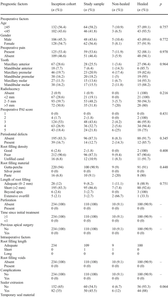
Table 1 Distribution of prognostic factors, inception cohort, study sample and their association with healing and non-healing and p values (univariate analysis)
Prognostic factors Inception cohort (n (%)) Study sample (n (%)) Non-healed (n (%)) Healed (n (%)) p Preoperative factors Age ≥45 132 (56.4) 64 (58.2) 7 (10.9) 57 (89.1) 0.757 <45 102 (43.6) 46 (41.8) 3 (6.5) 43 (93.5) Gender Male 106 (45.3) 48 (43.6) 5 (10.4) 43 (89.6) 0.772 Female 128 (54.7) 62 (56.4) 5 (8.1) 57 (91.9) Preoperative pain Present 125 (53.4) 59 (53.6) 7 (11.9) 52 (88.1) 0.970 Absent 109 (46.6) 51 (46.4) 3 (5.9) 48 (94.1) Tooth Maxillary anterior 67 (28.6) 28 (25.5) 1 (3.6) 27 (96.4) 0.964 Mandibular anterior 18 (7.7) 7 (6.4) 1 (14.3) 6 (85.7) Maxillary premolar 46 (19.7) 23 (20.9) 4 (17.4) 19 (82.6) Mandibular premolar 38 (16.2) 20 (18.2) 1 (5) 19 (95) Maxillary molar 27 (11.5) 15 (13.6) 1 (6.7) 14 (93.3) Mandibular molar 38 (16.2) 17 (15.5) 2 (11.8) 15 (88.2) Radiolucency Absent 2 (0.9) 1 (0.9) 0 (0) 1 (100) 0.216 <2 mm 67 (28.6) 21 (19.1) 0 (0) 21 (100) 2–5 mm 93 (39.7) 53 (48.2) 3 (5.7) 50 (94.3) >5 mm 72 (30.8) 35 (31.8) 7 (20) 28 (80) Preoperative PAI score
1 0 (0) 0 (0) 0 (0) 0 (0) 0.431 2 4 (1.7) 2 (1.8) 0 (0) 2 (100) 3 124 (53) 48 (43.6) 2 (4.2) 46 (95.8) 4 63 (26.9) 36 (32.7) 2 (5.6) 34 (94.4) 5 43 (18.4) 24 (21.8) 6 (25) 18 (75) Periodontal defects Absent 195 (83.3) 96 (87.3) 8 (8.3) 88 (91.7) 0.345 Present 39 (16.7) 14 (12.7) 2 (14.3) 12 (85.7)
Root filling density
Good 6 (2.6) 2 (1.8) 0 (0) 2 (100) 0.408
Poor 212 (90.6) 96 (87.3) 9 (9.4) 87 (90.6) Unfilled canal 16 (6.8) 12 (10.9) 1 (8.3) 11 (91.7) Root filling material
Gutta-percha 220 (94) 100 (90.9) 9 (9) 91 (91) 0.448
Silver point 0 (0) 0 (0) 0 (0) 0 (0)
Paste 16 (6.8) 10 (9.1) 2 (20) 8 (80)
Length of root filling
Adequate (0–2 mm) 28 (12) 9 (8.2) 1 (11.1) 8 (88.9) 0.751 Short (>2 mm) 195 (83.3) 95 (86.4) 7 (7.4) 88 (92.6) Beyond apex 6 (2.6) 3 (2.7) 0 (0) 3 (100) Extensive overfill 5 (2.1) 3 (2.7) 2 (66.7) 1 (33.3) Perforation Absent 234 (100) 110 (100) 10 (9.1) 100 (90.9) – Present 0 (0) 0 (0) 0 (0) 0 (0)
Time since initial treatment
≥1 234 (100) 110 (100) 10 (9.1) 100 (90.9) –
<1 0 (0) 0 (0) 0 (0) 0 (0)
Previous apical surgery
No 234 (100) 110 (100) 10 (9.1) 100 (90.9) –
Yes 0 (0) 0 (0) 0 (0) 0 (0)
Intraoperative factors Root filling length
Adequate 234 109 9 100
Short 0 1 1 0
Long 0 0 0 0
Root filling voids
Absent 234 (100) 110 (100) 10 (9.1) 100 (90.9) – Present 0 (0) 0 (0) 0 (0) 0 (0) Complications No 234 (100) 110 (100) 10 (9.1) 100 (90.9) – Yes 0 (0) 0 (0) 0 (0) 0 (0) Sealer extrusion No 152 (65) 60 (54.5) 4 (6.7) 56 (93.3) 0.064 Yes 82 (35) 50 (45.5) 6 (12) 44 (88)
This study solely focused on the outcomes of single-visit retreatment considering possible preoperative, intraoperative and postoperative factors affecting the results.
This retrospective study presented 90.9 % success rate as 98.2 % of the teeth were asymptomatic and fully functional after a mean observation time of 29 months. Age, gender, tooth type and periodontal defects had no effect on the out-come measures similar to other studies [4,7]. All patients with asymptomatic non-healed teeth were informed about the situ-ation. It is critical to communicate with the patients for
retaining their teeth whether with successful outcome or being asymptomatic with a notification of routine follow-ups of the involved teeth. Patients in this group and those in the study group were all in agreement for retaining their teeth and were willing to attend further follow-ups.
Root canal retreatment without apical periodontitis was reported to have a high healing rate (93–98 %) in previous studies [4,7,22,23] but due to high incidence of lesions in the teeth involved in the study group, only one tooth was free of apical periodontitis. Therefore, in this study, no conclusions
Table 2 Intraexaminer (T versus K) and inter-examiner Cohen’s kappa values based on PAI scores that were recorded from the same radiographs within 2 months interval (1 versus 2)
PAI Intra-examiner Inter-examiner
T 1–T 2 K 1–K 2 T 1–K 1 T 2–K 2 n (%) n (%) n (%) n (%) 1 33 (23.7) 31 (22.3) 32 (23) 29 (20.9) 2 35 (25.2) 35 (25.2) 34 (24.5) 37 (26.6) 3 23 (16.5) 25 (18) 25 (18) 24 (17.3) 4 29 (20.9) 29 (20.9) 29 (20.9) 30 (21.6) 5 19 (13.7) 19 (13.7) 19 (13.7) 19 (13.7)
Cohen’s kappa 0.936 (very good) 0.955 (very good) 0.964 (very good) 0.964 (very good) p <0.001** <0.001** <0.001** <0.001** **p < 0.01
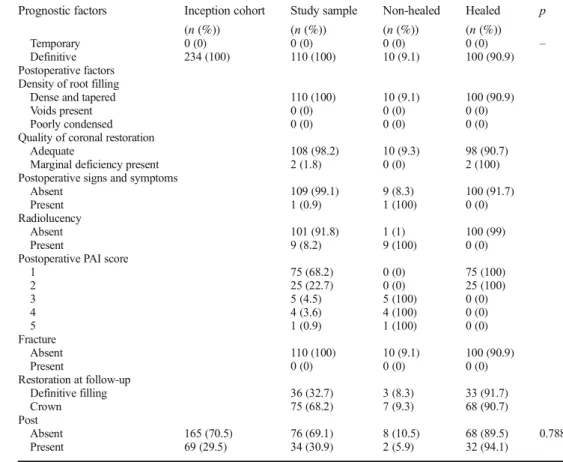
Table 1 (continued)
Prognostic factors Inception cohort (n (%)) Study sample (n (%)) Non-healed (n (%)) Healed (n (%)) p Temporary 0 (0) 0 (0) 0 (0) 0 (0) – Definitive 234 (100) 110 (100) 10 (9.1) 100 (90.9) Postoperative factors
Density of root filling
Dense and tapered 110 (100) 10 (9.1) 100 (90.9)
Voids present 0 (0) 0 (0) 0 (0)
Poorly condensed 0 (0) 0 (0) 0 (0)
Quality of coronal restoration
Adequate 108 (98.2) 10 (9.3) 98 (90.7)
Marginal deficiency present 2 (1.8) 0 (0) 2 (100) Postoperative signs and symptoms
Absent 109 (99.1) 9 (8.3) 100 (91.7)
Present 1 (0.9) 1 (100) 0 (0)
Radiolucency
Absent 101 (91.8) 1 (1) 100 (99)
Present 9 (8.2) 9 (100) 0 (0)
Postoperative PAI score
1 75 (68.2) 0 (0) 75 (100) 2 25 (22.7) 0 (0) 25 (100) 3 5 (4.5) 5 (100) 0 (0) 4 4 (3.6) 4 (100) 0 (0) 5 1 (0.9) 1 (100) 0 (0) Fracture Absent 110 (100) 10 (9.1) 100 (90.9) Present 0 (0) 0 (0) 0 (0) Restoration at follow-up Definitive filling 36 (32.7) 3 (8.3) 33 (91.7) Crown 75 (68.2) 7 (9.3) 68 (90.7) Post Absent 165 (70.5) 76 (69.1) 8 (10.5) 68 (89.5) 0.788 Present 69 (29.5) 34 (30.9) 2 (5.9) 32 (94.1)
could be drawn regarding the success rate of retreatments in apical periodontitis-free teeth. Yet, the presence of apical peri-odontitis, in this study, is an indispensable factor for the suc-cess rate of both root canal treatments and retreatments which was frequently reported in previous studies with a success rate in the range of 64–87 % [6,7,9,15,16,23,25]. Contrary to previous studies, in the presence of apical periodontitis with a reasoning of stronger predictive ability of other prognostic factors (presence of perforation, root filling quality, postoper-ative restoration) that were unique to retreatment [7], in this study, none of the prognostic factors that were reported to be primary predictors statistically affected the results. The size of the apical periodontitis was critical for the outcome of retreatments that was consistent with other studies [3,25].
Both in primary root canal treatments [16] and root canal retreatments [15], the size of the lesion has not been reported to be a significant predictor, providing that larger lesions need a longer observation time. Considering the observation time of this study, the impact of lesion size on the outcome may de-crease over time, which needs to be verified in long-term follow-up.
Preoperative perforation and root filling quality were pre-viously reported to be primary predictors [7], but in this study, no evidence was found supporting this statement. The reason for this might be the study group involved in this study that consisted of patients who applied to the university clinics for compromised root canal treatments they had received previ-ously that were poorly done as a consequence of insufficient
Table 3 Distribution of investigated preoperative, intraoperative and postoperative variables and their significance in Bhealed^ and Bnon-healed^ groups Number Healed (n = 100) (n (%)) Non-healed (n = 10) (n (%)) p Post hoc power Preoperative factors
Preoperative pain Absent 51 48 (94.1) 3 (5.9) 0.334a 0.304 Present 59 52 (88.1) 7 (11.9) Preoperative radiolucency <2 mm 21 21 (100) 0 0.025*b 0.715 2–5 mm 54 51 (94.4) 3 (5.6) >5 mm 35 28 (80) 7 (20) Preoperative PAI scores 3 50 48 (96) 2 (4) 0.014*b 0.792 4 36 34 (94.4) 2 (5.6) 5 24 18 (75) 6 (25) Min-max (median) 3–5 (4) 3–5 (5) Mean ± SD 3.7 ± 0.76 4.4 ± 0.84
Periodontal defects Absent 96 88 (91.7) 8 (8.3) 0.613a 0.142
Present 14 12 (85.7) 2 (14.3)
Root filling density Good 2 2 (100) 0 1.000b 0.067 Poor 96 87 (90.6) 9 (9.4)
Unfilled 12 11 (91.7) 1 (8.3)
Length of root-fill Adequate 11 9 (81.8) 2 (18.2) 0.072b 0.831 Short (>2 mm) 95 88 (92.6) 7 (7.4) Beyond apex 3 3 (100) 0 Extensive overfill 1 0 1 (100) Intraoperative factors
Sealer extrusion Yes 50 44 (88) 6 (12) 1.000a 0.157 No 60 56 (93.3) 4 (6.7) Postoperative factors Restoration at follow-up Definitive 36 33 (91.7) 3 (8.3) 1.000a 0.042 Crown 74 67 (90.5) 7 (9.5) Post Absent 76 68 (89.5) 8 (10.5) 0.721a 0.089 Present 34 32 (94.1) 2 (5.9) *p < 0.05 a
Fisher’s exact test
b
Fisher-Freeman Halton test
Table 4 Effect of preoperative radiolucency and length of root filling on the outcomes of single-appointment orthograde retreatment and odds ratio according to logistic regression model
p Odds ratio % 95 CI
Lower Upper Preoperative radiolucency (>5 mm) 0.012* 6.415 1.509 27.266 Length of root filling (adequate) 0.220 3.129 0.506 19.354
*
instrumentation, root canal filling or compromised coronal restoration. Therefore, the number of preoperative perforation cases in the archives was scarce and none were good enough for the indication of orthograde retreatment. Only one postop-erative perforation case could not be treated and therefore extracted suggesting a deleterious effect of perforation on the outcomes of orthograde retreatments.
The quality of previous root filling (root filling length and density) has been reported to be the most important outcome predictor [4,7]. The teeth with adequate previous root canal filling might be more resistant to orthograde retreatment [25,
26] due to possible extraradicular biofilm [21,24,28], apical cysts [14,20], foreign-body reactions [12,13] or undiagnosed root cracks. On the other hand, teeth with inadequate previous root canal filling are more susceptible to routine orthograde retreatment [1, 26]. Although adequate root canal filling length showed an OD of 3.13 (95 % CI 0.51–19.35), no sta-tistical significance was found between the quality of previous root canal filling and outcome of retreatments. This might be due to the approach of the previous dentist to root canal treat-ment causing many failed or failing cases to be involved of insufficiently shaped and filled root canals with only one or two 0.02 taper gutta-percha cones, ending around the middle third of the root canal. Hence, this could have affected any statistical difference consisting adequate previous root canal treatment cases.
The archive consisted of patients with inadequate pre-vious root canal treatments in terms of both root canal filling length and density. There were only nine teeth with adequate length that lacked the ideal density, whilst only two teeth had adequate density, yet both were overfilled. Although apical lesion size seemed to be the only predic-tor according to the results of this study, prolonged follow-up time and increased number of patients in the study group may reveal other predictors regarding the outcome and success rate of single-appointment ortho-grade root canal treatment.
Endodontic treatment requires multi-step procedures in-cluding root canal disinfection and a final functioning, aesthet-ic coronal restoration. The importance of coronal restorations was reported in previous studies as primary predictors regard-ing the outcome of endodontic treatments [16,19,31]. Since all treatments were carried out in single appointment, the neg-ative effect of temporary restorations was not an issue in this study [7]. The type of the definitive restoration was also found to be ineffective on the outcome of orthograde retreatments that was also in agreement with previous studies [4,7]. The single-appointment approach might also be the reason for high success rate of orthograde retreatments in this study regarding the previous studies [1,5,8,9,19,33,35]. Patients are being followed up for long-term observations on the outcome mea-sures reported in this study.
Conclusions
After 29 months of mean observation time, of the single-appointment orthograde retreatments of 110 endodontically treated teeth, 90.9 % of the teeth were healed, whereas 98.2 % remained asymptomatic and functional. The primary and only predictor seems to be the size of apical periodontitis (>5 mm).
Compliance with ethical standards
Conflict of interest The authors declare that they have no conflict of interest.
Funding The work was not supported by any fund source.
Ethical approval All procedures performed in studies involving hu-man participants were in accordance with the ethical standards of the institutional and/or national research committee and with the 1964 Helsinki Declaration and its later amendments or comparable ethical standards.
Informed consent Informed consent was obtained from all individual participants included in the study.
References
1. Ashraf H, Milani AS, Shakeri Asadi S (2007) Evaluation of the success rate of nonsurgical single visit retreatment. Iranian Endod J 2(2):69–72
2. Bergenholtz G, Lekholm U, Milthon R, Heden G, Odesjö B, Engstrom B (1979) Retreatment of endodontic fillings. Scand J Dent Res 87(3):217–224
3. Boucher Y, Matossian L, Rilliard F, Machtou P (2002) Radiographic evaluation of the prevalence and technical quality of root canal treatment in a French subpopulation. Int Endod J 35(3):229–238
4. Chugal NM, Clive JM, Spangberg LSW (2001) A prognostic mod-el for assessment of the outcome of endodontic treatment: effect of biologic and diagnostic variables. Oral Surg Oral Med Oral Pathol Oral Radiol Endod 91(3):342–352
5. de Chevigny C, Dao TT, Basrani BR, Marquis V, Farzaneh M, Abitbol S, Friedman S (2008) Treatment outcome in endodontics: the Toronto study—phases 3 and 4: orthograde retreatment. J Endod 34(2):131–137
6. Dugas NN, Lawrence HP, Teplitsky PE, Pharoah MJ, Friedman S (2003) Periapical health and treatment quality assessment of root-filled teeth in two Canadian populations. Int Endod J 36(3):181–192 7. Farzaneh M, Abitbol S, Lawrence HP, Friedman S (2004) Treatment outcome in endodontics: the Toronto Study. Phase II: initial treatment. J Endod 30(5):302–309
8. Farzaneh M1, Abitbol S, Friedman S (2004) Treatment outcome in endodontics: the Toronto Study. Phases I and II: orthograde retreatment. J Endod 30(9):627–633
9. Field JW, Gutmann JL, Solomon ES (2004) A clinical and radio-graphic retrospective assessment of the success rate of single visit root canal treatment. Int Endod J 37(1):70–82
10. Friedman S, Abitbol S, Lawrence HP (2003) Treatment outcome in endodontics: the Toronto Study. Phase 1: initial treatment. J Endod 29(12):787–793
11. Friedman S (2002) Considerations and concepts of case selection in the management of post-treatment endodontic disease (treatment failures. Endod Topics 1(1):54–78
12. Jurkak JJ, Bellizzi R, Loushine RJ (1993) Successful single visit endodontics during operation desert shield. J Endod 19(8):412–413 13. Nair PN, Sjögren U, Krey G, Sundqvist G (1990) Therapy-resistant foreign body giant cell granuloma at the periapex of a root-filled human tooth. J Endod 16(12):589–595
14. Nair PNR (2003) Non-microbial etiology: foreign body reaction maintaining post-treatment apical periodontitis. Endod Topics 6(1):114–134
15. Nair PNR (2003) Non-microbial etiology: periapical cyst sustains post-treatment apical periodontitis. Endod Topics 6(1):96–113 16. Ng YL, Mann V, Gulabivala K (2008) Outcome of secondary root
canal treatment: a systematic review of the literature. Int Endod J 41(12):1026–1046
17. Ng YL, Mann V, Rahbaran S, Lewsey J, Gulabivala K (2008) Outcome of primary root canal treatment: systematic review of the literature—part 2. Influence of clinical factors. Int Endod J 41(1):6–31
18. Ørstavik D, Pitt Ford TR (1998) Apical periodontitis: microbial infection and host responses. In: Ørstavik D, Pitt Ford TR, eds. Essential endodontology: prevention and treatment of apical peri-odontitis. Oxford: Blackwell Science, 1–8
19. Peters LB, Wesselink PR (2002) Periapical healing of endodonti-cally treated teeth in one and two visits obturated in the presence or absence of detectable microorganisms. Int Endod J 35(8):660–667 20. Ray HA, Trope M (1995) Periapical status of endodontically treated teeth in relation to the technical quality of the root filling and the coronal restoration. Int Endod J 28(1):12–18
21. Simon JH (1980) Incidence of periapical cysts in relation to the root canal. J Endod 6(11):845–848
22. Siqueira JF Jr (2003) Periapical actinomycosis and infection with Propionibacterium propionicum. Endod Topics 6(1):78–95
23. Sjögren U, Hagglund B, Sundqvist G, Wing K (1990) Factors af-fecting the long-term results of endodontic treatment. J Endod 16(10):498–504
24. Strindberg LZ (1956) The dependence of the results of pulp therapy on certain factors: an analytic study based on radiographic and clinical follow-up examination. Acta Odontol Scand 14(21):1–175 25. Sunde PT, Olsen I, Debelian GJ, Tronstad L (2002) Microbiota of periapical lesions refractory to endodontic therapy. J Endod 28(4): 304–310
26. Sundqvist G, Figdor D, Persson S, Sjögren U (1998) Microbiologic analysis of teeth with failed endodontic treatment and the outcome of conservative re-treatment. Oral Surg Oral Med Oral Pathol Oral Radiol Endod 85(1):86–93
27. Sundqvist G, Figdor D (2003) Life as an endodontic pathogen: ecological differences between the untreated and root-filled root canals. Endod Topics 6(1):3–28
28. Torabinejad M, Corr R, Handysides R, Shabahang S (2009) Outcomes of nonsurgical retreatment and endodontic surgery: a systematic review. J Endod 35(7):930–937
29. Tronstad L, Sunde PT (2003) The evolving new understanding of endodontic infections. Endod Topics 6(1):55–77
30. Trope M, Delano EO, Orstavik D (1999) Endodontic treatment of teeth with apical periodontitis: single vs. multi-visit treatment. J Endod 25(5):345–350
31. Trope M (1991) Flare up rate of single visit endodontics. Int Endod J 24(1):24–26
32. Van Meerbeek B, Vargas M, Inoue S, Yoshida Y, Peumans M, Lambrechts P, Vanherle G (2001) Adhesives and cements to pro-mote preservation dentistry. Oper Dent 26(6):119–144
33. Weiger R, Rosendahl R, Lost C (2000) Influence of calcium hy-droxide intracanal dressing on the prognosis of teeth with endodon-tically induced periapical lesions. Int Endod J 33(3):219–226 34. Wrong R (2004) Conventional endodontic failure and retreatment.
Dent Clin N Am 48(1):265–289
35. Yoldas O, Topuz A, Isci AS, Oztunc H (2004) Postoperative pain after endodontic retreatment: single vs. two visit treatment. Oral Surg Oral Med Oral Pathol Oral Radiol Endod 98(4):483–487