Astaxanthin alleviates oxidative damage in acute
pancreatitis via direct antioxidant mechanisms
Dilek Özbeyli1 , Esra Bihter Gürler2 , Hülya Buzcu3 , Özlem Tuğçe Çilingir-Kaya4 , Muhammet Emin Çam5 , Meral Yüksel6 1Department of Medical Pathological Techniques, Marmara University, Vocational School of Health Services, İstanbul, Turkey
2Department of Physiology, İstanbul Atlas University School of Medicine, İstanbul, Turkey 3Department of Physiology, University of Health Sciences School of Medicine, İstanbul, Turkey 4Department of Histology and Embryology, Marmara University School of Medicine, İstanbul, Turkey
5Department of Pharmacology, Marmara University School of Pharmacy, İstanbul, Turkey; University College London, Department of Mechanical Engineering,Torrington Place, London, UK
6Department of Medical Laboratory Techniques, Marmara University, Vocational School of Health Services, İstanbul, Turkey
ABSTRACT
Background/Aims: Astaxanthin (ATX) is a naturally occurring carotenoid and a potent antioxidant. Various anti-inflammatory effects of ATX have been examined. We aimed to investigate the protective effect of ATX and its mechanism in a cerulein-induced acute pancreatitis rat model.
Materials and Methods: The rats were randomized into 2 main groups as control (C) and acute pancreatitis group (AP). AP group was subsequently divided into subgroups as AP+vehicle (AP), AP+ATX, and ATX+peroxisome proliferator-activated receptor-alpha antagonist GW6471 (ATX+GW) groups. To induce AP, the rats were administered cerulein (50 µg/kg, intraperitonally [ip]) at 1 hour intervals, whereas the C group received saline. The AP group was treated with vehicle olive oil, ATX 40 mg/kg/orally, or GW6471 and ATX (GW1 mg/kg/ip; ATX; 40 mg/kg/peroral). Treatments were administered after the 1st cerulein injection. At the 7th hour after the final injection, the rats were killed and the pancreatic tissue was used for the determination of malondialdehyde (MDA), glutathione (GSH), and myeloperoxidase (MPO) ac-tivities and luminol-lucigenin chemiluminescence levels. Serum amylase, lipase, and histopathological analyses were performed.
Results: Elevated serum lipase and amylase levels in the vehicle-treated AP group (p<0.01) decreased in the ATX and ATX+GW groups (p<0.05). In the AP groups, GSH was reduced and MDA, MPO, luminol, and lucigenin levels were increased (p<0.05-0.001). ATX reversed these changes (p<0.05-0.001). The vehicle-treated group revealed significant severe cytoplasmic degeneration and vacuolization, whereas ATX ameliorated these destructions. GW6471 did not abolish the positive effects of ATX biochemically or histologically.
Conclusion: ATX has a potent protective effect on AP via its radical scavenging and antioxidant properties. Therefore, we believe that ATX may have therapeutic potential.
Keywords: Pancreatitis, inflammation, astaxanthin, free radical scavengers
Cite this article as: Özbeyli D, Gürler EB, Buzcu H, Çilingir-Kaya ÖT, Çam ME, Yüksel M. Astaxanthin alleviates oxidative damage in acute pancreatitis via direct antioxidant mechanisms. Turk J Gastroenterol 2020; 31(10): 706-12.
This study was presented at the 44th National Physiology Congress, Free Radical Scavenging Activity of Astaxanthin in Experimental Acute Pancreatitis Induced by Serulein, 01 - 04 November 2018 , Antalya, Turkey.
Corresponding Author: Esra Bihter Gürler; [email protected] Received: July 10, 2019 Accepted: December 6, 2019
© Copyright 2020 by The Turkish Society of Gastroenterology • Available online at turkjgastroenterol.org DOI: 10.5152/tjg.2020.19520
INTRODUCTION
Acute pancreatitis (AP) is an inflammatory disease char-acterized by a sudden inflammatory attack of the pancre-as. Acinar damage owing to the autodigestive processes stimulates an inflammatory response in the parenchyma. In most cases, it is generally mild and can also be accom-panied by interstitial pancreatic edema (1).
In the pathophysiology of AP, the key role of oxidative stress is well established. Free oxygen radicals induce acinar cell damage and trigger the inflammatory process, which leads to pancreatic edema and inflammatory cell accumulation (2). Previous experimental studies and clin-ical trials report that oxygen-derived metabolites and
lip-id peroxlip-idation products are increased in the early phase of AP, and their levels correlate with the severity of the disease. Thus, improving the antioxidant status could re-sult in a better clinical consequence in patients (3, 4). In addition, decrease in the plasma level of the antioxidant glutathione (GSH) is inversely proportional to the severity of disease (5).
Peroxisome proliferator-activated receptors (PPARs) are well-defined nuclear receptors that regulate the expres-sion of genes in various biological pathways, such as cel-lular metabolic pathways, inflammation, differentiation, and proliferation. The alpha, beta, and gamma subtypes of the receptor have different metabolic functions (6, 7).
PPAR-α is known to be a general modulator of inflam-mation and was 1st reported by Devchand et al. (6). In a recent study, endogenous PPAR-α ligands reduced the severity of pancreatic damage caused by AP induced by cerulein administration (8). Similarly, an agonist of PPAR-α was shown to reduce the pancreatic damage in a cerulein-induced pancreatitis model (9).
Several antioxidants neutralizing the harmful oxygen radicals act as direct scavengers, and others that regu-late the levels of antioxidant enzymes act indirectly. Me-ta-analysis of experimental studies suggests that antiox-idant therapy can be beneficial in AP (10, 11). However, the clinical benefits of antioxidants are still controversial owing to the limited number of the patients studied (12). Astaxanthin (ATX) is a naturally occurring carotenoid in the marine environment.Studies have reported about its powerful antioxidant and free radical scavenger effects (13, 14), and it is also known to be a PPAR-α receptor ag-onist (15).
The purpose of this study was to evaluate the protective effect and underlying mechanism of orogastric adminis-tration of ATX during the induction process of the dis-ease in a cerulein-induced rat model.
MATERIALS AND METHODS Animals
Female Sprague-Dawley rats (6–8 months old) obtained from the Marmara University Experimental Animals Research and Implementation Centre (Istanbul, Tur-key) were kept under controlled conditions of humidity (65%–70%), temperature (22°C±2°C), and under stan-dard light/dark (12 hour/12 hour) cycles. The rats were fed with standard rat pellets and water ad libitum. In compliance with the Turkish law on the use of animals in experiments, all the experimental protocols were ap-proved by the Marmara University Animal Care and Use
Committee (76. 2017.mar, and the principles and guide-lines developed by the New York Academy of Sciences were followed.
Experimental Design
The rats were divided randomly as control (C) and AP groups. The AP group was subsequently divided into 3 subgroups as vehicle-treated (AP), ATX- (Merck KgaA, Darmstadt, Germany) treated (ATX), and PPAR-α antag-onist GW6471- (Merck KgaA, Darmstadt, Germany) and ATX-treated (ATX+GW) groups.
To induce AP, cerulein (Sigma-Aldrich Co. LLC, Germany) was injected twice at 1 hour intervals (n=18), whereas the C group (n=6) received saline. After the 1st cerulein injec-tion, the AP group was treated with vehicle olive oil, ATX group was administered ATX, and ATX+GW group was treated with GW6471 and ATX. The antagonist GW6471 was applied 10 minutes before the ATX treatment (Fig-ure 1). Lipid-based formulations might enhance the oral bioavailability of ATX; therefore, an oil-based formulation was used in this study.
Cerulein was dissolved in saline (50 µg/kg) and intraper-itoneally (ip) injected. ATX mixed in olive oil was admin-istered via orogastric gavage (40 mg/kg) just after the 1st injection. GW6471, an antagonist of PPAR-α, was dis-solved in saline and injected once (1 mg/kg/ip).
At the 7th hour after the final injection, the rats were killed by cardiac puncture under anesthesia by thiopental so-dium (50 mg/kg/ip). The pancreatic tissue was removed, and half of the pancreatic tissue samples were fixed in 10% formaldehyde for histopathological analysis. The re-maining tissues were stored at –80°C for measurement of the pancreatic malondialdehyde (MDA), GSH, myeloper-oxidase (MPO) activities, and luminol and lucigenin che-miluminescence (CL) levels. Serum samples were collect-ed for amylase and lipase measurements.
MAIN POINTS
• Astaxanthin, a potent antioxidant significantly reduces oxidative stress and protects from pancreatic damage. • Inhibition of PPAR-α does not block the effect of ATX in
cerulein-induced AP.
• The antioxidant effects of astaxanthin are related to its scavenging activity and its increasing effect on antioxi-dant defense enzyme.
• Astaxanthin can be used as a valuable therapeutic
ap-proach in AP. Figure 1. Experimental protocol. GW6471 applied 10 minutes before
Biochemical Analyses Serum amylase and lipase
Blood was collected via cardiac puncture and centrifuged at 3,000 rpm for 10 minutes at 4°C. The serum amylase and lipase levels were determined using enzyme-linked immunosorbent assay kits (eLabscience, San Diego, CA, USA) in accordance with the manufacturer’s instructions and guidelines.
MDA and GSH levels
MDA levels, as a marker of lipid peroxidation, were deter-mined in the pancreatic tissues by monitoring thiobar-bituric-acid-reactive substance formation, which has a maximum absorbance at 532 nm as previously described (16). The pancreatic samples were homogenized in 10 volumes of ice-cold 10% trichloracetic acid and centri-fuged (3,000 rpm for 15 minutes at 4°C). The superna-tant was discarded and recentrifuged at 15,000 rpm at 4°C for 8 minutes. Lipid peroxide levels were expressed as MDA equivalents as nmol MDA/g tissue (16). GSH was measured using a spectrophotometric method via mod-ification of the Ellman procedure (17). Briefly, after cen-trifugation at 10,000 rpm for 10 minutes, 0.5 mL of the supernatant was added to 2 mL of 0.3 mol/L of Na2 H-PO4.2H2O solution, and then 0.2 mL solution of dithio-bisnitrobenzoate (0.4 mg/mL of 1% sodium citrate) was added to the mixture, and the absorbance at 412 nm was tested immediately after mixing. GSH levels were calcu-lated using an extinction coefficient of 1.36×10⁴ /M/cm. Results were expressed as GSH µmol/g tissue.
MPO
MPO, a heme protein expressed by phagocytic cells, is found predominantly in the azurophilic granules. Tissue MPO activity is used to predict the tissue polymorpho-nuclear leukocytes (PMNs) accumulation, especially in the inflamed tissues, and correlates with the number of PMNs determined histologically. MPO activity was mea-sured in 0.2–0.3 g of the pancreatic tissue samples. The pancreatic tissue was homogenized in 10 volumes of cold potassium phosphate buffer (pH 6.0, 50 mM of K2HPO4) containing hexadecyltrimethylammonium bromide (HET-AB; 0.5% w/v). The homogenate was centrifuged (10,000 rpm for 10 min at 4°C), and the supernatant was discard-ed. The pellet was homogenized with an equivalent vol-ume of 50 mM K2HPO4containing 0.5% w/v) HETAB and 10 mM. ethylenediamine tetraacetate. MPO activity was assessed by measuring the H2O2-dependent oxidation of o-dianisidine·2 HCl. A single unit of enzyme activity was set as the amount of MPO that caused a change in the
absorbance tested at 460 nm for 3 minutes. MPO activity was expressed as U/g tissue (18).
CL assay
We used the CL assay to determine the existence of reac-tive oxygen species (ROS) in the pancreatic tissue. It is a direct method to measure ROS where luminol and lucigen-in can be used as enhancers. First, the pancreatic tissue samples were added to the tubes, including hydroxyethyl piperazineethanesulfonic acid (20 mM; pH 7.2) solution in phosphate-buffered saline (PBS; 0.5 M). After the addition of luminol (5-amino-2,3-dihydro-1,4-phthalazinedione) or lucigenin (bis-N-methylacridiniumnitrate) probes (final concentration: 0.2 mM), CL levels were tested as mark-ers of ROS formation via a Junior LB 9509 luminometer (EG&G; Berthold, Germany). Lucigenin is sensitive to su-peroxide anion, whereas luminol is sensitive to peroxyni-trite, hydrogen peroxide, hydroxyl radical, hypochlorite, and lipid peroxyl radicals. Measurements were made at 1-min-ute intervals for 5 min1-min-utes. The results were calculated as the area under the curve and were adjusted for the wet tis-sue weight. All the results were expressed as relative light unit/mg tissue (rlu/mg) (19).
Histopathological Analysis
For light microscopic evaluations, the pancreatic tissue samples were fixed in 10% neutral buffered formalin for 48 hours, dehydrated in ascending alcohol series (70%, 90%, 96%, and 100%), cleared with xylene, and then em-bedded in paraffin. Paraffin sections of 4-µm thickness were cut by rotary microtome (Leica RM2125RT, Wetzlar, Deutschland) and stained with hematoxylin and eosin dye for histopathological evaluations. The sections were analyzed and photographed through a light microscope (Olympus BX51, Tokyo, Japan), which was attached to a digital camera (Olympus DP72, Tokyo, Japan). Each sec-tion was evaluated histopathologically using the modified technique according to the following criteria: vascular congestion, vacuolization in acinar cells, interstitial ede-ma, dilated ducts, leukocyte infiltration, and Langerhans islets disorders (Moreno) and scored as 0 (none), 1 (mild), 2 (moderate), and 3 (severe) (20).
Statistical Analysis
Statistical analysis was performed using analysis of vari-ance (ANOVA) followed by the post hoc Tukey’s test and Student’s t-test, and non-parametric histopathological data were analyzed by the Kruskal-Wallis and post hoc Dunn’s using GraphPad Prism 6.0. Differences were con-sidered significant if p<0.05. Values were expressed as mean±standard error of mean.
RESULTS
Effect of ATX on Serum Amylase and Lipase Levels Serum lipase and amylase levels were elevated in the vehicle-treated group compared with the control group (p<0.01). Lipase and amylase levels were decreased in the ATX group compared with the vehicle-treated group (p<0.05), and this decrease stayed constant in the ATX-+GW group (p<0.05) (Figure 2).
Effect of ATX on MPO Activity and MDA Level
In the vehicle-treated group, MPO activity and MDA level increased significantly compared with those in the con-trol group (p<0.001 and p<0.01, respectively, Figure 3A-B). Elevated MPO activity and MDA levels were reduced by ATX treatment (p<0.01), and reduced levels were maintained in the ATX+GW group (p<0.05).
Effect of ATX on GSH level
GSH levels decreased significantly in the vehicle-treated group compared with the control group (p<0.001). De-creased GSH level was elevated by the treatment of ATX (p<0.01), and significant increase in the levels with ATX
treatment was maintained in the ATX+GW group (p<0.5) (Figure 3c).
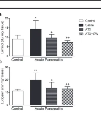
Effect of ATX on CL values
Both luminol and lucigenin levels were elevated in the saline-treated group compared with the control group (p<0.5 and p<0.01, respectively; Figure 4). The elevat-ed levels were significantly abolishelevat-ed by ATX treatment (p<0.5) with or without GW6471 (p<0.5 and p<0.01, re-spectively).
Histological Analyses
Well-organized acinar structures in the parenchymal tis-sue with regular morphology were observed in the histo-logical examination of the control group (Figure 5A). The vehicle-treated group revealed significantly severe cyto-plasmic degeneration and vacuolization, vascular
con-Figure 2. a, b. Lipase and amylase levels in sera. Lipase (a) and amylase (b) levels in the pancreatic tissues of the control and vehicle-treated
groups, astaxanthin (ATX) and ATX+GW-treated acute pancreatitis groups (n=6/group). Data are expressed as mean±standard error of mean; n=6 rats/group; **p<0.01 vs. control group; +p<0.05 vs.
vehicle-treated group.
Figure 3. a-c. Pancreatic tissue, malondialdehyde, myeloperoxidase activity, and glutathione levels. Data are expressed as mean±standard
error of mean; n=6 rats/group; **p<0.01, ***p<0.001 vs. control group; +p<0.05, ++p<0.01 vs. vehicle-treated group.
gestion, and inflammatory cell infiltration compared with those in the control group (Figure 5B). ATX treatment al-leviated these destructions (Figure 5C-D).
Concomitant with the improvements examined histolog-ically, the histological score was significant in all groups (p<0.001, Figure 6). In the vehicle-treated group, the his-tological score was increased significantly compared with that of the control group (p<0.001). The elevated score was reduced by ATX treatment, whereas the reduction in ATX treatment was constant in the ATX+GW group (p<0.001).
DISCUSSION
In this study, the effect of ATX on AP and its underly-ing mechanism of action were evaluated by means of a strong PPAR-α antagonist GW6471.
Oxidative stress occurs because of increased production of ROS and diminished antioxidant activity. The role of oxidative stress in inflammation has been reported in many studies (3, 4). Free oxygen radicals also play a role in the activation of AP and neutrophils and in the emer-gence of the inflammatory response (21). Once ROS was implicated in the pathophysiology of AP, several studies reported the ameliorative effect of many plant-derived natural products on the oxidative stress in AP (10, 22). In normal circumstances, ROS levels are balanced via the antioxidant defense system, which includes enzymat-ic and nonenzymatenzymat-ic antioxidants. These antioxidants neutralize the harmful free radicals and non-radicals. The imbalance between the oxidants and antioxidants caus-es structural damagcaus-es, such as lipid peroxidation, which leads to the inflammatory process (4, 5). ATX has a po-tent capacity for scavenging ROS owing to the presence Figure 4. a, b. Pancreatic luminol-enhanced (a)- and
lucigenin-enhanced (b) tissue chemiluminescence levels. Data are expressed as mean±standard error of mean; n=6 rats/group; *p<0.05, **p<0.01 vs.
control group; +p<0.05,++p<0.01 vs. vehicle-treated group.
Figure 6. Microscopic score in the pancreatic tissues of experimental groups. Data are expressed as mean±standard error of mean; n=6
rats/group; ***p<0.001 vs. control group; +++p<0.001 vs. vehicle-treated group.
Figure 5. a, b. Representative photomicrographs of experimental groups. Control (a), vehicle (b), astaxanthin (ATX) (c), ATX+GW (d) groups. (a): Acinar structures have regular morphology (arrow). (b): Severe vacuolization in acinar cell (arrowhead), acinar structure with irregular morphology (white arrow), severe fibrosis (asterisk),
inflammatory cell infiltration (white asterisk), severe vascular congestion (white arrowhead). (c): Acinar structure with regular morphology (arrow). (d): Acinar structure with regular morphology (arrow), slight inflammatory cell infiltration (notched-arrow), slightly
of 2 oxygenated groups in its molecular structure (13, 14). The biological activity of ATX was greater than that of other antioxidants because of its transmembranous nature (23) therefore, we used ATX in oil in this study. Our study confirmed the implication of reactive species at an early stage of cerulein-induced AP in rats and the ameliorative effect of ATX. In our study, parallel to the literature, MDA levels (24) and MPO activity (10, 5) were increased in the vehicle-treated group, whereas GSH lev-els decreased (24-26) in the AP group. In our study, MPO levels were decreased in the ATX group, which is consis-tent with a study by Zahng et al. (10). We also investigated MDA level, which is a sensitive marker of inflammation, in our groups. Multiple reports have revealed that ATX reverses the increased levels of MDA in inflammation (26, 27). The oxidative damage in the pancreatic tissue is the result of not only an increase in the ROS but also a decrease in the antioxidant capacity (25). Therefore, we investigated GSH, a tripeptide thiol and an important en-dogenous antioxidant defense molecule. Its intracellular concentration is an indicator of oxidative stress and holds a crucial part in the detoxification of ROS in the pancre-as. Our data revealed that AP induced a decrease in the pancreatic GSH level, confirming the association of stress with oxidative damage, which is reversed by ATX. Our findings are consistent with those of previous studies, showing that ATX inhibits GSH depletion and increases GSH levels (25, 28).
CL is an assay for ROS. The luminol probe use of this technique detects OH−, H
2O2, hypochlorite, peroxynitrite, and lipid peroxyl radicals, and lucigenin is specific for su-peroxide radicals. In conformity with the previous studies, our results showed an increased pancreatic production of ROS. ATX diminished the increased luminol and lucigenin levels in AP just like in other inflammatory diseases (14). In our study, decreases in the serum enzyme levels and in the histological score were consistent and supported the protective effects of ATX.
PPAR-α is a member of the nuclear receptor superfamily of ligand-dependent transcription factors related to ret-inoid, steroid, and thyroid hormone receptors. PPAR-α, which is one of the subtype of PPAR family, is a nucle-ar receptor with wide-ranging effects on genes involved in fatty acid and lipoprotein metabolism and inflamma-tion and highly expressed in the pancreas (29). A previ-ous study revealed that different PPAR-α agonists sup-pressed inflammation by decreasing pro-inflammatory cytokines (30) and inflammatory mRNA expression (31)
and reduced pancreatic damage. In this study, we in-vestigated the effect of ATX, which is suggested to be a PPAR-α agonist, on the cerulein-induced model of AP. Using GW6471, a selective PPAR-α antagonist, we in-vestigated whether PPAR-α inhibition alters the effect of ATX. In a recent study, the absence of the PPAR-α gene significantly increased cerulein-induced pancreati-tis and pancreatic injury (8). Furthermore, another recent study revealed that treatment with a PPAR-α agonist WY14643 ameliorated changes in lipase, amylase, and MPO activity and pathological scores in cerulean-induced AP model. The same study also demonstrated that these effects were completely abolished by co-administration of PPAR-α antagonist MK886. However, our results indi-cated that GW6471 did not abolish the positive impact of ATX biochemically or histologically. This result may be owing to ATX having a very strong antioxidant or protec-tive effect or ATX not acting via PPAR-α pathway. In conclusion, this study shows that inhibition of PPAR-α does not block the effect of ATX in cerulein-induced AP. ATX, a naturally occurring potent antioxidant, significant-ly reduces oxidative stress and protects from pancreatic damage. The antioxidant effect of ATX in the cerulein-in-duced AP model is not only related to its scavenging ac-tivity but also could be owing to its increasing effect on antioxidant defense enzyme expression. Thus, ATX can be used as a valuable therapeutic approach in AP.
Ethics Committee Approval: Ethics committee approval was re-ceived for this study from the Marmara University, Animal Care and Use Committee (Approval number: 76.2017.mar).
Informed Consent: N/A.
Peer-review: Externally peer-reviewed.
Author Contributions: Concept – D.Ö., E.B.G.; Design - D.Ö., E.B.G.; Supervision – D.Ö.; Resource - D.Ö.; Materials – D.Ö., Ö.T., Ç.K., M.Y.; Data Collection and/or Processing – D.Ö., H.B., Ö.T.Ç.K., M.E.Ç., M.Y.; Analysis and/or Interpretation - D.Ö., E.B.G.; Literature Search - D.Ö., E.B.G.; Writing - D.Ö., E.B.G., H.B., Ö.T.Ç.K., M.E.Ç., M.Y.; Critical Re-views – D.Ö., E.B.G.
Conflict of Interest: The authors have no conflict of interest to de-clare.
Financial Disclosure: The authors declared that this study has re-ceived no financial support.
REFERENCES
1. Lankisch P. Apte G, Banks, P. Acute pancreatitis. The Lancet 2015; 386: 85-96. [Crossref]
2. Yu JH, Kim H. Oxidative stress and inflammatory signaling in ceru-lein pancreatitis. World J Gastroenterol 2014; 20: 17324-9. [Crossref]
3. Abu-Zidan FM, Bonham MJ, Windsor JA. Severity of acute pancre-atitis: a multivariate analysis of oxidative stress markers and modi-fied Glasgow criteria Br J Surg 2000; 87: 1019-23. [Crossref] 4. Thareja S, Bhardwaj P, Sateesh J, Saraya A. Variations in the levels of oxidative stress and antioxidants during early acute pancreatitis Trop Gastroenterol 2009; 30: 26-31.
5. Polotow TG, Vardaris CV, Mihaliuc AR, et al. Astaxanthin supple-mentation delays physical exhaustion and prevents redox imbalanc-es in plasma and soleus musclimbalanc-es of Wistar rats. Nutrients 2014; 6: 5819-38. [Crossref]
6. Devchand PR, Keller H, Peters JM, Vazquez M, Gonzalez FJ, Wahli W. The PPAR-alpha-leukotriene B4 pathway to inflammation con-trol. Nature 1996; 384: 39-43. [Crossref]
7. Silva AKS, Peixoto CA. Role of peroxisome proliferator-activated receptors in non-alcoholic fatty liver disease inflammation. Cell Mol Life Sci 2018; 75: 2951-61. [Crossref]
8. Genovese T, Mazzon E, Di Paola R, et al. Role of peroxisome pro-liferator-activated receptor-alpha in acute pancreatitis induced by cerulein. Immunolog 2006; 118: 559-70. [Crossref]
9. Ding J, Zhou Z, Zhou X, et al. Attenuation of Acute Pancreatitis by Peroxisome Proliferator-Activated Receptor-alpha in Rats The Ef-fect on Toll-Like Receptor Signaling Pathways. Pancreas 2013; 42: 114-22. [Crossref]
10. Johnson CD. Antioxidants in acute pancreatitis. Gut 2007; 56: 1344-5. [Crossref]
11. Zhang H, Yang W, Li Y, et al. Astaxanthin ameliorates cerulein-in-duced acute pancreatitis in mice. Int. Immunopharmacol 2018; 56: 18-28. [Crossref]
12. Siriwardena AK, Mason JM, Balachandra S, et al. Randomised, double blind, placebo controlled trial of intravenous antioxidant (n-acetylcysteine, selenium, vitamin C) therapy in severe acute pan-creatitis. Gut 2007; 56: 1439-44. [Crossref]
13. Chang CS, Chang CL, Lai GH. Reactive oxygen species scav-enging activities in a chemiluminescence model and neuropro-tection in rat pheochromocytoma cells by astaxanthin, beta-car-otene, and canthaxanthin. Kaohsiung J Med Sci 2013; 29: 412-21. [Crossref]
14. Santocono M, Zurria M, Berrettini M, Fedeli D, Falcioni G. Influ-ence of astaxanthin, zeaxanthin and lutein on DNA damage and repair in UVA-irradiated cells. J Photochem Photobiol B 2006; 85: 205-15. [Crossref]
15. Jia Y, Kim JY, Jun HJ, et al. The natural carotenoid astaxanthin, a PPAR-α agonist and PPAR-γ antagonist, reduces hepatic lipid accu-mulation by rewiring the transcriptome in lipid-loaded hepatocytes. Mol Nutr Food Res 2012; 56: 878-88. [Crossref]
16. Casini A, Ferrali M, Pompella AS, Maellaro E, Comporti M. Lipid peroxidation and cellular damage in extrahepatic tissues of bromo-benzene-intoxicated mice. Am J Pathol 1986; 123: 520-31.
17. Beutler E. Glutathione in Red Blood Cell Metabolism A Manual of Biochemical Methods, Grune & Stratton, New York, 1975, pp. 112-4. 18. Bradley, Priebat D, Christensen R, Rothstein G. Measurement of cutaneous inflammation: estimation of neutrophil content with an enzyme marker. J Invest Dermatol 1982; 78: 206-9. [Crossref] 19. Kikuchi K, Nagano T, Hayakawa H, Hirata Y, Hirobe M. Detection of nitric oxide production from a perfused organ by a luminol-H2O2 system. Anal Chem 1993; 65: 1794-9. [Crossref]
20. Moreno C, Nicaise C, Gustot T, et al. Chemokine receptor CCR5 deficiency exacerbates cerulein-induced acute pancreatitis in mice. Am J Physiol 2006; 291: 1089 -99. [Crossref]
21. Genovese T, Mazzon E, Di Paola R, et al. Role of peroxisome pro-liferator-activated receptor-alpha in acute pancreatitis induced by cerulein. Shock 2006; 2: 161-7. [Crossref]
22. Anchi P, Khurana A. Bale S, Godugu C. The Role of Plant-derived Products in Pancreatitis: Experimental and Clinical Evidence 2017; 31: 591-623. [Crossref]
23. Mercke Odeberg J, Lignell A, Pettersson A, Höglund P. Oral bio-availability of the antioxidant astaxanthin in humans is enhanced by incorporation of lipid based formulations. Eur J Pharm Sci 2003; 19: 299-304. [Crossref]
24. Köksoy FN, Yankol Y, Şen Oran E, et al. Preventive effects of enoxaparin and hesperidin in cerulein-induced acute pancreatitis in rats. Turk J Gastroenterol 2013; 24: 495-501. [Crossref]
25. Guleken Z, Ozbeyli D, Acikel-Elmas M, et al. The effect of estrogen receptor agonists on pancreaticobiliary duct ligation induced exper-imental acute pancreatitis. J Physiol Pharmacol 2017; 68: 847-58. 26. Dokumacioglu E., Iskender H, Yenice G, et al. Effects of astax-anthin on biochemical and histopathological parameters related to oxidative stress on testes of rats on high fructose regime. Andrologia 2018; 50: e13042. [Crossref]
27. Alam MN, Hossain MM, Rahman MM, et al. Astaxanthin Prevent-ed Oxidative Stress in Heart and Kidneys of Isoproterenol-Adminis-tered Aged Rats. J Diet Suppl 2018; 15: 42-54. [Crossref]
28. Fındık H, Tumkaya L, Yılmaz A, et al. The protective effects of as-taxanthin against cisplatin-induced retinal toxicity. Cutaneous Ocul Toxicol 2019; 38: 59-65. [Crossref]
29. Escher P, Braissant O, Basu-Modak S, Michalik L, Wahli W, Des-vergne B. Rat PPARs: quantitative analysis in adult rat tissues and regulation in fasting and refeeding. Endocrinology 2001; 142: 4195-202. [Crossref]
30. Nakano Y, Uchiyama M, Arima T, et al. PPARα Agonist Suppress-es Inflammation after Corneal Alkali Burn by SupprSuppress-essing Proinflam-matory Cytokines, MCP-1, and Nuclear Translocation of NF-κB. Mol-ecules 2018; 24: 114. [Crossref]
31. Kim SM, Lee B, An HJ, et al. Novel PPARα agonist MHY553 alleviates hepatic steatosis by increasing fatty acid oxidation and decreasing inflammation during aging. Oncotarget 2017; 8: 46273-85. [Crossref]