556 © The Author 2015. Published by Oxford University Press on behalf of the European Orthodontic Society. All rights reserved.
For permissions, please email: [email protected]
Original article
Comparison of short-term effects of
mini-implant-supported maxillary expansion
appliance with two conventional expansion
protocols
Alev Yılmaz
*
, Ayça Arman-Özçırpıcı
**
, Seda Erken
***
and
Ömür Polat-Özsoy
****
*Department of Orthodontics, Faculty of Dentistry, Adnan Menderes University, Aydın, **Department of Orthodontics, Faculty of Dentistry, Başkent University, Ankara, ***Private practice, Sakarya, Turkey, ****Department of Orthodontics, Faculty of Dentistry, Başkent University, Ankara, Turkey
Correspondence to: Alev Yılmaz, Adnan Menderes Üniversitesi, Diş Hekimliği Fakültesi, Ortodonti Anabilim Dalı, Aydın 09100, TÜRKİYE. E-mail: [email protected]
Summary
Objective: This study evaluates the dentoskeletal effects of a mini-implant-supported maxillary
expansion (MISME) appliance in comparison with two types of conventional expansion methods.
Methods: Records of 42 patients with bilateral or unilateral posterior crossbite were included in
this study. The patients were divided into three groups. In group 1, four miniscrews were placed to the palatal region and an acrylic expansion device was bonded on these screws. A bonded maxillary expansion appliance was used in group 2, while a banded expansion appliance was used in group 3. Measurements from cephalometric, postero-anterior radiographs, and dental casts taken before and after expansion were evaluated statistically.
Results: ANB angle increased significantly in group 1 and 3. MISME group also showed an increase
of SNA angle. Measurements regarding the vertical dimension did not change with MISME but significant posterior rotation was found in group 2 and 3. Overbite value showed a significant decrease in group 2 and 3, but remained stable in group 1. The nasal, maxillary, maxillary intermolar widths showed significant increases in all groups. The difference between MISME group and other groups in maxillary width was significant indicating more skeletal expansion in MISME group. The maxillary molars showed significant buccal tipping in group 2 and 3, while lingual tipping of molars was found in MISME group.
Conclusions: MISME can be a better alternative to bonded expansion particularly in patients with
vertical growth patterns and lack of anchorage teeth.
Introduction
Maxillary deficiency is a common problem in orthodontic patients and is usually accompanied by bilateral or unilateral posterior cross-bite, narrow nasal cavity, and crowding (1, 2). Expansion of the maxilla or the maxillary arch is an accepted method of treatment
that was first outlined by Angle (3) in 1860 and popularized by Haas (4) 100 years later. Rapid maxillary expansion (RME) has become a routine procedure in orthodontic clinics and numerous studies con-firmed its effectiveness in orthopaedic widening of the maxilla (5–8). Various RME appliances have been widely used in adolescents with constricted maxillary arches such as tooth-borne Hyrax,
doi:10.1093/ejo/cju094 Advance Access publication 6 January 2015
tooth-tissue-borne Haas, or bonded RME appliances (8–12). The conventional RME appliances widen the maxillary arch mainly by separating the maxillary halves opening the midpalatal suture. Along with the desired orthopaedic effect of midpalatal suture splitting RME unavoidably elicits an orthodontic effect of buccal movement or tipping of the posterior teeth supporting the appliance (4, 7, 13). Tipping and extrusion of the posterior teeth and alveolar bending usually enhances bite opening, posterior rotation of the mandible, and also increases the tendency to relapse due to the resistance to deformation from surrounding structures (5, 13). Tooth-borne expanders, in which the forces are concentrated at the dentoalveo-lar area, may cause iatrogenic effects to the periodontal tissues and cause root resorption, buccal dehiscences, and gingival recession at the buccal aspects of the supporting teeth (12, 14). Haas (5), sug-gested that more bodily movement and less dental tipping were produced when acrylic palatal coverage was added to support the appliance. Studies have demonstrated that both tooth-tissue-borne (Haas) or bonded RME appliances reduce the undesired effects of tooth-borne devices, but still lead to limited effects on the maxillary basal bone with significant dental tipping and relapse potential (7, 9, 15, 16).
Bone-borne transpalatal distractors which have been suggested to avoid all these aforementioned problems, require an invasive sur-gery and besides their high cost, carry a risk of infection and root damage (17–19). Recently, implant-supported or assisted expansion devices have been suggested as an alternative method for applying forces directly to the maxilla (20–23). These methods have some disadvantages, such as the need for a minimally invasive procedure for insertion of the screws, extra cost, and failure of the implants. Selection of the palatinal region for insertion of the supporting mini-screws decreases the risk of root damage and the rates of miniscrew failure, because of its superior bone quality and attached mucosa. It is also reported that screw-supported expansion appliances can also be used for patients with missing deciduous teeth or premolars with underdeveloped roots (24, 25).
The aim of this study is to evaluate the effects of a mini-implant-supported maxillary expansion (MISME) appliance that incorpo-rates four palatal mini-implants for bone anchorage in comparison with bonded and banded maxillary appliances.
Material and methods
The sample consisted of 20 female and 22 male total of 42 patients treated in Department of Orthodontics, Faculty of Dentistry ………… University. All patients or parents consented to the treatment pro-cedure and this retrospective study was approved by ………… University Institutional Review Board (Project no: D-KA10/13).
Patients with indication of maxillary expansion due to transversal maxillary deficiency with unilateral or bilateral posterior crossbite
were included in the study group. The first group of patients con-sisted of 14 patients (eight males and six females) who treated with an acrylic expansion appliance bonded on four screws, inserted in anterior palate. The second and third group comprised of 14 patients (eight males, six females in group 2, six males and eight females in group 2) treated with a bonded and a banded maxillary expansion appliance, respectively. The mean chronological age at the beginning of the treatment was 13.2 ± 2.1 in group 1, 12.1 ± 2.1 in group 2, and 13.4 ± 1.7 in group 3 (Table 1). Distribution of malocclusions of patients in each group is given in Table 2.
In group I, four titanium miniscrew implants (Turquoise, Medikodental, Istanbul, Turkey) measuring 1.6 mm in diameter and 7 mm in length were placed under local anaesthesia by the two authors (AAÖ, AY) of this study to provide skeletal anchorage. Before placement of the implants the palatal region was rinsed with chlorhexidine (0.12 per cent). The two anterior palatal implants were placed in the anterior palate bilaterally, 3–4 mm lateral to the suture and 3–4 mm posterior to the incisive foramen. Two posterior implants were inserted in the palatal alveolus bilaterally, between the projection of the second premolar and first molar roots. Moreover, care was given to provide enough space for the expansion screw between the implants and not to damage the roots of neighbouring teeth. A self-drilling method was used for implant placement with an approximately 60–70 degrees of angulation to the long axis of the teeth (Figure 1a).
After placement of the implants, impressions and dental casts were obtained with the mini-implants in place. On the cast, the screw heads were blocked out with wax, and the acrylic expansion appliance was constructed. A maxi or midi jackscrew was embed-ded in the acrylic between the first premolars as close as possible to the palate, with the resin covering the mini-implants and the sur-rounding palatal surface. The biggest expansion screw, which can be placed in between the implants, was selected in order to provide greater amounts of expansion.
The acrylic appliance was connected to the screw heads using cold-curing, methyl methacrylate free acrylic resin (Ufi Gel hard; Voco GmbH, Cuxhaven, Germany). Small holes were made on the appliance for the excess resin to flow out (Figure 1b).
Strict instructions were given to the patients regarding oral hygiene and no medication was prescribed.
Table 1. Chronological ages at the beginning of treatment (T1) and duration of expansion (days). X: average, s: standard deviation, min–
max: minimum and maximum values.
Group I (n:14) Group II (n:14) Group III (n:14) P
Age (years)
X± s 13.2 ± 2.1 12.1 ± 2.1 13.4 ± 1.7 0.177
Median (min–max) 12.8 (8.2–15.7) 12.0 (8.1–15.3) 13.4 (10.6–15.6)
Duration of expansion (days)
D ± s 87.3 ± 45.9 55.3 ± 27.0 75.5 ± 35.1 0.079
Median (min–max) 71.0 (44–206) 45.5 (28–119) 70.5 (21–150)
Table 2. Distribution of malocclusions of patients in each group.
Group I Group II Group III
Class I 6 11 5
Class II 3 2 7
Class III 5 1 2
In group 2, a bonded expansion appliances with acrylic cover-age was constructed on the cast. The acrylic part of the appliance extended over occlusal and middle third of the vestibular surfaces of all posterior teeth. The thickness of the occlusal acrylic surface was limited to the freeway space and was in contact with all lower
teeth. The appliance bonded to the upper posterior teeth with glass ionomer luting cement (Ketac Cem radiopaque; 3m ESPE, Neuss, Germany). Holes were opened for the escape of excess cement dur-ing cementation (Figure 2a).
In group 3, after molar bands were placed on the upper left and right first molars, impressions and dental casts were obtained. Two banded hyrax expansion appliance (Leone; Sesto Fiorentino, Firenze, Italy) was constructed on the cast and bonded to the molar teeth with glass iono-mer luting cement (Ketac Cem radiopaque; 3m ESPE) (Figure 2b).
Expansion appliances were activated with a semirapid protocol in all groups. After bonding of the expansion appliances, the patient’s parents were instructed to activate it by turning the screw one turn in the morning and another turn in the evening in the first 7–10 days. After the confirmation of the opening of the suture via occlusal radi-ographs, the activation was continued once in every 3 days until the desired expansion was achieved as suggested by Işeri and Ozsoy in 2004 (26). Each turn of the screw produced 0.2 mm of expansion.
Duration of the expansion depended on the amount of expansion needed. No overcorrection of the transversal relationship was done in MISME group, whereas expansion was continued until overcor-rection was achieved in the bonded and banded groups.
Cephalograms, postero-anterior radiographs, and dental casts were obtained at the beginning of treatment (T1) and at the end of desired expansion (T2).
Figure 1. (a) Palatal implants, (b) MISME appliance, (c) MISME appliance after expansion. MISME, mini-implant-supported maxillary expansion.
Figure 2. (a) Bonded maxillary expansion appliance used in the study. (b) Banded maxillary expansion appliance used in the study.
Cephalometric, PA, and cast analysis
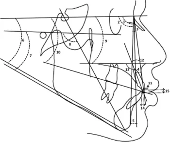
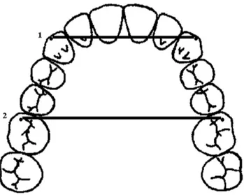
Lateral cephalometric and postero-anterior radiographs were taken with the same cephalometer (Sirona, Siemens, Germany). In accord-ance with the As Low As Reasonably Achievable (ALARA) princi-ples, the lowest dosage that can provide the appropriate image was chosen considering patient’s age and size. The radiographs were traced and measured by one blind investigator (AY) in random order. In instances of bilateral structures, a single average tracing was made. The reference landmarks were marked simultaneously on the T1 and T2 films of each subject to obtain maximal agree-ment in the landmark determination. Fifteen measureagree-ments (10 angular, 1 linear) on cephalometric radiographs (Figure 3), six linear measurements on postero-anterior radiographs (Figure 4), and four measurements (one angular, three linear) on dental models (Figures 5 and 6), at a total of 27 measurements were made for each patient. The intercanine and intermolar widths were measured directly on the casts with a digital caliper, whereas the degree of tipping of the molars (molar angulation) and palatal width at the gingival height was determined from photocopy images taken after trimming the posterior of the casts up to the cusp tips of the first molars (Figure 6).
Statistical analysis
The primary aim of this study was to compare by the differences in maxillary molar angulation among groups. Total sample size of 42 (14 per group) achieve 85 per cent power to detect a difference of 3.2 degrees between the null hypothesis that both group means are 41.3 and the alternative hypothesis that the mean of another group is 38.1 with estimated group standard deviations of 3.19 and 2.78 and with a significance level (alpha) of 0.025 using a two-sided Mann–Whitney test assuming that the actual distribution is double exponential. The difference of 3.2 degrees was taken from litera-ture (27). Sample size estimation was performed by using NCSS and PASS 2000 software.
Statistical analysis was performed using SPSS for Windows, ver-sion 11.5 (SPSS Inc., Chicago, Illinois, USA). Whether the continu-ous variables were normally distributed or not were determined by using Shapiro–Wilk test. Homogeneity of variances was evaluated
by Levene test. Data were shown as mean ± standard deviation. While, the differences between pre- and post-treatment measure-ments regarding for normally distributed parameters were analyzed by Bonferroni-adjusted paired samples t-test. Wilcoxon sign rank test was used for not normally distributed parameters (treatment duration, U1-NA, U1-PP, overjet, overbite, incisors apex, and pala-tal width). The mean differences among groups regarding for nor-mally distributed parameters were compared by one-way analysis of variance (ANOVA). Kruskal–Wallis test was used for not normally distributed data. When the P value from the one-way ANOVA or Kruskal–Wallis test are statistically significant to know which group differ from which others by using post hoc Tukey Tukey’s honest sig-nificant difference or Conover’s non-parametric multiple comparison test were used. Intraclass correlation coefficient and 95 per cent confi-dence intervals were calculated for evaluation of reliability. A P value less than 0.05 was considered statistically significant. But all possible multiple comparisons, the Bonferroni correction was applied for con-trolling type I error.
To calculate the error of measurements, cephalometric/posteroante-rior films and study casts of 15 randomly selected patients were retraced and remeasured 2 weeks later by the same clinician. Intraclass correla-tion coefficients were found to be close to 1.00, thus the difference between the first and the second measurements was insignificant.
Results
Clinically successful amount of expansion was achieved in all patients (see online supplementary Figures 1 and 2). There were no
Figure 4. PA measurements used in the study. 1. Nasal cavity width, 2. Maxillary width, 3. Maxillary intermolar width, 4. Mandibular intermolar width, 5. Mandibular width, 6. Incisors apex.
Figure 3. Lateral cephalometric measurements used in the study. 1. SNA, 2. SNB, 3. ANB, 4. Nperp-A, 5. Nperp-Pg, 6. GoGn-SN, 7. FMA, 8. Y axis, 9. SN/ PP, 10. SN/OP, 11. U1-NA (mm), 12. U1-NA (degree), 13. U1-PP, 14. Overjet, 15. Overbite.
Figure 5. Dental cast measurements used in the study. 1. Intercanine width, 2. Intermolar width.
Figure 6. 1. Maxillary molar angulation, 2. Palatal width at gingival height.
dropouts or appliance failures. Only in one patient in group 1, the appliance needed to be remade because of a problem of the expan-sion screw and the treatment duration was elongated. No negative side effects were recorded.
Descriptive statistics for the chronological age and treatment duration, and cephalometric, PA, and model measurements at T1, T2 and treatment changes (T2-T1) obtained with maxillary expan-sion are given in Tables 1 and 3, respectively.
The duration of expansion was 87.3 ± 45.9 in group 1, 55.3 ± 27.0 in group 2 and 75.5 ± 35.1 in group 3 and showed no statistically sig-nificant difference between groups (Table 1).
Skeletal sagittal measurements demonstrated significant increases in ANB angle in screw-supported and banded expansion groups. SNA angle showed a significant increase demonstrating forward movement of the maxilla only in group I. While the increases in Nperp-A measurement were not statistically significant, a significant decrease in Nperp-Pg distance was found in group II and III. There was no statistically significant difference between groups concern-ing skeletal sagittal measurements except Nperp-Pg measurement (Table 3).
Skeletal vertical parameters showed significant increases in bonded and banded expansion groups indicating posterior rotation of the mandible. However, vertical measurements did not change in MISME group. Statistically significant differences were found between MISME and other groups regarding the vertical measures (Table 3).
None of the dentoalveolar measurements demonstrated a signifi-cant change in group I. Signifisignifi-cant increase was found in overjet only in the third group. Overbite value showed a slight non-significant increase in group I, while there was a significant decrease in group II and III, and significant differences between MISME and the other two groups were found. Upper incisors showed a retrusion accord-ing to NA line and palatal plane, in bonded and banded expansion groups (Table 3).
PA measurements showed significant increases in the nasal width, maxillary width, and the maxillary intermolar width in all groups. The difference between MISME and other groups was statistically significant in maxillary width measurement. Mandibular width measurements increased significantly in group I and group III, while mandibular intermolar width measurements increased significantly only in group I. Incisor apex measurement showed a significant increase in all groups and significant difference between all groups (Table 3).
Model measurements also revealed expansion in the maxillary dental arch. Both the intermolar and intercanine width increased sig-inificantly in all groups. The maxillary molar angulation increased significantly in the second and third groups demonstrating buccal tipping of the first molars. The first group showed significant palatal tipping of the maxillary first molars. There was a significant differ-ence between the MISME and the other groups. The palatal width at the gingival height increased significantly in the first and second groups. The differences between group 1–3, and 2–3 were significant (Table 3).
Discussion
Introduction of bone anchorage via orthodontic implants has made a great revolution in orthodontics. Therefore many reports were pub-lished on orthodontic absolute anchorage systems incorporating mini-screws, reflecting their increasing popularity and importance (28–30). MISME is an appliance that incorporates four miniscrews for bone anchorage. Many intraoral sites can be used for mini-implant placement; however, the palate is a frequently used site because it is easily accessible, is relatively safe to work on, is less susceptible to inflammation and has good bone quantity (31). Previous studies indicate that cortical bone thickness has a strong impact on primary stability and overall success rates of implants. The midpalatal area (32), anterior paramedian area (31), palatal area between the level of the first and second premolars (33) were reported to be the most favourable areas for implant placement. There are studies indicating that the thickest bone is located 3–4 mm distal to the incisive fora-men and 3 mm paramedian to the palatal suture (14, 34).
A self-drilling method was preferred during implant placement because of easier application and higher primary stability (35, 36). Angular placement of 60–70 degrees to the long axis of teeth was performed in order to avoid root damage and enable more cortical bone contact for better primary stability (37).
The primary stability of the implants is also proportional to increased length and diameter (38). Orthodontic miniscrews (1.6 mm collar diameter and 7 mm length) used in the presented appliance were shorter and/or thinner than the palatal implants, used in other bone-anchored palatal appliances (21, 30). However, two bilaterally placed implants at the anterior and two at the posterior provided sufficient anchorage without any complications. The head of the miniscrews was approximately 3.5 mm and secured with the acrylic part, which also enhanced the stability of the appliance. The MISME appliance may be classified as a bone-tissue-borne appliance as the
Table
3.
The descripti
ve statistics of cephalometric. P
A and dental cast measurements at the beginning of the treatment (T1) and at the end of expansion (T2), significance of treatment c
hanges
in groups and statistical comparison of treatment c
hanges between groups.
X: average, D: dif ference, s: standard deviation. Parameter Group I Group II Group III P T1 X ± s T2 P T1 X ± s T2 X ± s P T1 X ± s T2 X ± s P Cephalometric measurements ( n = 14)
Skeletal measurements SNA (°)
77.32 ± 5.07 78.11 ± 5.41 0.009 77.61 ± 4.46 77.71 ± 4.61 0.664 81.11 ± 3.87 81.61 ± 3.58 0.068 0.168 SNB (°) 76.14 ± 3.68 75.75 ± 4.15 0.174 75.14 ± 5.19 74.18 ± 4.41 0.065 77.71 ± 4.41 77.11 ± 4.10 0.112 0.565 ANB (°) 1.25 ± 3.26 2.36 ± 3.64 0.006 2.46 ± 2.61 3.54 ± 2.14 0.046 3.39 ± 3.00 4.50 ± 2.81 0.004 0.997 Nperp-A (mm) −3.19 ± 4.29 −2.26 ± 3.66 0.020 −2.86 ± 4.54 −2.50 ± 4.61 0.231 1.25 ± 3.41 1.89 ± 3.72 0.039 0.426 Nperp-Pg (mm) −7.94 ± 6.83 −7.61 ± 5.85 0.666 −8.00 ± 8.00 −10.07 ± 7.42 0.004 −2.50 ± 8.98 −4.00 ± 9.18 0.008 0.023 † GoGn-SN (°) 38.82 ± 6.14 38.82 ± 6.54 1.000 37.86 ± 3.62 39.54 ± 3.45 <0.001 34.61 ± 5.05 36.64 ± 5.40 <0.001 <0.001 †,‡ FMA (°) 32.32 ± 6.60 32.04 ± 6.45 0.503 30.96 ± 4.20 33.64 ± 4.95 0.005 26.75 ± 5.23 28.71 ± 5.58 <0.001 0.002 †,‡ Y axis (°) 61.04 ± 4.74 61.14 ± 4.47 0.795 61.18 ± 3.71 62.86 ± 3.44 <0.001 58.54 ± 3.57 60.14 ± 4.27 <0.001 0.006 †,‡ SN/PP (°) 8.25 ± 2.85 8.39 ± 2.88 0.770 8.68 ± 4.10 8.86 ± 4.47 0.513 8.54 ± 2.22 8.57 ± 2.38 0.909 0.959 SN/OP (°) 18.54 ± 4.83 18.71 ± 5.47 0.814 18.68 ± 4.32 19.36 ± 3.23 0.163 17.36 ± 3.89 18.00 ± 3.57 0.092 0.770
Dentoalveolar measurements U1i-NA (mm)
6.09 ± 2.92 5.54 ± 2.19 0.170 4.61 ± 2.00 3.93 ± 1.92 0.009 4.18 ± 1.40 3.25 ± 1.44 0.011 0.872 U1-NA (°) 23.75 ± 6.87 21.61 ± 5.68 0.039 21.64 ± 6.61 19.04 ± 5.42 <0.001 19.89 ± 5.54 17.46 ± 6.04 <0.001 0.894 U1-PP (°) 109.89 ± 5.16 108.75 ± 3.87 0.270 108.25 ± 6.59 104.36 ± 10.25 0.023 109.43 ± 6.25 107.68 ± 6.61 0.009 0.575 Overjet (mm) 2.54 ± 3.29 3.36 ± 2.45 0.024 2.68 ± 3.83 3.75 ± 2.52 0.258 2.54 ± 2.36 3.32 ± 2.27 0.005 0.549 Overbite (mm) 0.22 ± 2.65 0.49 ± 2.66 0.329 2.14 ± 2.59 0.93 ± 2.85 0.009 1.14 ± 1.47 −0.29 ± 2.19 0.002 <0.001 †,‡ PA measurements ( n = 14) Nasal width (mm) 29.65 ± 2.65 33.36 ± 2.89 <0.001 29.64 ± 2.66 32.32 ± 3.53 <0.001 31.82 ± 3.47 34.18 ± 3.93 <0.001 0.127 Maxillary width (mm) 64.04 ± 3.79 69.58 ± 4.01 <0.001 65.79 ± 3.70 68.93 ± 3.76 <0.001 67.00 ± 4.75 70.50 ± 4.55 <0.001 0.006 †,‡ Max. intermolar width (mm) 58.14 ± 4.28 63.94 ± 3.05 <0.001 58.54 ± 3.19 65.00 ± 3.80 <0.001 60.68 ± 4.35 67.43 ± 3.71 <0.001 0.617 Mandibular width (mm) 86.49 ± 5.91 87.26 ± 5.99 <0.001 86.68 ± 6.02 87.25 ± 5.95 0.007 88.21 ± 5.07 88.64 ± 5.02 0.068 0.421 Mand. intermolar width (mm) 62.77 ± 5.30 63.56 ± 4.87 <0.001 63.32 ± 4.84 63.46 ± 4.46 0.537 63.61 ± 3.37 64.04 ± 3.58 0.040 0.084 Incisors apex 6.81 ± 1.79 14.63 ± 2.68 0.002 7.96 ± 2.27 10.43 ± 2.67 0.003 7.39 ± 1.11 12.04 ± 2.82 0.002 <0.001 †,‡,¶
Dental cast measurements (
n = 12) Max. intermolar width (mm) 46.56 ± 2.87 52.12 ± 2.83 <0.001 48.96 ± 2.25 55.66 ± 2.44 <0.001 48.26 ± 2.56 55.42 ± 2.15 <0.001 0.075 Max. intercanine width (mm) 31.91 ± 2.66 37.10 ± 2.27 <0.001 33.30 ± 3.16 37.50 ± 3.87 <0.001 33.24 ± 3.34 37.55 ± 4.22 <0.001 0.488 Max. molar angulation (°) 151.42 ± 14.27 154.96 ± 12.85 <0.001 154.67 ± 9.85 142.96 ± 8.81 0.005 153.42 ± 10.83 143.21 ± 12.61 <0.001 <0.001 †,‡ Palatal width (mm) 31.53 ± 3.59 36.75 ± 2.19 0.002 32.91 ± 2.78 38.51 ± 4.12 0.002 32.82 ± 3.44 34.60 ± 1.25 0.136 0.002 ‡,¶
According to the Bonferroni Correction
P
< 0.017 was considered as statistically significant.
Statistically significant
P
values are shown in boldface.
†Group I versus group II (
P
< 0.05),
‡Group I versus group III (
P
< 0.05),
¶Group II versus group III (
P
< 0.01).
miniscrews provide bone anchorage while the acrylic palatal cover-age adds tissue support to the appliance. Moreover the forces exerted on the screws are purely compressive in nature, applying a ‘push-in’ force vector not a ‘pull-out’one, which increases the stability.
Bioglass-coated aluminum oxide implants (39), titanium plates with osteosynthesis screws (18), and onplants with miniscrews (22) have been used to anchor bone-borne distractors. Disadvantages of these applications are their invasiveness, higher risk of infection, and expensive cost. On the other hand, MISME can be applied easily by an orthodontist and easily be constructed in a clinic laboratory. Lee
et al. (23), Garib et al. (21), and Wilmes et al. (24) demonstrated successful results with palatal implant-assisted maxillary expansion devices. These appliances were tooth-bone-borne hybrid appliances and the patients still received a banded hyrax appliance. Increased patient comfort and oral hygiene compared to conventional tooth-borne or bone-tooth-tooth-borne appliances are the other advantages of MISME. This appliance may also be used when the patient is missing permanent posterior teeth or otherwise the health of the anchorage teeth can be compromised.
In this study, the treatment results of three maxillary expansion appliances were examined via cephalometric, PA radiographs, and model analysis. From the parameters demonstrating the sagittal position of the maxilla (SNA, Nperp-A), the increase in SNA was statistically significant only in MISME group. Forward movement of maxilla during the expansion period may be an advantage particu-larly in Class III cases with maxillary retrusion. Five of the subjects included in the study sample presented skeletal Class III malocclu-sion at the initiation of the treatment and spontaneous correction of the sagittal relationship was clinically evident. Haas (4) was the first to mention the forward positioning of the maxilla after expansion. Thereafter, some studies collaborated these results (9, 40), whereas other studies did not show forward displacement, instead noted vari-able sagitttal behaviour that was clinically insignificant (13, 41).
ANB angle showed a significant increase in MISME and banded expansion group. A significant sagittal movement of the mandible was not found in MISME group, so the increase of ANB angle may be due to the anterior movement of the maxilla. Significant decrease in Nperp-Pg measurements and significant posterior rotation of the mandible may be result in increase of ANB angle in banded group.
Given the assumption that RME opens the bite, many clinicians consider an anterior open bite or a steep mandibular plane angle to be an outright contraindication to RME use. In support of this assumption, the literature is replete with studies that affirm the notion that RME opens the bite and also moves the maxilla down-ward and fordown-ward (5, 9). Bonded RME appliances with full occlusal coverage have been reported to have advantages in controlling the vertical dimension but they still have a significant bite-opening effect (7, 10). In this study, while skeletal vertical measurements indicating posterior rotation of the mandible showed no change in MISME group, significant increases were found in bonded and banded expan-sion groups. The differences between MISME and both bonded and banded groups were statistically significant.
The average decrease in overbite was 1.21 mm in bonded and 1.43 mm in banded expansion group. In MISME group, negative overbite values were noted in four of the patients at T1, seven of the positive values were only between 0 and 1.1. Eight of the subjects were high angle with GoGn-SN angle more than 39. Eventhough high-risk patients or patients who are considered as poor candidates for expansion treatment were selected; the change in overbite was not significant. Again, if the data were examined individually only three patients showed a decrease in overbite, whereas six showed an increase. Overbite values remained stable in other five patients.
As the MISME appliance is a bone-tissue-borne one, the tipping or extrusion of maxillary teeth is prevented providing usage in open bite cases.
Upper incisors showed a retrusion in bonded and banded expan-sion groups, while dentoalveolar measurements did not change in MISME group. Although upper incisors showed a significant retru-sion, overjet significantly increased in group III. This may be due to posterior rotation of mandible.
The PA measurements revealed significant increases in the nasal, maxillary, and maxillary intermolar widths in all groups. Increase in the width of the nasal cavity after RME has been demonstrated by using PA cephalograms and computed tomography studies (4, 7, 8). Treatment results with MISME point to significant skeletal (5.54 mm) and dental (5.80 mm) expansion in a parallel manner. In bonded and banded expansion groups the greatest widening was found in the dentoalveolar area and the widening effect gradually decreased through the upper structures in a triangular pattern, as reported in previous studies with conventional tooth-borne RME appliances (5, 7, 13, 42).
A small amount of palatal tipping of maxillary molars (3.5 degrees) was found in MISME group while molar angulation meas-urements showed 11.75 and 10.25 degrees buccal tipping in bonded and banded groups, respectively. Kiliç et al. (16) reported that both tooth-borne and acrylic-bonded expanders produced significant buc-cal tipping of the supporting teeth, but the amount of tipping was less in the tooth-tissue-borne appliance. Christie et al. (8) in a cone beam computed tomography (CBCT) study also demonstrated significant buccal tipping with a bonded expansion appliance. Lagravére et al. (22), in a CBCT study, compared the effects of a bone-anchored expansion device with a conventional one and found similar results with two appliances. They also noted more dentoalveolar response than skeletal in both groups, whereas Tausche et al. (20) reported more skeletal response than dental with a bone-borne RME appli-ance. The palatal tipping of the molars may be due to the lack of tooth support of the MISME appliance. The movement of the molars without buccal tipping may reduce the risk of negative periodontal sequelae such as gingival recession or root resorption.
While the maxillary intermolar width and palatal width meas-ured on the dental cast found very similar in screw-supported group; intermolar width increased much more than the palatal width in banded expansion group. This finding clearly demonstrates that the dental expansion is more significant compared to the skeletal expan-sion in this group.
The increases of intermolar (5.56 mm) and intercanine (5.20 mm) widths are similar in screw-supported group indicating a paral-lel dentoalveolar expansion in the antero-posterior direction. The MISME incorporates two anterior and two posterior miniscrews for anchorage and this could be the reason of the parallel dental arch expansion. In bonded and banded groups, the posterior denti-tion showed the greatest expansion with a gradually lessening effect towards the anterior dental arch as reported previously (11).
A number of researchers have considered the stability of con-ventional RME therapies. These studies claim that the degree of relapse might be related to such factors as age of the patient, rate of expansion, design of the device, length of the retention phase, sever-ity of the maxillary collapse and response of the midpalatal suture and surrounding structures of the maxilla, cooperation during the retention period, and adaptation of the soft tissues (13, 42, 43). Researches have proven the stability of RME, but overcorrection and a retention phase of 3 months is suggested. The buccolingual tipping should be corrected and the overexpansion be reduced in the fixed appliance stage. The MISME appliance was activated until the
desired expansion was attained whereas expansion was continued until overcorrection was achieved in the bonded and banded expan-sion groups. Overcorrection of the transversal relationship was not required as the molars expanded in a bodily fashion without buccal tipping in MISME expansion group. Therefore the transver-sal changes are not directly comparable to the short-term changes obtained with tooth-borne expanders in which overexpansion is inevitable to overcome the relapse potential.
At the end of the expansion period all appliances were left in place for retention. After 3–6 months the expansion appliance was removed and then fixed appliance therapy was initiated in bonded and banded expansion groups. Banded expansion appliances may seem like not preventing the initiation of fixed orthodontic therapy but because of buccal tipping of first molars it is not possible to make an accurate levelling with the appliance in place. The MISME appliance allows usage of fixed appliances at the same time so there is no need to wait for a retention period. Elimination of the retention phase will also shorten the total treatment duration. The appliance was removed 3–6 months after initiation of fixed appliance therapy, when rigid rectangular arch wires were applied. Removal of the appliance was quite easy under topical and/or infiltrative anaesthesia and only a slight soft tissue irritation was observed which was later solved by routine oral maintenance.
MISME appliance should be preferred in patients with missing anchorage teeth, cases with severe skeletal discrepancies and particu-larly in high-angle cases. It may also be used in adult orthognathic surgery cases together with necessary corticotomies to achieve the skel-etal expansion. MISME provides a better alternative for RME in adult cases as it is easily tolerated by the patients because of its smaller size.
In this study, the short-term effects of a MISME appliance were evaluated in comparison with conventional RME appliances. Given the promising results of this new system, prospective studies with larger sample sizes, integrating three-dimensional images and long-term results should be conducted.
Conclusion
The MISME showed successful expansion of the maxilla without side effects such as buccal tipping of molars and bite opening. Similar increases in dental and skeletal widths provided a parallel expansion of the maxilla. The bone-tissue-borne appliance can be considered as a simple and economic alternative to transpalatal distraction. Usage of MISME is especially suggested in patients with missing posterior teeth and high-angle cases with decreased overbite values.
Supplementary material
Supplementary material is available at European Journal of
Orthodontics online.
References
1. Harrison, J.E., Ashby, D. (2002) Orthodontic treatment for posterior crossbites. Cochrane Database of Systematic Reviews, 1, CD000979. 2. Ramires, T., Mais, R.A. and Barone, J.R. (2008) Nasal cavity changes and
the respiratory standard after maxillary expansion. Brazilian Journal of Otorhinolaryngology, 74, 763–769.
3. Angle, E.H. (1860) Treatment of irregularities of the permanent teeth. Dental Cosmos, 1, 540–544.
4. Haas, A.J. (1961) Rapid expansion of the maxillary dental arch and nasal cavity by opening the mid palatal suture. The Angle Orthodontist, 31, 73–90.
5. Hass, A.J. (1965) The treatment of maxillary deficiency by opening the mid-palatal suture. The Angle Orthodontist, 35, 200–217.
6. Berger, J.L., Pangrazio-Kulbersh, V., Borgula, T. and Kaczynski, R. (1998) Stability of orthopedic and surgically assisted rapid palatal expansion over time. American Journal of Orthodontics and Dentofacial Orthope-dics: Official Publication of the American Association of Orthodontists, Its Constituent Societies, and the American Board of Orthodontics, 114, 638–645.
7. Memikoglu, T.U. and Işeri, H. (1999) Effects of a bonded rapid maxillary expansion appliance during orthodontic treatment. The Angle Orthodon-tist, 69, 251–256.
8. Christie, K.F., Boucher, N. and Chung, C.H. (2010) Effects of bonded rapid palatal expansion on the transverse dimensions of the maxilla: a cone-beam computed tomography study. American Journal of Orthodon-tics and Dentofacial Orthopedics: Official Publication of the American Association of Orthodontists, Its Constituent Societies, and the American Board of Orthodontics, 137, S79–S85.
9. Asanza, S., Cisneros, G.J. and Nieberg, L.G. (1997) Comparison of Hyrax and bonded expansion appliances. The Angle Orthodontist, 67, 15–22. 10. Akkaya, S., Lorenzon, S. and Uçem, T.T. (1999) A comparison of sagittal
and vertical effects between bonded rapid and slow maxillary expansion procedures. European Journal of Orthodontics, 21, 175–180.
11. Davidovitch, M., Efstathiou, S., Sarne, O. and Vardimon, A.D. (2005) Skeletal and dental response to rapid maxillary expansion with 2- versus 4-band appliances. American Journal of Orthodontics and Dentofacial Orthopedics: Official Publication of the American Association of Ortho-dontists, Its Constituent Societies, and the American Board of Orthodon-tics, 127, 483–492.
12. Garib, D.G., Henriques, J.F., Janson, G., de Freitas, M.R. and Fernandes, A.Y. (2006) Periodontal effects of rapid maxillary expansion with tooth-tissue-borne and tooth-borne expanders: a computed tomography evalu-ation. American Journal of Orthodontics and Dentofacial Orthopedics: Official Publication of the American Association of Orthodontists, Its Constituent Societies, and the American Board of Orthodontics, 129, 749–758.
13. Wertz, R.A. (1970) Skeletal and dental changes accompanying rapid mid-palatal suture opening. American Journal of Orthodontics, 58, 41–66. 14. Odenrick, L., Karlander, E.L., Pierce, A. and Kretschmar, U. (1991) Surface
resorption following two forms of rapid maxillary expansion. European Journal of Orthodontics, 13, 264–270.
15. Erverdi, N., Okar, I., Kücükkeles, N. and Arbak, S. (1994) A comparison of two different rapid palatal expansion techniques from the point of root resorption. American Journal of Orthodontics and Dentofacial Orthope-dics: Official Publication of the American Association of Orthodontists, Its Constituent Societies, and the American Board of Orthodontics, 106, 47–51.
16. Kiliç, N., Kiki, A. and Oktay, H. (2008) A comparison of dentoalveolar inclination treated by two palatal expanders. European Journal of Ortho-dontics, 30, 67–72.
17. Mommaerts, M.Y. (1999) Transpalatal distraction as a method of maxil-lary expansion. The British Journal of Oral & Maxillofacial Surgery, 37, 268–272.
18. Matteini, C. and Mommaerts, M.Y. (2001) Posterior transpalatal distrac-tion with pterygoid disjuncdistrac-tion: a short-term model study. American Jour-nal of Orthodontics and Dentofacial Orthopedics: Official Publication of the American Association of Orthodontists, Its Constituent Societies, and the American Board of Orthodontics, 120, 498–502.
19. Gerlach, K.L. and Zahl, C. (2003) Transversal palatal expansion using a palatal distractor. Journal of Orofacial Orthopedics = Fortschritte der Kieferorthopädie: Organ/Official Journal Deutsche Gesellschaft für Kief-erorthopädie, 64, 443–449.
20. Tausche, E., Hansen, L., Hietschold, V., Lagravère, M.O. and Harzer, W. (2007) Three-dimensional evaluation of surgically assisted implant bone-borne rapid maxillary expansion: a pilot study. American Journal of Orthodontics and Dentofacial Orthopedics: Official Publication of the American Association of Orthodontists, Its Constituent Societies, and the American Board of Orthodontics, 131, S92–S99.
21. Garib, D.G., Navarro, R., Francischone, C.E. and Oltramari, P.V. (2008) Rapid maxillary expansion using palatal implants. Journal of Clinical Orthodontics: JCO, 42, 665–671.
22. Lagravére, M.O., Carey, J., Heo, G., Toogood, R.W. and Major, P.W. (2010) Transverse, vertical, and anteroposterior changes from bone-anchored maxillary expansion vs traditional rapid maxillary expansion: a randomized clinical trial. American Journal of Orthodontics and Dentofa-cial Orthopedics, 137, 304.e1–304.e12.
23. Lee, K.J., Park, Y.C., Park, J.Y. and Hwang, W.S. (2010) Miniscrew-assisted nonsurgical palatal expansion before orthognathic surgery for a patient with severe mandibular prognathism. American Journal of Orthodon-tics and Dentofacial Orthopedics: Official Publication of the American Association of Orthodontists, Its Constituent Societies, and the American Board of Orthodontics, 137, 830–839.
24. Wilmes, B., Ngan, P., Liou, E.J., Franchi, L. and Drescher D. (2014) Early class III facemask treatment with the hybrid hyrax and Alt-RAMEC pro-tocol. Journal of Clinical Orthodontics: JCO, 48, 84–93.
25. Ludwig, B., Glas, B., Bowman, S.J., Drescher, D. and Wilmes, B. (2010) Miniscrew-supported Class III treatment with the Hybrid RPE Advancer. Journal of Clinical Orthodontics: JCO, 44, 533–9; quiz 561.
26. Işeri, H. and Ozsoy, S. (2004) Semirapid maxillary expansion–a study of long-term transverse effects in older adolescents and adults. The Angle Orthodontist, 74, 71–78.
27. Pangrazio-Kulbersh, V., Wine, P., Haughey, M., Pajtas, B. and Kaczynski, R. (2012) Cone beam computed tomography evaluation of changes in the naso-maxillary complex associated with two types of maxillary expand-ers. The Angle Orthodontist, 82, 448–457.
28. Miyawaki, S., Koyama, I., Inoue, M., Mishima, K., Sugahara, T. and Takano-Yamamoto T. (2003) Factors associated with the stability of tita-nium screws placed in the posterior region for orthodontic anchorage. American Journal of Orthodontics and Dentofacial Orthopedics: Official Publication of the American Association of Orthodontists, Its Constituent Societies, and the American Board of Orthodontics, 124, 373–378. 29. Liou, E.J., Pai, B.C. and Lin, J.C. (2004) Do miniscrews remain
station-ary under orthodontic forces? American Journal of Orthodontics and Dentofacial Orthopedics: Official Publication of the American Associa-tion of Orthodontists, Its Constituent Societies, and the American Board of Orthodontics, 126, 42–47.
30. Polat-Özsoy, Ö., Kırcelli, B.H., Arman-Özçırpıcı, A., Pektaş, Z.O. and Uçkan, S. (2008) Pendulum appliances with 2 anchorage designs. Ameri-can Journal of Orthodontics and Dentofacial Orthopedics, 133, 339. e9–339.e17.
31. Moon, S.H., Park, S.H., Lim, W.H. and Chun, Y.S. (2010) Palatal bone density in adult subjects: implications for mini-implant placement. The Angle Orthodontist, 80, 137–144.
32. Kyung, H.M., Park, H.S., Bae, S.M., Sung, J.H. and Kim, I.B. (2003) Devel-opment of orthodontic micro-implants for intraoral anchorage. Journal of Clinical Orthodontics: JCO, 37, 321–8; quiz 314.
33. Baumgaertel, S. (2009) Quantitative investigation of palatal bone depth and cortical bone thickness for mini-implant placement in adults. Ameri-can Journal of Orthodontics and Dentofacial Orthopedics: Official
Pub-lication of the American Association of Orthodontists, Its Constituent Societies, and the American Board of Orthodontics, 136, 104–108. 34. Rungcharassaeng, K., Caruso, J.M., Kan, J.Y., Kim, J. and Taylor, G.
(2007) Factors affecting buccal bone changes of maxillary posterior teeth after rapid maxillary expansion. American Journal of Orthodontics and Dentofacial Orthopedics: Official Publication of the American Associa-tion of Orthodontists, Its Constituent Societies, and the American Board of Orthodontics, 132, 428.e1–428.e8.
35. Kim, J.W., Ahn, S.J. and Chang, Y.I. (2005) Histomorphometric and mechanical analyses of the drill-free screw as orthodontic anchorage. American Journal of Orthodontics and Dentofacial Orthopedics: Official Publication of the American Association of Orthodontists, Its Constituent Societies, and the American Board of Orthodontics, 128, 190–194. 36. Chen, Y., Shin, H.I. and Kyung, H.M. (2008) Biomechanical and
histologi-cal comparison of self-drilling and self-tapping orthodontic microimplants in dogs. American Journal of Orthodontics and Dentofacial Orthopedics: Official Publication of the American Association of Orthodontists, Its Con-stituent Societies, and the American Board of Orthodontics, 133, 44–50. 37. Deguchi, T., Nasu, M., Murakami, K., Yabuuchi, T., Kamioka, H. and
Takano-Yamamoto, T. (2006) Quantitative evaluation of cortical bone thickness with computed tomographic scanning for orthodontic implants. American Journal of Orthodontics and Dentofacial Orthopedics: Official Publication of the American Association of Orthodontists, Its Constituent Societies, and the American Board of Orthodontics, 129, 721.e7–721.12. 38. Deguchi, T., Takano-Yamamoto, T., Kanomi, R., Hartsfield, J.K., Jr, Rob-erts, W.E. and Garetto, L.P. (2003) The use of small titanium screws for orthodontic anchorage. Journal of Dental Research, 82, 377–381. 39. Turley, P.K., Shapiro, P.A. and Moffett, B.C. (1980) The loading of
bio-glass-coated aluminium oxide implants to produce sutural expansion of the maxillary complex in the pigtail monkey (Macaca nemestrina). Archives of Oral Biology, 25, 459–469.
40. Sandikçioğlu, M. and Hazar, S. (1997) Skeletal and dental changes after maxillary expansion in the mixed dentition. American Journal of Ortho-dontics and Dentofacial Orthopedics: Official Publication of the can Association of Orthodontists, Its Constituent Societies, and the Ameri-can Board of Orthodontics, 111, 321–327.
41. Garib, D.G., Henriques, J.F., Carvalho, P.E. and Gomes, S.C. (2007) Longi-tudinal effects of rapid maxillary expansion. The Angle Orthodontist, 77, 442–448.
42. da Silva Filho, O.G., Montes, L.A. and Torelly, L.F. (1995) Rapid maxillary expansion in the deciduous and mixed dentition evaluated through poster-oanterior cephalometric analysis. American Journal of Orthodontics and Dentofacial Orthopedics: Official Publication of the American Associa-tion of Orthodontists, Its Constituent Societies, and the American Board of Orthodontics, 107, 268–275.
43. Ballanti, F., Lione, R., Baccetti, T., Franchi, L. and Cozza, P. (2010) Treat-ment and posttreatTreat-ment skeletal effects of rapid maxillary expansion investigated with low-dose computed tomography in growing subjects. American Journal of Orthodontics and Dentofacial Orthopedics: Official Publication of the American Association of Orthodontists, Its Constitu-ent Societies, and the American Board of Orthodontics, 138, 311–317.