Abstract
i
ntroductionMaintaining flap viability has been a challenge for reconstructive surgeons. Ischemia-hypoxia and ischemia-reperfusion injury are the main reasons behind the flap necrosis.[1,2] Thus,
increasing resistance of the flap to ischemic conditions which is called preconditioning has been aimed to improve flap viability. Strategies for preconditioning can be summarized as surgical and nonsurgical. The surgical method is the staged elevation of the flap and delaying the final adaptation.[3]
Nonsurgical methods are consisting of pharmacological agents, growth factor implications, hyperthermia, and hypothermia. Limitations of pharmacological agents and growth factor implications are their toxicity, side effects, and cost.[4-8]
Hypothermia is the condition of body temperature to be under 35°C.[9,10] Hypothermia has been used in medicine for
locally and systemic applications. Cold intravenous infusions
are used for systemic hypothermia. It is shown that systemic hypothermia is protective against cell damage in hypoxic conditions such as cardiac surgery.[11-13] In addition, it is
reported that hypothermia is beneficial in ischemia-reperfusion injury. Thus, application of hypothermia modalities in the clinic might be used as a cheaper and nontoxic preconditioning method when it is compared with reported methods.
Background: Preconditioning is the improving the overall viability of the flaps before surgery. Hypothermia is one of preconditioning methods. In literature, the effect of short time hypothermia in skin flap viability has been studied. However, there is no information about the effects of long-term application of hypothermia on skin flap viability. In this study, we investigated the effect of long-term local hypothermia on flap viability and new vessel formation on random pattern skin flaps. Materials and Methods: Thirty-six adult male Sprague-Dawley rats were used. The flap model was, 3 cm × 9 cm sized random pattern skin flap. Three groups were composed as control group, continuous hypothermia induction group with ice bags, and intermittent hypothermia induction with chloroethyl spray. Flaps were raised on the 15th day
of hypothermia sessions. Flap viability was measured in the software program. Microangiography and blood vascular endothelial growth factor (VEGF) levels were assessed for the detection of new vessel formation. Results: Average flap viabilities were found to be 64.87% in Group I, 57.69% in Group II, and 62.22% in Group III. The difference between Group II and other groups were statistically significant. When microangiographies were examined macroscopically, diameters, and amount of vascular branches of vessels in Group II were found to be higher than other groups. The difference between blood VEGF levels day 1 values among groups was not statistically significant. When day 4 values were compared to baseline values difference in Group III was statistically significant. At days 7 and 15, differences between groups and corresponding baseline values were not statistically significant. Conclusion: Continuous long-term application of hypothermia with ice-water bags causes a significant increase in neovascularization in random pattern skin flaps without an increase in skin flap viability. Hence, we can say that 2 weeks of hypothermia on random pattern skin flaps is not an efficient preconditioning method in clinical use.
Keywords: Hypothermia, neovascularization, preconditioning, vascular endothelial growth factor
Access this article online
Quick Response Code:
Website:
http://www.turkjplastsurg.org
DOI:
10.4103/tjps.tjps_82_19
Effect of Long‑term Intermittent Hypothermia on Random Skin
Flap Viability and New Vessel Formation
Ibrahim Baris Caglar, Burak Ozkan, Abbas Albayati, Ahmet Cagri Uysal, Nilgun Markal Ertas
Department of Plastic, Reconstructive and Aesthetic Surgery, Baskent University, Ankara, Turkey
Address for correspondence: Dr. Burak Ozkan,
Department of Plastic, Reconstructive and Aesthetic Surgery, Baskent University Fevzi Cakmak Cd 53. Sokak 48. Bahcelievler/Cankaya, Ankara 06900, Turkey. E‑mail: [email protected]
This is an open access journal, and articles are distributed under the terms of the Creative Commons Attribution‑NonCommercial‑ShareAlike 4.0 License, which allows others to remix, tweak, and build upon the work non‑commercially, as long as appropriate credit is given and the new creations are licensed under the identical terms.
For reprints contact: [email protected]
How to cite this article: Caglar IB, Ozkan B, Albayati A, Uysal AC,
Ertas NM. Effect of long-term intermittent hypothermia on random skin flap viability and new vessel formation. Turk J Plast Surg 2020;28:200-4.
Submission: 07-10-2019, Revision: 05-11-2019, Acceptance: 07-11-2019, Publication: 28-09-2020.
Hypothermia applications in the literature have focused on the effects of systemic or single-stage local administrations.[14]
There is no study about the effect of local hypothermia on flap viability in the literature. We aimed to investigate the effects of long-term intermittent hypothermia on random pattern flap viability.
m
aterialsandm
ethodsThe experiment started after the approval of Baskent University ethic committee. 36 Sprague Dawley rats were used for the study. Four groups were formed as follows:
• Group I: Control group, only flap elevated without cold application.
• Group II: Cold pack was placed on the dorsum of the rats 30 min a day for 14 days. Dorsal McFarlane flap was elevated at day 15.
• Group III: Chloroethyl ice spray (Chloraethyl spray, Walter Ritter, Hamburg-Germany) was used for hypothermia induction seven times a day with 5 min intervals for 14 days. Dorsal McFarlane Flap was elevated on day 15. All rats were anesthetized with intraperitoneally administered 40 mg/kg ketamine and 5 mg/kg xylazine hydrochloride before hypothermia induction and surgical procedures.
Local hypothermia induction and assessment of skin temperature
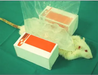
Dorsum of the rats was shaved after anesthesia. Ice water bag which was filled with the same amount of water and ice kept constant at 4°C–8°C once a day for 30 min was brought into contact with the back of the rat in Group II. Each rat was placed between two supports for preventing slipping of the ice bag and full contact with the dorsal skin where flaps would elevated were maintained [Figure 1]. Skin temperature of dorsal skin and ears measured with a touchless infrared digital thermometer in every 5 min. The temperature was constant between 11°C and 14°C of the back and normal temperature in the ears.
Chloraethyl spray, Walter Ritter, Hamburg-Germany was applied to shaved back of rat a total of seven times at 5-min intervals in each session for Group III. Each spraying was carried out for 4–6 s until a white layer was formed on the skin of the rat. Immediately, after spray application, the back skin temperature was measured 2°C–5°C with touchless infrared temperature, which increased to 12°C–18°C within 10 s and returned to normal values after 40–60 s [Figure 2].
Flap model
Caudally based 3 cm × 9 cm size random pattern McFarlane skin flap was elevated from the dorsum of the rat. Flaps were replaced to the donor site. Flap viability was assessed 1 week later.
Assessment of flap viability
Flaps were photographed (Canon EOS 20D camera with 50 mm f:1.8 II lens and Canon Speedlite 43OEX external flash) at the postoperative 7th day. A 2 cm × 2 cm white paper sheet
was placed next to rats for calibration. Images were assessed in Adobe Photoshop Cs5 program. Pixels of the viable and necrotic part of the flaps were measured, and flap viability percentage was calculated.
Blood vascular endothelial growth factor level measurement
Commercially available human vascular endothelial growth factor (VEGF) Immunoassay kit (Biosource, California, USA) was used for VEGF concentration analysis and the analysis of VEGF165 isoform. Blood levels of VEGF were studied on days 1-4-7 and 15 to investigate new vessel formation in all rats in all groups. A volume of 1 ml of blood were taken with 22G needle from rats’ tail vein for each measurement. On the 1st day, blood was collected before hypothermia and formed
basal values for the groups. On the 15th day, blood was collected
24 h after the last hypothermia before flap elevation. No blood was collected from the flap-elevated rats, as the flap elevation itself would create hypoxia and affect VEGF levels. Therefore, the blood levels of Group I were evaluated only on the 1st day.
Microangiographic assessment
All rats were anesthetized with 40 mg/kg ketamine before the procedure. Blood was replaced with intracardially injected barium oxide. After sacrification of the rat, rats were placed to-32°C freeze for 1 day. Flaps were harvested and radiologic images were taken with digital mammography. Images were
Figure 1: Ice water bag placement of the back side of the rat for local
uploaded to Adobe Photoshop program for vessel count. Lateral thoracic artery and deep circumflex iliac arteries were marked. New vessel formation in four regions around these vessels and the center of the flap were counted [Figure 3].
Statistical analysis
The ratio of living areas to total surface area and VEGF blood levels between the groups were statistically investigated. Levene test was used to determine whether the distribution of independent variables was homogeneous. In the comparisons where the variables were homogeneous, t-test was used for statistical analysis. The Kruskal–Wallis test was used for statistical analysis in comparisons where the variables were not homogeneous. P < 0.05 was considered statistically significant.
r
esultsFlap viability percentage
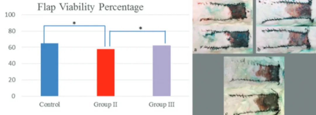
The ratio of living areas to the total flap area was 64.87% in Group I, 57.69% in Group II, and 62.22% in Group III [Figure 4]. The mean viable percentage in Group II
was lower than the other groups and the difference was statistically significant when compared to Group I (P = 0.006) and Group III (P = 0.029). There was no statistically significant difference between Group I and Group III (P > 0.05).
Blood vascular endothelial growth factor levels
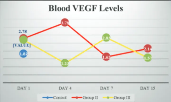
On the 1st day blood VEGF levels were: Group I: 1.82 pg/ml,
Group II: 2.78 pg/ml, and Group III: 2.69 pg/ml. The difference between the 1st day values in the groups was not statistically
significant (P > 0.05). On day 4, Group II was 3.78 pg/ ml and Group III was 1.23 pg/ml. At the 4th day values, the
difference between Group II and Group III was statistically significant (P < 0.001). When the 4th day values were compared
with the basal values of the groups (1st day), there was no
statistically significant difference in Group I (P > 0.05), but the decrease in Group III was statistically significant (P < 0.05). Day 7 values were Group II: 1.62 pg/ml, Group III: 2.83 pg/ml; 15th-day values were found as Group II: 2.14 pg/ml, and Group III:
1.57 pg/ml. The difference between 7 and 15-day values was not statistically significant between the groups and when compared with the basal values of the groups (P > 0.05). There was no statistically significant increase in hypothermia groups (Group II and Group III) compared to basal levels (P > 0.05). The difference between 7 and 15-day values was not statistically significant between the groups and when compared to the basal values of the groups (P > 0.05) [Figure 5].
Microangiographic vessel count
When microangiographies were investigated macroscopically, enlargement and expansion were shown in axial vessels [Figure 6]. The mean number of vessels in the flap was 11.6 in Group I, 35.25 in Group II, and 13.2 in Group III. The difference between Group II and Group I (P = 0.024) and Group III (P = 0.032) was statistically significant. The number of vessels in Group III was higher than in Group I, but the difference was not statistically significant (P > 0.05) [Figure 7]. The mean number of vessels in the middle region of the flap was 0.6 in Group I, 6.75 in Group II, and 1 in Group III. The difference between Group II and Group I was statistically significant (P = 0.029), but the difference with Group III was not statistically significant (P > 0.05).
Figure 3: Axial vessels seen in microangiography and vessel counting regions. Arrowhead: Lateral thoracic artery. Arrow: Deep circumflex iliac artery. Numbers 1, 2, 3, 4: Distal branching of the axial artery. Number 5: The center of the flap
Figure 4: Flap viability chart. The mean viable percentage in Group II was lower than the other groups and the difference was statistically significant when compared to Group I and Group III (*P < 0.05). On the right examples of skin flaps were shown. (a) Group I (control group). (b) Group II (ice water group). (c) Group III (spray group)
d
iscussionIn order to reduce patient morbidity and increase surgical success, studies on the development of flap surgery are continuously performed. Various methods have been tried to prevent or reduce the amount of flap necrosis. The best known of these applications called “preconditioning” are surgical delay, ischemic preconditioning, hypothermia, hyperthermia, growth factors, and pharmacological agents. Although some applications increase flap viability, there are significant disadvantages such as high cost, need for technical equipment, additional surgical intervention, side effects, and nonpractical application. In order to be able to apply the flap viability
widely in the clinic, it should not have toxic effects, be easy to apply and inexpensive, and do not need advanced technology. Thus, hypothermia seems safer and cheaper compared to other preconditioning methods.
In the literature, studies about hypothermia are generally directed to systemic or short-term local hypothermia applications.[15-17]
Systemic hypothermia is the cooling of the whole body to about 32°C–35°C. The aim of systemic hypothermia is to reduce cell damage by decreasing the rate of metabolism when the tissues remain hypoxic such as hypoxic encephalopathy and cardiac arrest.[12,18,19] Local hypothermia is the cooling of a part
of the body to 4°C–8°C for 30 min. Harder et al. observed that local hypothermia was protective against ischemia and ischemia-reperfusion injury and increased flap viability.[2]
Kubulus et al. concluded that hypothermia applied 24 h before flap surgery increased flap viability[20] Xie YC et al. showed
increased angiogenesis in hypothermia induced rats.[21] In other
studies, hypothermia has been shown to trigger angiogenesis.[22,23]
Short term hypothermia was induced with ice water bags in these studies; however, there is no study on the effect of long-term intermittent hypothermia application in the literature. Thus, we aimed to investigate both flap viability and the effect of long-term hypothermia on new vessel formation with microangiography and blood VEGF which is a marker of angiogenesis.
When the viability of the dorsal skin flap was evaluated as surface area, the viability percentage of Group II was lower than the other groups and the difference was statistically significant. There was no statistically significant difference between Group I and Group III. The decrease in Group II can be considered as an impairment of cellular functions as a result of long-term hypothermia. Another possibility is that the 2-week process is insufficient for investigating hypothermia effects on flap viability.
When blood VEGF levels were examined, there was no statistically significant increase in hypothermia groups compared to basal levels. The first factor suggesting that the increase in VEGF level was not observed was that blood levels of VEGF were studied, not tissue. The tissue level of VEGF is more significant than the blood level.[24] Hypoxic stimuli
have been shown to increase blood levels of VEGF.[25,26] While
VEGF is elevated in tissue, it may not be elevated in blood yet. Therefore, the absence of an increase in blood values cannot be interpreted as not being synthesized in the tissue. The half-life of VEGF in plasma has been shown to be 13–33 min.[27] In our
study, blood VEGF levels were studied 24 h after hypothermia application. Since hypothermia was administered daily for 15 days, this stimulus was predicted to keep VEGF levels constantly high. However, VEGF may not be evaluated due to its short rise and fall after cold administration. Increased blood VEGF levels may be detected in an earlier administration. When microangiograms were examined macroscopically, it was observed that the vessel diameters and branching amounts of Group II were higher than the other groups. The difference in neovascularization between Group I and Group III is not significant. When the number of vessels in microangiography Figure 5: Graphical representation of vascular endothelial growth factor
levels by groups and days
Figure 7: Graph of mean vessel numbers in microangiography (*P < 0.05) Figure 6: Left lateral thoracic artery territory in (a) An angiography sample from Group II (b) An angiography sample from Group I (control group)
b a
was evaluated, it was determined that the vessel amount of Group II was higher than the other groups. The number of vessels in Group III was higher than in Group I, but the difference was not statistically significant. The findings support the hypothesis that long-term hypothermia leads to new vessel formation more than intermittent hypothermia. Controversially, flap viability in Group II was found lower. This paradoxical result might be explained as the formation of the new vessels against the hypoxic gradient, duration of hypothermia. It is known that local hypothermia causes vasoconstriction, which leads to hypoxia in tissues which is a trigger for angiogenesis.[28] New vessels are formed according to the
hypoxic gradient. When the hypoxia is not at the same level all over the flap, the newly formed vessels are aligned to the region where the hypoxia is less. In this way, the vessels acquire a structure parallel to the flap axis and can feed the distal flap region where hypoxia is most severe. However, the hypoxia caused by hypothermia in the dorsal skin flap is the same in general and does not show a gradient in our study. In the microangiography of the new vessels formed by hypothermia, not parallel to the flap axis, extending from the edges to the middle is shown to extend. Such a vessel increase cannot feed the distal ischemic area and prevent necrosis when the flap is randomly raised. Considering the new vascular structures caused by hypothermia, there is an increase in the number and diameter of the existing axial vessels. This suggests that the area fed by the axial vessel is enlarged. If the flap is planned axially such as lateral thoracic artery based, it may be possible to increase the surface area of the flap live.
c
onclusionIt was shown that intermittent local hypothermia for 2 weeks had no effect on flap viability and blood VEGF levels. Increased viability and increased blood VEGF levels might be observed in longer term applications or axially planned flaps.
Financial support and sponsorship
Nil.
Conflicts of interest
There are no conflicts of interest.
r
eFerences1. Bakhach J. 1984-1994: Ten years of skin flaps. Recent advances in experimental surgery. Ann Chir Plast Esthet 1995;40:583-95.
2. Harder Y, Amon M, Laschke MW, Schramm R, Rücker M, Wettstein R,
et al. An old dream revitalised: Preconditioning strategies to protect
surgical flaps from critical ischaemia and ischaemia-reperfusion injury. J Plast Reconstr Aesthet Surg 2008;61:503-11.
3. Myers MB, Cherry G. Mechanism of the delay phenomenon. Plast Reconstr Surg 1969;44:52-7.
4. Huemer GM, Wechselberger G, Otto-Schoeller A, Gurunluoglu R, Piza-Katzer H, Schoeller T. Improved dorsal random-pattern skin flap survival in rats with a topically applied combination of nonivamide and nicoboxil. Plast Reconstr Surg 2003;111:1207-11.
5. Carroll SM, Carroll CM, Stremel RW, Heilman SJ, Steffen JM, Tobin GR, et al. Vascular delay and administration of basic fibroblast growth factor augment latissimus dorsi muscle flap perfusion and
function. Plast Reconstr Surg 2000;105:964-71.
6. Carroll CM, Carroll SM, Schuschke DA, Barker JH. Augmentation of skeletal muscle flap survival using platelet derived growth factor. Plast Reconstr Surg 1998;102:407-15.
7. Zhang F, Fischer K, Komorowska-Timek E, Guo M, Cui D, Dorsett-Martin W, et al. Improvement of skin paddle survival by application of vascular endothelial growth factor in a rat TRAM flap model. Ann Plast Surg 2001;46:314-9.
8. Zhang F, Richards L, Angel MF, Zhang J, Liu H, Dorsett-Martin W,
et al. Accelerating flap maturation by vascular endothelium growth
factorin a rat tube flap model. Br J Plast Surg 2002;55:59-63.
9. Swan H. Clinical hypothermia: A lady with a past and some promise for the future. Surgery 1973;73:736-58.
10. Koytman A, Long BJ. Therapeutic Hypothermia. Available from: http:// emedicine.medscape.com/article/812407-overview. [Last accessed on 2019 Jul 26].
11. Niklasch DM. Induced mild hypothermia and the prevention of neurological injury. J Infus Nurs 2010;33:236-42.
12. Seder DB, Fraser GL, Riker RR. Temperature modulation for neuroprotection after acute brain injury. Crit Connect 2009;7:16. 13. Sahuquillo J, Vilalta A. Cooling the injured brain: How does moderate
hypothermia influence the pathophysiology of traumatic brain injury. Curr Pharm Des 2007;13:2310-22.
14. Yunoki M, Nishio S, Ukita N, Anzivino MJ, Lee KS. Hypothermic preconditioning induces rapid tolerance to focal ischemic injury in the rat. Exp Neurol 2003;181:291-300.
15. Shaw WW, Ko CY, Ahn CY, Markowitz BL. Safe ischemia time in free-flap surgery: A clinical study of contact-surface cooling. J Reconstr Microsurg 1996;12:421-4.
16. Bastiaanse J, Nanhekhan LV, Slaaf DW, Boeckx WD, oude Egbrink MG. Preservation of rat cremaster muscle microcirculation after prolonged cold storage and transplantation. J Surg Res 2006;131:41-8.
17. Iwamoto S, Takasu A, Sakamoto T. Therapeutic mild hypothermia: Effects on coagulopathy and survival in a rat hemorrhagic shock model. J Trauma 2010;68:669-75.
18. Yuan HB, Huang Y, Zheng S, Zuo Z. Hypothermic preconditioning increases survival of purkinje neurons in rat cerebellar slices after an
in vitro simulated ischemia. Anesthesiology 2004;100:331-7.
19. Bernard SA, Jones BM, Horne MK. Clinical trial of induced hypothermia in comatose survivors of out-of-hospital cardiac arrest. Ann Emerg Med 1997;30:146-53.
20. Kubulus D, Amon M, Roesken F, Rücker M, Bauer I, Menger MD. Experimental cooling-induced preconditioning attenuates skin flap failure. Br J Surg 2005;92:1432-8.
21. Xie YC, Li CY, Li T, Nie DY, Ye F. Effect of mild hypothermia on angiogenesis in rats with focal cerebral ischemia. Neurosci Lett 2007;422:87-90.
22. Xue Y, Petrovic N, Cao R, Larsson O, Lim S, Chen S, et al. Hypoxia-independent angiogenesis in adipose tissues during cold acclimation. Cell Metab 2009;9:99-109.
23. Asano A, Morimatsu M, Nikami H, Yoshida T, Saito M. Adrenergic activation of vascular endothelial growth factor mRNA expression in rat brown adipose tissue: Implication in cold-induced angiogenesis. Biochem J 1997;328 (Pt 1):179-83.
24. Egginton S. Temperature and angiogenesis: The possible role of mechanical factors in capillary growth. Comp Biochem Physiol A Mol Integr Physiol 2002;132:773-87.
25. Hudlicka O, Tyler KR. Angiogenesis. London: Academic Press; 1986. p. 726-9.
26. Detmar M, Brown LF, Berse B, Jackman RW, Elicker BM, Dvorak HF,
et al. Hypoxia regulates the expression of vascular permeability factor/
vascular endothelial growth factor (VPF/VEGF) and its receptors in human skin. J Invest Dermatol 1997;108:263-8.
27. Houck KA, Ferrara N, Winer J, Cachianes G, Li B, Leung DW. The vascular endothelial growth factor family: Identification of a fourth molecular species and characterization of alternative splicing of RNA. Mol Endocrinol 1991;5:1806-14.
28. Johnson JM, Kellogg DL Jr. Local thermal control of the human cutaneous circulation. J Appl Physiol (1985) 2010;109:1229-38.