Ankara Üniv Vet Fak Derg, 53, 5-13, 2006
Radiographic and ultrasonographic evaluation of the
upper urinary tract diseases in dogs: 22 cases
M. Doğa TEMİZSOYLU1, Ali BUMİN2, Mahir KAYA2, Zeki ALKAN2
1Department of Surgery, Faculty of Veterinary Medicine, Akdeniz University, Burdur; 2Department of Surgery, Faculty of Veterinary Medicine, Ankara University, Ankara .
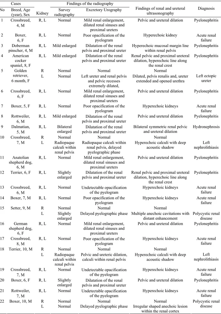
Summary: The objectives of the present study were to describe the complementary use of radiography and ultrasonography in the diagnosis of upper urinary tract disorders in dogs, and to compare ultrasonographic findings with the survey and contrast radiographic findings in the evaluation of canine upper urinary tract diseases. The study materials were composed of 22 dogs of various breed, age, and sex with upper urinary tract diseases. Pyelonephritis (9 cases), acute renal failure (7 cases), policystic renal disease (2 cases), bilateral hydronephrosis (one case), radiopaque nephrolith (2 cases), unilateral ectopic ureter (one case) were diagnosed radiographically and ultrasonographically. The results of the present study indicate that ultrasonography was more sensitive than radiography in diagnosis of upper urinary diseases but was incapable in qualitative evaluation of renal functions and in examination of the ureters. Survey radiographs had little value in the diagnosis of pyelonephritis, disorders of renal pelvis, ureters except for the identification of radiopaque nephrolith. It was determined that EU was valuable for determination of the ectopic ureter, hydronephrosis, and evaluation of renal function.
Key words: Dog, radiography, ultrasonography, upper urinary system.
Köpeklerde üst üriner sistem hastalıklarının radyografik ve ultrasonografik değerlendirmesi: 22 olgu
Özet: Bu çalışmada köpeklerde üst üriner sistem hastalıklarının tanısında radyografi ve ultrasonografi kullanımını tartışmak ve ultrasonografik bulgular ile direkt ve indirekt radyografik bulguları karşılaştırmak amaçlandı. Çalışma materyalini üst üriner sistem hastalığı olan değişik ırk, cinsiyet ve yaşta 22 köpek oluşturdu. Radyografik ve ultrasonografik muayene sonucunda pyelonefritis (9 olguda), akut renal yetmezlik (7 olguda), polikistik renal hastalık (2 olguda), hidronefrozis (1 olguda), radyoopak nefrolit (2 olguda) ve unilateral ektopik üreter (1 olguda) tanısı konuldu. Bu çalışmanın sonunda köpeklerin üst üriner sistem hastalıklarının tanısında ultrasonografinin radyografiden daha hassas bir teknik olduğu ancak böbreklerin fonksiyonel olarak değerlendirilmesi ve üreterlerin incelenmesinde ise ultrasonografinin yetersiz kaldığı görüldü. Direkt radyografiler, radyoopak böbrek taşlarının belirlenmesi dışında pyelonefritis, pelvis renalis bozuklukları ve üreterlerin değerlendirilmesinde çok az bilgiye sahipti. Ektopik üreter, hidronefrozis ve böbrek fonksiyonlarının değerlendirilmesinde EU’nin değerli bir teknik olduğu belirlendi.
Anahtar sözcükler: Köpek, radyografi, ultrasonografi, üst üriner sistem.
Introduction
Survey and contrast radiography are frequently utilized for diagnostic evaluation of the urinary system in dogs. Radiographic imaging may allow assessment of a relationship existing between the urinary tract diseases and clinical signs. The kidneys are usually visible on survey radiographs, but the ureters are not (2, 5, 9, 13, 19). Survey radiography provides information regarding renal size, location, number, and radiographic density. However, survey radiographs may not provide adequate morphologic information when the patient is emaciated or has retroperitoneal fluid (5, 6, 9, 23). Excretory urography (EU) is a radiographic contrast-enhanced procedure used to enhance visualization of the renal parenchyma and to provide visualization of the structures not normally identified on survey radiographs, i.e., pelvic recesses, renal pelves, and ureters (5, 6, 9, 19, 23). EU
also provides limited qualitative evaluation of renal blood flow and function as well as gamuts of potential pathophysiologic cause based on renal opacification patterns (23). However, a nondiagnostic study may result if renal function is severely compromised, and the systemically administered contrast agent may cause further damage or elicit systemic adverse reactions (13, 23). Ureters are seen as thin opaque lines approximately 1 to 2 mm in diameter that extend caudally from the renal hilus to the urinary bladder on 2 to 5 minutes radiographs during EU. Ureters are not normally identified throughout their entire length on single radiographs because of peristaltic emptying of the radiopaque urine (19).
Two-dimensional, gray scale ultrasonography can complement data obtained from other diagnostic modalities, including survey and contrast radiography (7, 9, 10, 13, 15).
Table 1. Radiographic and ultrasonographic findings in 22 dogs with upper urinary tract diseases. Cases Findings of the radiography
No Breed, Age (year), Sex Kidney
Survey radiography
Excretory Urography Findings of renal and ureteral ultrasonography
Diagnosis 1 Crossbreed,
4, M
R, L Normal Mild renal enlargement, dilated renal sinuses and
proximal ureters
Pelvic and ureteral dilation Pyelonephritis 2 Boxer,
6, F
R, L Normal Poor opacification of the pyelogram
Hyperechoic kidney Acute renal failure 3 Doberman
pinscher, 4, M
R, L Mild enlarged Dilatation of the renal pelvis and proximal ureter
Hyperechoic mucosal margin line within renal pelvis
Pyelonephritis 4 American
cocker spaniel, 8, F
R, L Mild enlarged Dilatation of the renal pelvis and proximal ureter
Renal pelvic and proximal ureteral dilation, hyperechoic line along
the renal crest
Pyelonephritis
R Normal Normal Normal
5 Golden retriever,
6 month, F L Normal Left ureter and renal pelvis and pelvic recesses extremely dilated,
Dilated, pelvis renalis and, ureter extended and opened urethra
Left ectopic ureter 6 Crossbreed,
6, F
R, L Normal Mild renal enlargement, dilated renal sinuses and
proximal ureters
Pelvic and ureteral dilation Pyelonephritis 7 Boxer, 5, F R, L Normal Poor opacification of the
pyelogram
Hyperechoic kidneys Acute renal failure 8 Rottweiler,
6, M
R, L Mild enlarged Dilatation of the renal pelvis and proximal ureter
Pelvic and ureteral dilation Pyelonephritis 9 Dalmatian,
5, M
R, L Bilateral
enlarged pelvis and proximal ureter Dilatation of the renal
Bilateral symmetric renal pelvic
and ureteral dilation Hydronephrosis
R Normal Normal Normal
10 Crossbreed,
7, M L Radiopaque calculi within
renal pelvis
Radiopaque calculi within renal pelvis, delayed
pyelographic phase
Hyperechoic calculi with deep
acoustic shadow nephrolithiasis Left 11 Anatolian
shepherd dog, 6, M
R, L Normal Mild renal enlargement, dilated renal sinuses and
proximal ureters
Pelvic and ureteral dilation Pyelonephritis 12 Terrier, 6, F R, L Slightly
enlarged pelvis and proximal ureter Dilatation of the renal Renal pelvic and proximal ureteral dilation, hyperechoic line along the renal crest
Pyelonephritis 13 Crossbreed,
6, M
R, L Normal Undetectable opacification of the pyelogram
Hyperechoic kidneys Acute renal failure 14 Boxer, 7, M R, L Normal Poor opacification of the
pyelogram Hyperechoic kidneys Acute renal failure
R Normal Normal Normal
15 Setter, 9, M
L Slightly enlarged
Delayed pyelographic phase Multiple anechoic cavitations with distant enhancement Polycystic renal disease 16 German shepherd dog, 6, F
R, L Normal Mild renal enlargement, dilated renal sinuses and
proximal ureters
Pelvic and ureteral dilation Pyelonephritis 17 Crossbreed,
8, M
R, L Normal Poor opacification of the pyelogram
Hyperechoic kidneys Acute renal failure
R Normal Normal Normal
18 Terrier, 10, M
L Radiopaque calculi within
renal pelvis
Pelvic and ureteric dilation,
calculi within renal pelvis Hyperechoic calculi with deep acoustic shadow
Left nephrolithiasis 19 Crossbreed,
7, M
R, L Normal Undetectable opacification of the pyelogram
Hyperechoic kidneys Acute renal failure 20 Boxer, 6, F R, L Slightly
enlarged pelvis and proximal ureter Dilatation of the renal
Pelvic and ureteral dilation Pyelonephritis 21 Rottweiler,
7, M
R, L Normal Undetectable opacification of the pyelogram
Hyperechoic kidneys Acute renal failure
R Normal Normal Normal
22 Boxer, 10, M
L Normal Delayed pyelographic phase Irregular shaped anechoic lesion within the renal cortex
Polycystic renal disease R: Right, L: Left, F: Female, M: Male.
In the diagnosis of renal disease in the dog, ultrasonography has many advantages over conventional radiographic procedures. Although ultrasonography does not provide the qualitative functional information that is available through EU, it is an effective method of defining the extent and nature of renal masses as well as general parenchymal architecture. Although ultraso-nographic appearance is not microscopically specific for type of disease, its use can limit the possible diagnosis considered and can aid nonsurgical localization of focal disease for biopsy (25). Ultrasonography often permits differentiation of basic tissue characteristics within an organ that can then be used to further define diseases of excretory pathway (12, 25). Renal ultrasonography, as a step in the evaluation of animals with clinical signs of renal disease, generally is performed after physical examination and the acquisition of laboratory results (13, 15, 21, 25). Clinical indication for renal ultrasonography include palpable parenchymal irregularity or abnormal size, inadequate visualization of the kidneys on survey radiographs because of poor intra-abdominal radiograp-hic contrast apparent abdominal pain and laboratory evidence of urinary tract disease (hematuria, proteinuria, and high serum urea nitrogen and serum creatinin concentration). Ultrasonography also is applicable in debilitated patients or nondiagnostic as a result of poor kidney opacification. Additional complementary information may be obtained by this noninvasive method before initiation of more invasive diagnostic procedure (exploratory laparotomy and biopsy) (21, 25). The greatest advantage of the ultrasonography is its ability to provide superior assessment of internal renal parenchymal architecture. An additional major advantage of sonographic examination of the kidneys is that it is not hampered by the lack of retroperitoneal fat or the presence of retroperitoneal or abdominal fluid or ingesta or fluid in superimposed bowel (13, 23). Ultraso-nography can be used to differentiate nonradiopaque uroliths from other soft tissue attached or free luminal filling defects of the excretory pathway (5).
With the above as a basis, the purposes of the study reported here were to describe the complementary use of radiography and ultrasonography in the diagnosis of upper urinary tract disorders in dogs, and to compare ultrasonographic findings with the survey and contrast radiographic findings in the evaluation of canine upper urinary tract diseases.
Material and Methods
The material of this retrospective study was derived from the case records of 22 dogs (Table 1) with upper urinary tract diseases at the Surgery Clinics of the Faculty of Veterinary Medicine, in Akdeniz and Ankara Universities.
The survey radiographs were evaluated for determination of the size, shape and position of the
kidneys if possible, and to note whether there are any abnormalities elsewhere in the abdominal cavity; to ensure that exposure factors and other technical aspects are correct before contrast media are used; to examine the urinary system for visible lesions; to determine whether calculi are present, since these can be masked contrast media; to ensure that abdomen and intestinal tract are free of material which could decrease detail, e.g. ascitic fluid, faecal material. EU was performed, as described (5, 6, 9, 19), to evaluate renal parenchymal and excretory pathway architecture, to delineate the kidneys when abdominal contrast was reduced, and to qualitatively assess renal function. The excretory urograms were evaluated for renal size, architectural abnormalities, and renal functional capacity (22), ureters evaluation on 2 to 5 minutes radiographs during EU.
Ultrasound images were obtained by using with real-time ultrasonographic equipment using either convex or linear array transducers of 5.0 to 7.5 MHz frequencies, depending on the size of the patient and sonographic images were printed with a video printer. All ultrasonograms were made with the dog in dorsal recumbency. The dogs were prepared for examination by clipping hair from the scanning site, cleansing of the skin with surgical spirit to remove debris and by application of liberal amounts of coupling gel to exclude air bubbles, which degrade image quality. Ultrasonography was performed, as described (5, 13, 19, 21), to evaluate kidney echogenicity, to determine focal or diffuse abnormalities of the renal parenchyma or abnormalities of the renal pelvis, collecting system and proximal ureters.
Aiding physical examination findings, urinalysis, and bacterial urine culture of the cases carried out the final diagnosis but patient clinical and laboratory findings were extracted from the case records.
Results
The radiographic and ultrasonographic findings for the 22 dogs are summarized in Table 1. Twenty-two dogs had diagnosed renal and ureteral diseases. Nine dogs had pyelonephritis. In survey radiography; the seven of these nine dogs had bilateral slightly renal enlargement. Two of them had normal renal appearance but urinalysis and abdominal palpation suggested renal diseases. In EU, five of these dogs had incomplete filling of pelvic recesses, dilatation of the renal pelvis and proximal ureter. Four of these dogs had a normal to slightly diminished homogenous density during the nephrogram phase, and mild to moderate dilation of diverticulae, renal sinus and proximal ureters. The ultrasonographic findings of 9 dogs with pyelonephritis were renal pelvic and proximal ureteral dilation and a hyperechoic mucosal margin line within the renal pelvis and proximal ureters (Figure 1).
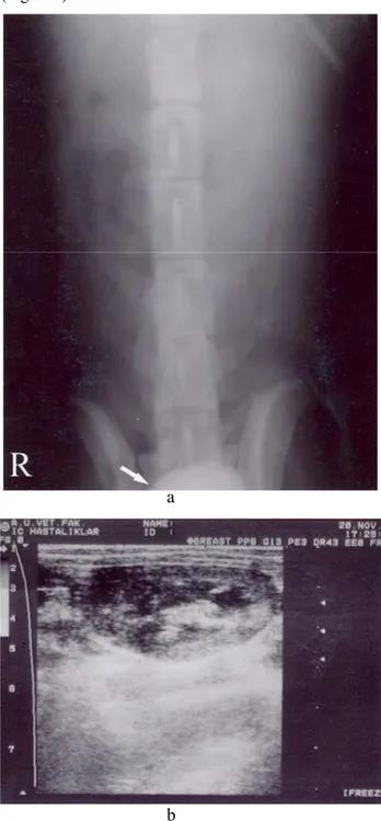
Acute renal failure (ARF) was diagnosed in seven dogs. Kidneys of these dogs were seen normal in survey radiography. In EU, nephrographic opacification was poor and did not fade with time. One hundred-twenty minute after intravenous injection of contrast medium for EU, four dogs with ARF had poor opacification of the pyelogram and three of them had undetectable opacification of the pyelogram. On ultrasonography, kidneys were normal sized but with hyperechoic pattern (Figure 2).
a
b
a
Figure 1. Longitudinal sonograms of right (a) and left kidney (b), and ventrodorsal view of 10 minutes after (c) intravenous injection of contrast medium for EU, and in a 6-year-old female crossbreed dog. On sonograms, there were mild dilations of the renal pelvises and there was a hyperechoic zone in the renal pelvis in right (a) and left (b) kidneys. The EU (c) revealed that renal enlargement, which is smooth regular, and the renal sinuses and proximal ureters were dilated.
c
b
Figure 2. Ventrodorsal view of 120 minutes after (a) intravenous injection of contrast medium for EU, and longitudinal sonogram of the left kidney (b) in a 5-year-old female Boxer. EU revealed poor nephrographic opacification, and delayed pyelographic and cystographic phase (white arrow). Sonogram of the left kidney (b) revealed that kidney was normal in size and appeared hyperechoic pattern.
Bilateral hydronephrosis was diagnosed in one dog and kidneys appeared bilaterally enlarged in survey radiography. Both kidneys symmetrically dilated and severe pelvic dilation were present in pyelograghic phase. Ultrasonography revealed severe dilation of renal pelvis, diverticula and proximal ureter. Renal parenchyma of both kidneys was thin (Figure 3).
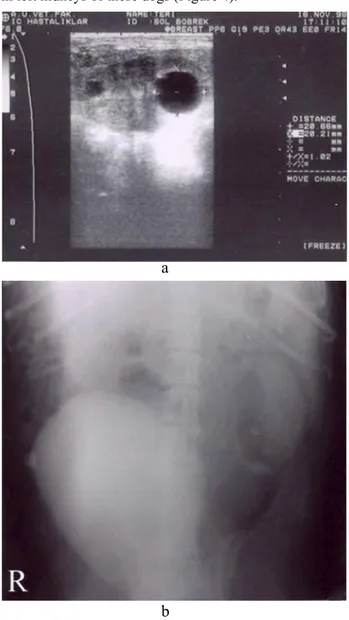
Two dogs had polycystic renal diseases. In survey radiography, while right kidneys were seen normal, left kidneys were slightly enlarged. Early stages of EU revealed multiple foci of relative radiolucency separated by septae, which were contrast enhancement, and pyelographic phase was delayed in left kidneys of the cases. Ultrasonographic examination of the kidneys showed a 20.66-20.21 mm diameter rounded, anechoic with deep acoustic enhancement cysts within the cortex in left kidneys of these dogs (Figure 4).
a
b
a
Figure 3. Longitudinal sonograms of right (a) and left kidney (b), and ventrodorsal view of 20 minutes after (c) intravenous injection of contrast medium for EU, and in a 5-year-old male Dalmatian. On sonograms revealed dilatation of the renal pelvis and proximal ureters in right (a) and left (b) kidneys. There were thin rim of functional renal parenchyma remains and acoustic enhancement distal to the pelvis. The left and right renal pelvis and pelvic recesses were dilated on the EU (c), indicating hydronephrosis, and the kidneys were enlarged (greater than 3.5 times the length of L2).
c
b
Figure 4. Ventrodorsal view of 20 minutes after (a) intravenous injection of contrast medium for EU, and longitudinal sonogram of left kidney (b) in a 9-year-old male Setter. EU revealed that pyelographic phase was delayed in left kidney (a). On sonogram of the left kidney revealed a 20.66-20.21mm diameter, anechoic with deep acoustic enhancement cysts within the cortex in left kidneys (b).
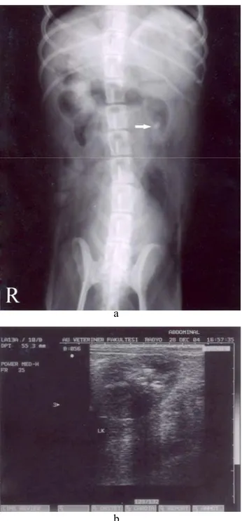
Two dogs were affected by left nephrolithiasis. A calcific density within the left renal pelvis was seen in survey radiography. EU revealed delayed pyelographic phase and pelvic and ureteric dilation in the left kidneys of these dogs. Ultrasonographic examination of the affected kidneys disclosed hyperechoic renal calculi with deep acoustic shadows (Figure 5).
a a
b b
Figure 6. Ventrodorsal view of 20 minutes after (a) intravenous injection of contrast medium for EU, and longitudinal sonogram of the left ureter at the level of the bladder trigone (b) in a 6-month-old female Golden retriever. EU revealed that the left ureter was extremely dilated (white arrow) and terminated in the proximal urethra. Sonogram of the ureter (b) revealed that, ureter was opened into the urethra (white arrow) Figure 5. Ventrodorsal view of 10 minutes after (a) intravenous
injection of contrast medium for EU, and longitudinal sonogram of left kidney (b) in a 10-year-old male Terrier. EU revealed a calcific density shaped like a renal pelvis (white arrow). On sonogram there were hyperechoic region within renal pelvis with acoustic shadow.(b)
Left ectopic ureter was diagnosed in a dog. In survey radiography both kidneys and ureter were seen normal. EU revealed that left ureter was extremely dilated, as were the left renal pelvis and pelvic recesses and terminated in the urethra. On sonography, left ureter and renal pelvis and pelvic recesses were extremely dilated. It was disclosed that the dilated left ureter opened proximal urethra (Figure 6).
Discussion and Conclusion
Radiography and ultrasonography provide addition-nal information to that obtained by physical examination and laboratory analysis (2, 19). Survey radiographic procedures with contrast medium and ultrasonography can contribute much information toward the diagnosis and ureteral diseases in dogs (2, 5, 9, 22, 23).
Radiographic imaging may allow assessment of a relationship existing between the upper urinary tract disease and clinical signs (13, 22). The external boundaries of kidneys can usually be identified on survey radiographs. This identification permits assessment of the size, shape, and radiographic opacity of the kidneys, which may aid in the diagnosis of disease progress but ureters are not visualized in survey radiography. However, kidney may not be adequately visualized if sufficient retroperitoneal fat is absent or if the following is present: retroperitoneal fluid, ingesta or fluid in superimposed bowel, abdominal fluid (5, 9, 23). In this study, survey radiography provided satisfactory information in 9 of 22 dogs. These abnormal survey radiographic findings were slight to mild renal enlargement and radiopaque calculi within the renal pelvis in two cases. The 13 of 22 cases had no abnormal survey radiographic findings.
EU is defined as sequential radiographic imaging that includes the opacification of the kidneys, renal pelves and recess, and the ureters following the intravenous administration of iodinated contrast medium. Pyelonephritis presents as a normal to slightly diminished homogenous density during the nephrogram phase. Mild to moderate dilation of diverticulae, renal sinus, and proximal ureters as well as blunting of the pseudopapillae may be seen in the pyelographic phase (2, 3, 5, 13, 20, 21). Nine dogs with pyelonephritis in the present study had similar EU findings. EU is useful for defining anatomic structures and for qualitatively assessing the function of the kidneys. It is a common way of verifying and localizing upper urinary tract diseases, and it may be used to assess the reversibility of disease. Although EU is not a quantitative measurement of renal function, it may be used to assess the relative function of kidneys and loosely interpreted to assess the phatophysiologic mechanisms of renal failure (9, 13, 19, 23). In our 7 of the 22 cases had ARF and the immediate ventrodorsal view of the EU revealed a normal nephrogram. The 20-minute view of the EU revealed that no collecting structures have become apparent not that any contrast is seen in the bladder. The density of the kidney parenchyma is nearly the same as the immediate view and cystograms phases of the EU were seen in 120 minutes after intravenous injection of contrast medium for EU. The opacity of the pyelogram is depends on both
the filtration of the contrast medium from the blood and concentration of the contrast medium within the tubules. Hydronephrosis presents smoothly enlarged kidney and uniformly distended renal sinus in EU. In severe cases of hydroneprosis, the diverticulae smoothly and evenly expanded (5, 14, 19, 21-23). Both kidneys symmetrically dilated and severe pelvic dilation were present in pyelograghic phase of the case. The renal pelves and proximal ureteral dilations were eminently detected and the pyelogram was markedly delayed due to the decreased rate of glomerular filtration in a case with hydronephrosis. Renal cysts may be solitary or multiple, some times involving both kidneys (5, 16, 19, 26). The density during the nephrogram phase will generally be irregular due to obliteration of normal renal tissue by the cysts. Classically, there is a fine rim of normally dense renal tissue surrounding the non-opacifed cyst. The pyelogram may show distortion of the collecting structures if the cysts produce pressure upon them (5, 13, 23). Two dogs of 22 cases had polycystic renal disease and cysts had not apparently seen in EU. In the diagnosis of the renal cysts, EU does not provide specific finding. Mineral opacity may be due to the presence of renal calculi in dogs (9, 12, 22). Increased renal opacity may result from parenchymal mineralization or calculus formation within the pelvis. Radiographic detection of renal calculi is dependent on mineral content and size of calculi (12, 19, 22). Our nephrolithiasis cases had radiopaque left renal calculi. These were diagnosed by means of survey radiography and EU. Pyelographic phase was delayed and it could be due to partial obstruction of pelvic outflow. The abnormal location of the ureter that is mostly encountered is ectopic ureter, in which the distal portion of the ureter terminates at a point other than bladder trigone. The most common site of abnormal ureteral termination is the vagina, followed in relative frequency by the urethra, bladder neck, and uterus. The EU does not only provide insight on ectopic ureters, regardless where they terminate, but it also provides a physiologic means to fill the urinary bladder with positive-contrast medium to assess urethral sphincter continence (1, 11, 13, 24). One dog had left ectopic ureter and EU revealed that left ureter was extremely dilated, as were the left renal pelvis and pelvic recesses and terminated in the proximal urethra.
The internal architecture of the kidney can be visualized regardless of the renal function status by ultrasonography (5, 8, 9, 13). Ultrasonographic examination of seven cases with ARF revealed only hyperechoic kidneys suggesting ethylene glycol toxicity. Ultrasonography did not effective in qualitative assessment of kidney function. Ultrasonography is more sensitive than EU in detecting pyelonephritis. Renal
pelvic and proximal dilation and hyperechoic line along the renal crest have been described as the major finding. A uniformity echogenic renal cortex, focal hypoechoic or hyperechoic areas within the renal cortex, and focal lesions within the medulla have been observed (5, 18, 19, 21). Nine dogs with pyelonepritis revealed more specific findings by ultrasonography than EU similar as previous literature. The sonographic features of simple cysts include: a round-ovoid contour; echo-free contents; smooth, sharply demarcated thin walls with a distinct far-wall border; strong distal echo enhancement (21, 23). Renal cysts were more sensitively detected ultrasonographically than EU because renal cysts located in renal parenchyma. Dilation of renal pelvis and proximal ureter may be caused by hydronephrosis or pyelonephritis (14, 21). The differential diagnosis of pelvic dilations may be difficult by ultrasonographically. It should be emphasized that EU is the most sensitive method for detecting subtle renal pelvic dilation. Our hydronephrosis case was diagnosed both ultrasonographically and EU as this case was in advanced stages of hydronephrosis. Renal calculi produce intense hyperechoic foci with strong acoustic shadowing both radiopaque and radiolucent calculi on ultrasonography (4, 12, 13, 21). Our cases had radiopaque nephroliths and so these calculi were detected both radiographically and ultrasonographically. The distal ureters can be detected with ultrasonography only if they are dilated because of ectopia, urethritis, obstruction, or other congenital conditions (17, 19). In our case with left ectopic ureter, left dilated ureteral termination within the proximal urethra was apparently detected ultrasonographically.
The results of the present study indicate that ultrasonography was more sensitive than radiography in diagnosis of upper urinary diseases but was incapable in qualitative evaluation of renal functions and in examination of the ureters. Survey radiographs had little value in the diagnosis of pyelonephritis, disorders of renal pelvis, ureters except for the identification of radiopaque nephrolith. It was determined that EU was valuable for determination of the ectopic ureter, hydronephrosis, and evaluation of renal function. It was concluded that radiography and ultrasonography were both complementary techniques to diagnose upper urinary tract disorders in dogs. However, in certain disorders one may present superior diagnostic advantages.
References
1. Acar SE, Mahzunlar H, Şadalak D, Barlan M, Yücel R (2003): Bir köpekte rastlanılan unilateral intramural ektopik üreter olgusu ve intravesiküler diversiyon-neoureterostomi tekniği ile tedavisi. Vet Cer Derg, 9, 54-58.
2. Alkan Z (1999): Veteriner Radyoloji. 250-260. Mina Ajans, Ankara.
3. Barber DL, Finco DR (1979): Radiographic findings in induced bacterial pyelonephritis in dogs. J Am Vet Med Assoc, 175, 1183-1189.
4. Brown NO, Parks J L, Greene RW (1977): Canine urolithiasis: Retrospective analysis of 438 cases. J Am Vet Med Assoc, 170, 414-418.
5. Burk RL, Ackerman N (1996): The Abdomen. 319-389, 401-408. In: RL Burk, N Ackerman (Eds), Small Animal Radiology and Ultrasonography. A Diagnostic Atlas and Text. W. B. Saunders Company. Philadelphia.
6. Carlise CH (1977): Radiological examination of the upper urinary tract. Aust Vet Pratic, 7, 97-106.
7. Cartee RE, Selcer BA, Patton CS (1980): Ultrasonographic diagnosis of renal disease in small animals. J Am Vet Med Assoc, 176, 426-430.
8. Feeney DA, Johnston GR, Walter PA (1985): Two-dimensional gray scale abdominal ultrasonography: General interpretation and abdominal masses. Vet Clin North Am Small Anim Pract, 26,74-81.
9. Feeney DA (2002): The Kidney and Ureters. 556-570. In: DE Thrall (Ed), Textbook of Veterinary Diagnostic Radiology. W. B. Saunders Company, Philadelphia. 10. Felkai CS, Vörös K, Fenyves B (1995): Lesion of the
renal pelvis and proximal ureter in various nephro-urological conditions: An ultrasonographic study. Vet Radiol Ultrasoun, 36, 397-401.
11. Hager DA, Blevins WE (1986): Ectopic ureter in a dog: extension from the kidney to the urinary bladder and to the urethra. J Am Vet Med Assoc, 189, 309-310.
12. Johnston GR, Walter PA, Feeney DA (1986): Radiographic and ultrasonographic features of uroliths and other urinary tract filling defects. Vet Clin North Am Small Anim Pract, 16, 261-292.
13. Johnston GR, Walter PA, Feeney DA (1995): Diagnostic Imaging of the Urinary Tract. 230-276. In: CA Osborne and DR Finco (Eds), Canine and Feline Nephrology and Urology. Williams & Wilkins, Baltimore.
14. Knottenbelt DC (1988): Unilateral hydronephrosis in a dog. Aust Vet J, 65, 400-402.
15. Konde LJ (1985) Sonography of the kidney. Vet Clin North Am Small Anim Pract, 15, 1149-1158.
16. Konde LJ, Park RD, Wrigley RH, Lebel JL (1986): Comparison of radiography and ultrasonography in the evaluation of renal lesions in the dog. J Am Vet Med Assoc, 188, 1420-1425.
17. Lamb CR, Gregory SP (1994): Ultrasonography of the ureterovesicular junction in the dog: A preliminary report. Vet Rec, 134, 36-38.
18. Macdougall DF, Cook T, Steward AP, Cattell V (1986): Canine chronic renal disease: prevalence and types of glomerulonephritis in the dog. Kidney Int, 29, 1144-1151. 19. Mahaffey MB, Barber EL (1992): Radiographic and
Ultrasonographic Evaluation of the Urinary Tract. 53-79.In: EA Stone and JA Barsanti (Eds), Urologic Surgery of The Dog and Cat. Lea and Febiger. Philadelphia. 20. Neuwirth L, Mahaffey M, Crovell, W, Selcer B,
excretory urography and ultrasonography for detection of experimentally induced pyelonephritis in dogs. Am J Vet Res, 54, 660-669.
21. Nyland TG, Mattoon JS, Wisner ER (1995): Ultrasonograpy of the Urinary Tract and Adrenal Glands. 95-125. In: TG Nyland and JS Mattoon (Eds), Veterinary Diagnostic Ultrasound. Eds: W.B. Saunders Company, Philadelphia.
22. Pugh CR, Rhode WH, Biery DN (1993): Contrast studies of the urogenital system. Vet Clin North Am Small Anim Pract, 23, 281-306.
23. Rivers BJ, Johnston GR (1986): Diagnostic-imaging strategies in small animal nephrology. Vet Clin North Am Small Anim Pract, 26, 1505-1517.
24. Smith CW, Stowater JL., Kneller SK (1980): Bilateral ectopic ureter in a male dog with urinary incontinence. J Am Vet Med Assoc, 170, 1022-1024.
25. Walter PA, Feeney DA, Johnston GR, O’Leary TP (1987): Ultrasonographic evaluation of renal parenchymal diseases in dogs: 32 cases (1981-1986). J Am Vet Med Assoc, 191, 999-1007.
26. White SD, Rosychuk RAW, Schultheiss P, Scott KV (1998): Nodular dermatofibrosis and cystic renal disease in three mixed-breed dogs and a boxer dog. Vet Dermatol, 9, 119-126.
Geliş tarihi : 09.03.2005 / Kabul tarihi : 25.04.2005 Address for correspondance:
Yard. Doç. Dr. Mustafa Doğa Temizsoylu Akdeniz Üniversitesi Veteriner Fakültesi, Cerrahi Anabilim Dalı,


