Therapeutic evaluation of interleukin 1-beta antagonist
Anakinra against traumatic brain injury in rats
Askin Esen Hasturk, M.D.,1 Erdal Resit Yilmaz, M.D.,2 Erhan Turkoglu, M.D.,2 Hayri Kertmen, M.D.,2 Bahriye Horasanli, M.D.,3 Nazli Hayirli, MSc.,4 Imge Berrin Erguder, M.D.,5 Oya Evirgen, M.D.4 1Department of Neurosurgery, Oncology Training and Research Hospital, Ankara;
2Department of Neurosurgery, Diskapi Yildirim Beyazit Training and Research Hospital, Ankara; 3Department of Neurology, Baskent University Faculty of Medicine, Ankara;
4Department of Histology and Embryology, Ankara University Faculty of Medicine, Ankara; 5Department of Biochemistry, Ankara University Faculty of Medicine, Ankara
ABSTRACT
BACKGROUND: The aim of this study was to evaluate the therapeutic efficiency of Anakinra, an IL-1β antagonist with anti-inflam-matory effects, in an experimental model of traumatic brain injury (TBI).
METHODS: Fifty-four rats underwent TBI after a weighted object was dropped onto a metal disc secured to their skulls. Animals were randomized into 3 main groups: control (n=18), TBI + saline (n=18; six animals per time-point) with samples obtained at the first, sixth and twenty-fourth h postoperatively, and TBI + Anakinra (n=18; six animals per time-point) with brain samples obtained at the first, sixth and twenty-fourth h postoperatively. Brain tissue and blood serum were extracted for the analysis of IL-1β, malondial-dehyde, glutathione peroxidase, superoxide dismutase, and catalase levels. Tissue sections were evaluated histopathologically under a light microscope.
RESULTS: After trauma, tissue and serum IL-1β levels were significantly elevated and after Anakinra administration, these levels substantially decreased. Glutathione peroxidase, superoxide dismutase, and catalase activity decreased following TBI and Anakinra ad-ministration proved effective in increasing the activity of these antioxidant enzymes. Histopathological analysis confirmed that Anakinra might protect the brain tissue and nerve cells from injury.
CONCLUSION: Results demonstrate that Anakinra reduces the development of inflammation and tissue injury events associated with TBI.
Key words: Antioxidant; anakinra; interleukin-1; neuroprotection; traumatic brain injury.
potentially avoidable event. This secondary neuronal death is determined by a large number of cellular, molecular, and bio-chemical cascades. One such cascade thought to contribute to the evolution of this secondary damage is the local inflam-matory response in the injured brain tissue.[2] Microglial cells
have been suggested to be the source of cytotoxic cytokines, such as tumor necrosis factor alpha (TNF-α) and interleukin 1β (IL-1β), killing oligodendrocytes. In fact, increased synthe-sis and/or secretion of IL-1β is detectable at the injury site within one h after spinal cord injury (SCI). IL-1β is a member of the interleukin 1 cytokine family. The gene encoding IL-1β, along with eight other IL-1 family genes, form a cytokine clus-ter on chromosome 2.[3] IL-1β is produced by activated
mac-rophages as a pro-protein, which is proteolytically processed to its active form by caspase 1 (CASP1/ICE). This cytokine is an important mediator of the inflammatory response and involves in a variety of cellular activities, including cell prolif-eration, differentiation, and apoptosis.[3]
Address for correspondence: Askin Esen Hasturk, M.D. Onkoloji Eğitim ve Araştırma Hastanesi, Beyin ve Sinir Cerrahisi Bölümü, 06200 Ankara, Turkey
Tel: +90 312 - 336 09 09 E-mail: [email protected]
Qucik Response Code Ulus Travma Acil Cerrahi Derg 2015;21(1):1-8
doi: 10.5505/tjtes.2015.57894 Copyright 2015
TJTES
INTRODUCTION
Traumatic brain injury (TBI) immediately causes direct me-chanical damage to the brain, referred to as the primary damage,[1] resulting in the immediate death of a number of
neurons that cannot be recovered or regenerated. However, neurons continue to die for hours after TBI, representing a
IL-1 is produced in response to inflammatory stimuli and mediates various physiologic responses, including inflamma-tory and immunologic reactions. Anakinra, an antagonist ex-pressed in many tissues and organs, has been shown to com-petitively inhibit the binding of IL-1 to the IL-1 type receptor.
[4] In patients with rheumatoid arthritis, this natural IL-1
receptor antagonist is not found in effective concentrations to counteract elevated IL-1 concentrations. Thus, Kineret is not considered a disease-modifying anti-rheumatic drug, but rather a biological response modifier due to its ability to se-lectively target the pathologic element of the disease.[5]
In the current study, the following endpoints of the inflam-matory response were determined: histological damage; cytokine expression (IL-1β); measurement of lipid peroxida-tion and oxidative stress (glutathione peroxidase, malondi-aldehyde, and superoxide dismutase) and these various fac-tors were used to evaluate whether Anakinra administration could protect brain tissue following traumatic brain injury.
MATERIALS AND METHODS
Fifty-four adult male Wistar albino rats weighing 300-350 g were used in this study. Animal care and all experiments were in accordance with the European Communities Council Di-rective of November 24, 1986 (86/609/EEC) on the protec-tion of animals for experimental use. All experimental pro-cedures used in this research were approved by the ethical committee of the Ministry of Health, Ankara Education and Research Hospital.
Surgical Procedure and Sample Preparation
All rats were kept under environmentally controlled condi-tions at 22-25°C with appropriate humidity, a 12-h light cycle, and free access to food and water. The surgical procedure was performed under general anesthesia induced by intra-peritoneal (IP) xylazine (10 mg/kg; Bayer, Istanbul, Turkey) and Ketamine hydrochloride (50 mg/kg; Parke-Davis, Istanbul, Turkey). A rectal probe was inserted and the animals were positioned on a heating pad in order to ensure that their body temperature was maintained at 37°C. A moderate brain-in-jury model, described by Marmarou et al. and modified by Ucar et al., was applied for head trauma.[6,7] Briefly, rats wereplaced in prone position and a midline incision was made on the head to expose the coronal and lambdoid sutures. A me-tallic disc in 10 mm diameter and 3 mm thickness was fixed to the cranium using bone wax, and a lead object weighing 450 g was allowed a free fall from a height of 70 cm through a copper tube onto the metal disc. The head of the animals was supported on a 10-cm foam bed providing confirmation of impact. After surgery, 1.0 cc of saline was subcutaneously administered to replace the blood volume lost during surgery and the wound was closed in layers with silk sutures. All ani-mals were anesthetized with the above-mentioned agents at 24 h after trauma and their brains were extracted without any
damage. Neural tissue samples were obtained by excising the left frontoparietal lobes from the boundary of the interhemi-spheric fissure and were subjected to biochemical analyses. The remaining parts of the brains were maintained in formal-dehyde solution for histopathological analysis.
Experimental Groups
Rats were randomly allocated into the following 3 main groups and subgroups: Control: (n=18, 6 rats per time window); rats
un-derwent skin incisions only and non-traumatic brain samples were obtained at the first, sixth and twenty-fourth h after surgery.
TBI + Saline: (n=18, 6 rats per time window); rats were
sub-jected to TBI and received a single IP dose of 1 mL/kg saline. Traumatic brain tissue samples were obtained at the first, sixth and twenty-fourth h after surgery.
TBI + Anakinra: (n=18, 6 rats per time window); rats were
subjected to TBI and received a single IP dose of Kineret (Anakinra, Swedish Orphan Biovitrum AB, Stockholm, Swe-den) immediately following TBI. Traumatic brain tissue sam-ples were then obtained at the first, sixth and twenty-fourth h after surgery.
Cytokine Assay
Serum and tissue IL-1β concentrations were determined us-ing the double-antibody sandwich enzyme-linked immunosor-bent assay (R & D systems, Minneapolis, MN, USA) according to the manufacturer’s instructions.
Measurement of Lipid Peroxidation and
Oxidative Stress
Glutathione Peroxidase Analysis
Glutathione peroxidase (GSH-Px) activity was measured fol-lowing changes in NADPH absorbance at 340 nm.[8] In the
activity calculations (IU, international unit), extinction coef-ficients of NADPH were used for GSH-Px. Results were ex-pressed as IU/mg protein.
Malondialdehyde Analysis
Malondialdehyde (MDA) is formed from the breakdown of polyunsaturated fatty acids and serves as an important and reliable index for determining the extent of peroxidation re-actions.[9] Tissue MDA levels were determined by a method
based on a reaction with thiobarbituric acid (TBA). Briefly, samples were mixed with two volumes of cold saline solu-tion containing 0.001% butylated hydroxytoluene (BHT) (200 μl of 0.01% BHT solution in methanol) and 0.07% sodium dodecyl sulfate (SDS) (20 μl of 7% SDS). One ml of sample was then added to 500 μl of 0.01 NH2SO4 and 500 μl of the TBA reagent (0.67% TBA in 50% acetic acid) to precipitate protein. Samples were heated in boiling water for 60 min, and after cooling, an equal volume (2 ml) of n-butanol was added
to each test tube and mixed. The mixture was centrifuged at 4,000 rpm for 10 min at room temperature. The absor-bance of the organic layer in a 1-ml cell was read at 535 nm (Molecular Devices Corporation, Sunnyvale, CA, USA). MDA concentrations were expressed as nanomoles per milligram wet tissue weight.
Superoxide Dismutase Analysis
Total superoxide dismutase (SOD) (Cu–Zn and Mn, EC 1.15.1.1) activity was determined according to the method of Sun et al.[10] The principle of the method is based on the
inhibition of nitroblue tetrazolium (NBT) reduction by the xanthine–xanthine oxidase system as a superoxide generator. Activity was assessed in the ethanol phase of the supernatant after 1.0 ml ethanol/chloroform mixture (5/3, v/v) was added to the same volume of sample and centrifuged. One unit of SOD was defined as the enzyme amount causing 50% inhibi-tion in the NBT reducinhibi-tion rate. SOD activity was expressed as U/mg protein.
Catalase Analysis
Catalase (CAT) activity was determined by the method de-scribed by Aebi.[11] The principle of CAT activity is based on
the determination of the rate constant (k, sec-1) or of the hydrogen peroxide decomposition rate at 240 nm. Results were expressed as kU/g of protein.
Histopathological Procedures
For histological examination, brain tissue samples were fixed at 10% neutral buffered formalin, dehydrated through a graded series of ethanol and embedded in paraffin. 5μm thick sections stained with hematoxylin-eosin were analyzed and photographed with light microscopy (Olympus CX21, Olympus America Inc., Melville, NY, USA). In all groups, a semi quantitative scoring system, ranging between 0 and 3, was used for grading both histopathological changes (vascular congestion, PMNL infiltration, gliosis/satellitosis and spongio-sis) and neuronal degeneration signs (cytoplasmic eosinophil-ia and nuclear pyknosis) in the brain tissues of each animal. Histopathological changes were evaluated by two observers
blinded to the groups and scored as follows: 0: absent; 1: mild; 2: moderate; 3: common. Histopathological scores for each group was calculated averaging the scores of each animal in groups.[12]
Statistical Analysis
Data were analyzed using the Statistical Package for Social Sciences (SPSS) software version 19.0 for Windows (SPSS Inc., Chicago, IL). Non-parametric tests were applied and the Mann-Whitney U test was used to compare two independent groups while the Kruskal-Wallis test was used to compare more than two groups. The Wilcoxon Signed Ranks Test was used to compare two dependent groups while the Friedman Test was used to compare more than two groups. Bonferroni correction for multiple tests was used for post-hoc compari-sons. All differences associated with a chance probability of 0.05 or less were considered statistically significant. Continu-ous variables were presented as mean±SD.
RESULTS
Tissue and Serum IL-1β Analysis
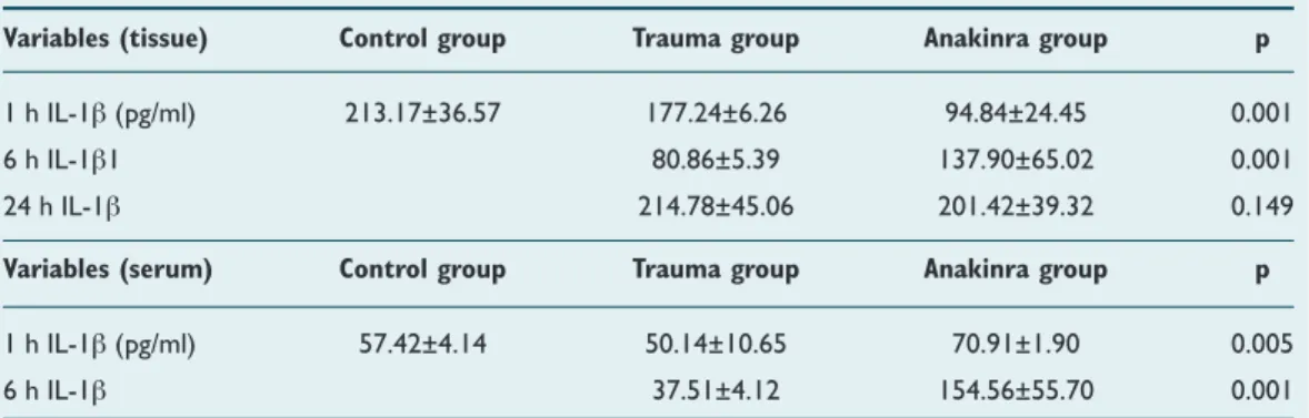
Table 1 summarizes the changes in tissue and serum levels of IL-1β. Mean tissue and serum level of IL-1β were the same although tissue and serum samples were obtained at different time periods. There were statistically significant differences between the trauma (177.24±6.24 pg/ml), con-trol (213.17±36.57 pg/ml), and Anakinra (94.8 4±24.45 pg/ ml) groups at the first h after trauma with regard to mean tissue IL-1β activity (p=0.001). After the sixth h, increased tissue IL-1β activity in trauma- and Anakinra-treated groups was shown at 180.86±5.89 pg/ml and 137.90±65.02 pg/ml, respectively. There was also a statistically significant differ-ence determined between the groups (p=0.001). Trauma (214.78±45.06 pg/ml), control (213.17±36.57 pg/ml), and Anakinra (201.42±39.32 pg/ml) groups were compared at the twenty-fourth h after trauma with regard to mean tis-sue IL-1β activity and no statistically significant difference was found (p=0.149), even though tissue levels of IL-1β in Anakinra-treated groups continued to decline. There were,
Table 1. Summarizing IL-1β changes in the tissue and serum
Variables (tissue) Control group Trauma group Anakinra group p
1 h IL-1β (pg/ml) 213.17±36.57 177.24±6.26 94.84±24.45 0.001
6 h IL-1β1 80.86±5.39 137.90±65.02 0.001
24 h IL-1β 214.78±45.06 201.42±39.32 0.149
Variables (serum) Control group Trauma group Anakinra group p
1 h IL-1β (pg/ml) 57.42±4.14 50.14±10.65 70.91±1.90 0.005
6 h IL-1β 37.51±4.12 154.56±55.70 0.001
however, statistically significant differences between trauma (50.15±4.88 pg/ml), control (60.05±2.19 pg/ml) and Anakinra-treated (52.25±2.01 pg/ml) groups one h after trauma with regard to mean serum IL-1β activity (p=0.003). Additionally, six h after trauma, a statistically significant increase (p=0.001) in serum IL-1β activity was observed in the Anakinra-treated group (154.56±55.70 pg/ml).
Tissue and Serum MDA Analysis
Mean tissue MDA levels in the trauma group were 7.16±1.02 nmol/mg, 11.66±2.94 nmol/mg, and 15.95±02.98 nmol/mg at the first, sixth and twenty-fourth h, respectively. Mean tis-sue levels in the Anakinra group were 7.20±4.39 nmol/mg, 11.46±4.00 nmol/mg, and 9.37±4.44 nmol/mg at the first, sixth and twenty-fourth h after trauma, respectively. A statistically significant difference was observed when mean serum MDA levels in trauma and Anakinra groups were compared with the control group (10.25±2.12 nmol/mg) at all time points (p=0.008). Mean serum MDA levels in the trauma groups were 0.10±0.04 nmol/mg and 0.07±0.04 nmol/mg at the first and sixth h, respectively. Mean tissue levels in the Anakinra group were 0.04±0.01 nmol/mg and 0.05±0.01 nmol/mg at
the fist and sixth h after trauma, respectively. A statistically significant difference was seen when mean serum MDA levels in trauma and Anakinra groups were compared with the con-trol group (0.04±0.01 nmol/mg) at all time-points (p=0.005) (Table 2).
Tissue and Serum GPx Analysis
Mean tissue GPx levels at the first h following trauma for trauma (0.28±0.12 IU/mg), control (0.27±0.11 IU/mg), and Anakinra groups (0.21±0.25 IU/mg) were not significantly dif-ferent (p=0.899). Six hours following TBI, mean tissue GPx levels were 0.25±0.12 IU/mg in the trauma groups, 0.41±0.33 IU/mg in the Anakinra groups, and 0.27±0.11 IU/mg in the control group; there were no statistically significant differenc-es between groups at this time-point (p=0.05). After twenty-four h, mean tissue GPx continued to decline in the Anakinra-treated group (0.31±0.19 IU/mg), but this reduction was not determined to be statistically significant (p=0.189). Mean se-rum GPx levels in trauma groups were 0.0041±0.0007 IU/mg and 0.0033±0.002 IU/mg at the first and sixth h, respectively. The mean serum GPx levels in the Anakinra group were 0.0029±0.0011 IU/mg and 0.001±0.00 IU/mg at the first and
Table 2. Biochemical alterations in the tissue and serum among groups
Variables (tissue) Control Trauma Anakinra p
1 h MDA (nmol/mg) 10.25±2.12 7.16±1.02 7.20±4.39 0.008 6 h MDA 10.25±2.12 11.66±2.95 11.46±4.00 0.567 24 h MDA 10.25±2.12 15.95±2.98 9.37±4.44 0.008 1 h GPx (IU/mg) 0.27±0.11 0.28±0.12 0.21±0.25 0.899 6 h GPx 0.27±0.11 0.25±0.12 0.41±0.33 0.049 24 h GPx 0.27±0.11 0.39±0.13 0.31±0.14 0.189 1 h SOD (U/mg) 14.51±1.74 10.05±0.52 7.63±1.84 0.001 6 h SOD 14.51±1.74 17.82±1.21 10.14±4.00 0.003 24 h SOD 14.51±1.74 19.92±4.25 13.46±1.46 0.144 1 h CAT (IU/mg) 3.48±0.91 4.67±1.69 10.63±3.55 0.001 6 h CAT 3.48±0.91 4.06±0.47 4.83±1.07 0.007 24 h CAT 3.48±0.91 7.11±1.85 1.95±0.11 0.004
Variables (serum) Control Trauma Anakinra p
1 h MDA (nmol/mg) 0.04±0.01 0.10±0.04 0.04±0.01 0.004 6 h MDA 0.04±0.01 0.07±0.04 0.05±0.01 0.005 1 h GPx (IU/mg) 0.003±0.001 0.004±0.007 0.002±0.001 0.061 6 h GPx 0.003±0.001 0.005±0.002 0.002±0.001 0.003 1 h SOD (U/mg) 0.06±0.01 0.08±0.02 0.05±0.03 0.064 6 h SOD 0.06±0.01 0.06±0.01 0.06±0.03 0.805 1 h CAT (IU/mg) 0.28±0.06 0.52±0.12 0.30±0.09 0.003 6 h CAT 0.28±0.06 0.16±0.07 0.30±0.22 0.008
All values are expressed as mean±SD or median (IQR), where applicable.
sixth h following TBI, respectively. A statistically significant difference was observed in mean serum GPx levels when the trauma and Anakinra groups were compared to the control group (0.0032±0.0013 IU/mg) at all time-points (p=0.003). However, there was no statistically significant difference in serum GPx levels between the three time-points of the Anakinra-treated groups (p=0.513) (Table 2).
Tissue and Serum Superoxide Dismutase Analysis
Mean tissue SOD levels in the trauma groups were 10.05±0.52 U/mg, 17.82±7.11 U/mg, and 19.92 ± 4.25 nmol/mg at the first, sixth, and twenty-fourth h, respectively. The mean tis-sue SOD levels in the Anakinra group were 7.63±1.84 U/mg, 10.14±4.00 U/mg, and 13.46±1.46 U/mg at the first, sixth, and twenty-fourth h after trauma, respectively. Mean tissue SOD levels of the trauma and Anakinra groups were then compared with those of the control group (14.51±2.74 U/ mg) and a statistically significant difference was found at all time-points (p=0.003). Mean serum SOD levels in the con-trol group were 0.08±0.02 U/mg and 0.08±0.01 U/mg at the first and sixth h after sham surgery, respectively. Mean serum SOD levels in the Anakinra group were 0.05±0.03 U/mg and0.06±0.03 U/mg at the first and sixth h after trauma, respec-tively. When mean tissue SOD levels in trauma and Anakinra groups were compared with the control group (0.06±0.01 U/mg), no statistically significant differences were found be-tween groups at any of the time-points (p=0.805) (Table 2).
Tissue and Serum Catalase Analysis
Mean tissue CAT levels in the trauma group were 4.67±1.69 IU/mg, 4.06±0.47 IU/mg, and 7.11±1.85 IU/mg at the first, sixth, and twenty-fourth h, respectively. Mean tissue CAT lev-els in the Anakinra group were 10.63±3.55 IU/mg, 4.83±1.07 IU/mg, and 1.95±0.11 IU/mg 1, 6, and 24 h after trauma, re-spectively. When mean tissue CAT levels in the trauma and Anakinra groups were compared with the control group (3.48±0.91 IU/mg), a statistically significant difference was found between groups at all time-points (p=0.004). Mean serum CAT levels in the trauma group were 0.52±0.12 IU/ mg and 0.16±0.07 IU/mg at the first and sixth h, respectively. Mean serum CAT levels in the Anakinra group were 0.30±0.09 IU/mg and 0.30±0.26 IU/mg at the first and sixth h after trau-ma, respectively. No statistically significant difference was observed when CAT levels in the Anakinra group were com-(a)
(c)
(b)
(d)
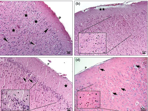
Figure 1. Photomicrograph of the control and trauma groups (a) Control group: The parenchymal features of the cor-tex morphology appeared normal, P: Piamater, Stars: neurophil, Arrowheads: neurons, Arrows: blood vessels, x10, Bar:50 µm; (b-d) Trauma 1,6,24 hours, Stars: edema in neurophil, Arrows: blood vessels with perivascular astrocytic foot process swelling x10, Bar: 50 µm, inlets: Arrowheads: pyramidal neurons showing cytoplasmic eosinophilia and pyknotic nucleus with perineural satellitosis and cleared areas formed by swollen astrocytic extensions, x40, Bar: 20 µm, Hematoxylin-eosin staining.
pared to serum levels in the control group (0.28±0.06 IU/mg) at any of the time-points (p=0.56) (Table 2).
Histopathological Assessment
All rats of the control group showed no nerve tissue damage in the brain cortex. Neurons and neuroglia cells had normal morphologic features (Fig. 1a). In the first, sixth and twenty-fourth h trauma groups, cortical neurons showed cytoplas-mic eosinophilia so called eosinophilic neuron and pyknotic nucleus with no discernible nucleolus. Perineural satellitosis accumulating of more than one glia cell around eosinophilic neurons were also observed. The perineural and perivascular
spaces were prominent due to the swelling of astrocytic foot processes. In addition, blood vessels showed congestion and stasis. The fibrillary matrix of the cerebral cortex (neurophil) showed spongiosis due to the edema of cellular extensions of the neurons and glia cells (Fig. 1b-d). In the Anakinra ap-plied groups, first, sixth and twenty-fourth h after trauma also showed same signs of damage of nerve tissue including neu-rons and neurophil of the brain cortex (Figs. 2a-f). Neuronal degeneration (eosinophilic neurons with pyknotic nucleus) and satellitosis scores of the Anakinra groups were found to be better than trauma groups at the sixth and twenty-fourth h. Moreover, vascular congestion scores were determined to
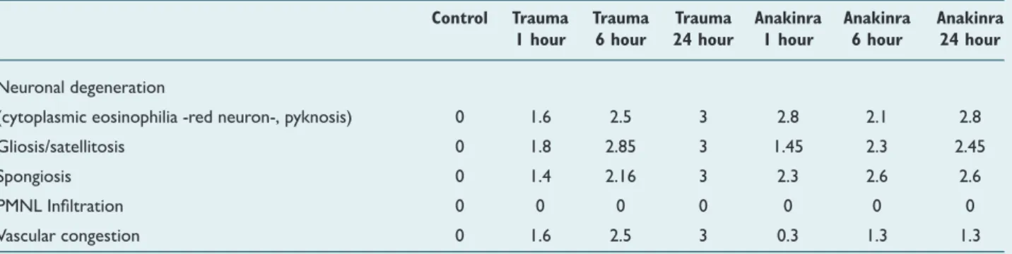
Table 3. Mean values of histopathological score distribution among groups after traumatic brain injury
Control Trauma Trauma Trauma Anakinra Anakinra Anakinra
1 hour 6 hour 24 hour 1 hour 6 hour 24 hour
Neuronal degeneration
(cytoplasmic eosinophilia -red neuron-, pyknosis) 0 1.6 2.5 3 2.8 2.1 2.8
Gliosis/satellitosis 0 1.8 2.85 3 1.45 2.3 2.45
Spongiosis 0 1.4 2.16 3 2.3 2.6 2.6
PMNL Infiltration 0 0 0 0 0 0 0
Vascular congestion 0 1.6 2.5 3 0.3 1.3 1.3
0-3 (0: absent, 1: mild, 2: moderate, 3: common).
Figure 2. Photomicrograph of brain sections from the Anakinra-treated group 1 h after trauma (a) Arrow: Gliosis around blood vessels; Star: neurophil, x10, Bar: 50 µm inlets: Arrowhead: Satellitosis; Wavy arrow: eosinophilic neuron; Arrow: microglia x40, Bar: 20 µm (b) Stars: vacuolization and edema in neurophil; Wavy arrows: neurons with eosinophilic cytoplasm and pyknotic nucleus, x40, Bar: 20 µm, Hematoxylin-eosin staining. Photomicrograph of a brain section from the Anakinra-treated group 6 h after trauma (c) Proliferation of glial cells between injured neurons, x40, Bar: 20 µm (d) Arrowheads: perineural satellitosis; Wavy arrows: numerous neurons with eosinophilic cytoplasm and pyknotic nucleus, x40, Bar: 20 µm, Hematoxylin-eosin staining. Photomicrograph of the Anakinra-treated group 24 h after trauma, (e, f) Arrowheads: perineural satellitosis; Wavy arrows: neurons with eosinophilic cytoplasm and pyknotic nucleus, x40, Bar: 20 µm, Hematoxylin-eosin staining. (a) (d) (b) (e) (c) (f)
be lower than the trauma groups at all time points and PMNL infiltration was not seen in trauma and Anakinra groups at all time points (Table 3).
DISCUSSION
Neuronal damage is thought to continue for several days af-ter the initial ischemia expansion occurring within the first 24 h of TBI.[13] The expression of proinflammatory cytokines at
the site of injury, including IL-1β and TNF-α, regulate precise cellular events that occur within the first few hours of TBI and persist in the neural tissue for several days with cytokines also being detectable on microglia, perivascular macrophages, and astrocytes.[14,15] Most important secondary factors leading to
further neuronal death are lipid peroxidation, apoptosis, and development of reactive oxygen species.[16-20] In our study,
cellular and biochemical changes were observed in order to assess the actual outcome of TBI in relation to short-term histological damage and cytokine expression. Fassbender et al. have analyzed trauma-induced release of IL-1β in brain tissue perfusates and reported that IL-1β was unexpectedly detected within 60 min of injury. Moreover, extracellularly se-creted IL-1β protein was found to gradually increase, peaking at day two, and decrease thereafter. Therefore, IL-1β release may represent a precondition for the orchestrating role of this mediator in the inflammatory response cascade.[21] In this
study, we also demonstrated that serum and tissue levels of IL-1β were elevated at the sixth h after TBI. As shown in our results, Anakinra was found to be more effective in reducing tissue levels of IL-1β in the first h after trauma. This finding is not surprising since the release of IL-1β in brain tissue occurs within less than one h of trauma. Rothwell et al. have shown that neuronal inflammation, inducing pro-cytokine IL-1β, plays a key role in this process involving glial cells as well as in-vading immune cells. Moreover, induced cytokines have been suggested to indicate the extent of central nervous system in-jury.[14,22] In the present study, morphological features relevant
with neuronal injury and higher histopathological scores fol-lowing brain trauma in rats were demonstrated. In contrast, administration of Anakinra led to an improvement of nerve tissue and cell morphology after TBI through a reduction in oxidative stress. Furthermore, this amelioration was revealed to be associated with decreased tissue and serum levels of IL-1β. On the contrary, no significant reduction in MDA was observed, indicating a lack of lipid peroxidation inhibition in brain sections and serum levels obtained from Anakinra-treated rats. This observation is in disagreement with a study conducted by Marini et al. demonstrating that inhibition of lipid peroxidation has reduced IL-1β expression and pro-tected neuronal tissue from damage.[20] Finally, the
evalua-tion with light microscopy showed that the histopathological damage scores of neuronal degeneration, satellitosis, vascular congestion and spongiosis were reduced in rats treated with Anakinra at the twenty-fourth h after TBI. The findings of the current study suggested that IL-1β played a detrimental role in the development and severity of post-traumatic injury and
that this damaging effect could be attenuated by blocking this cytokine’s signaling pathway.
Treatment with Anakinra inhibits IL-1β and protects the brain tissue and neuronal cells from damage in TBI. However, a few of the possible mechanisms by which Anakinra attenu-ates neurological injury were demonstrated in this study and a better understanding of the mechanisms of Anakinra might lead to future clinical strategies aimed at treating TBI. Conflict of interest: None declared.
REFERENCES
1. Yakovlev AG, Faden AI. Mechanisms of neural cell death: implications for development of neuroprotective treatment strategies. NeuroRx 2004;1:5-16. CrossRef
2. Bartholdi D, Schwab ME. Methylprednisolone inhibits early inflamma-tory processes but not ischemic cell death after experimental spinal cord lesion in the rat. Brain Res 1995;672:177-86. CrossRef
3. Hayashi M, Ueyama T, Nemoto K, Tamaki T, Senba E. Sequential mRNA expression for immediate early genes, cytokines, and neurotroph-ins in spinal cord injury. J Neurotrauma 2000;17:203-18. CrossRef
4. Courcoul A, Vignot E, Chapurlat R. Successful treatment of Erdheim-Chester disease by interleukin-1 receptor antagonist protein. Joint Bone Spine 2014;81:175-7. CrossRef
5. Nandi P, Kingsley GH, Scott DL. Disease-modifying antirheumatic drugs other than methotrexate in rheumatoid arthritis and seronegative arthritis. Curr Opin Rheumatol 2008;20:251-6. CrossRef
6. Marmarou A, Foda MA, van den Brink W, Campbell J, Kita H, Deme-triadou K. A new model of diffuse brain injury in rats. Part I: Pathophysi-ology and biomechanics. J Neurosurg 1994;80:291-300. CrossRef
7. Ucar T, Tanriover G, Gurer I, Onal MZ, Kazan S. Modified experimen-tal mild traumatic brain injury model. J Trauma 2006;60:558-65. CrossRef
8. Paglia DE, Valentine WN. Studies on the quantitative and qualitative characterization of erythrocyte glutathione peroxidase. J Lab Clin Med 1967;70:158-69.
9. Tator CH, Fehlings MG. Review of the secondary injury theory of acute spinal cord trauma with emphasis on vascular mechanisms. J Neurosurg 1991;75:15-26. CrossRef
10. Sun Y, Oberley LW, Li Y. A simple method for clinical assay of superoxide dismutase. Clin Chem 1988;34:497-500.
11. Aebi H, Wyss SR, Scherz B, Skvaril F. Heterogeneity of erythrocyte cata-lase II. Isolation and characterization of normal and variant erythrocyte catalase and their subunits. Eur J Biochem 1974;48:137-45. CrossRef
12. Mena H, Cadavid D, Rushing EJ. Human cerebral infarct: a proposed histopathologic classification based on 137 cases. Acta Neuropathol 2004;108:524-30. CrossRef
13. Yilmaz ER, Kertmen H, Gürer B, Kanat MA, Arikok AT, Ergüder BI, et al. The protective effect of 2-mercaptoethane sulfonate (MESNA) against traumatic brain injury in rats. Acta Neurochir (Wien) 2013;155:141-9. 14. Song C, Zhang Y, Dong Y. Acute and subacute IL-1β administrations
differentially modulate neuroimmune and neurotrophic systems: possible implications for neuroprotection and neurodegeneration. J Neuroinflam-mation 2013;10:59. CrossRef
15. Cheong CU, Chang CP, Chao CM, Cheng BC, Yang CZ, Chio CC. Etan-ercept attenuates traumatic brain injury in rats by reducing brain TNF-α contents and by stimulating newly formed neurogenesis. Mediators In-flamm 2013;2013:620837. CrossRef
OLGU SUNUMU
İnterlökin 1-beta inhibitörü Anakinra’nın sıçanlarda travmatik
beyin hasarına karşı terapötik etkinliğinin değerlendirmesi
Dr. Aşkın Esen Hastürk,1 Dr. Erdal Reşit Yılmaz,2 Dr. Erhan Türkoğlu,2 Dr. Hayri Kertmen,2
Dr. Bahriye Horasanlı,3 Uzm. Bio. Nazlı Hayırlı,4 Dr. Imge Berrin Ergüder,5 Dr. Oya Evirgen4
1Onkoloji Eğitim ve Araştırma Hastanesi, Beyin ve Sinir Cerrahisi Bölümü, Ankara;
2Dışkapı Yıldırım Beyazıt Eğitim ve Araştırma Hastanesi, Beyin ve Sinir Cerrahisi Bölümü, Ankara; 3Başkent Üniversitesi Tıp Fakültesi, Nöroloji Anabilim Dalı, Ankara;
4Ankara Üniversitesi Tıp Fakültesi, Histoloji ve Embriyoloji Anabilim Dalı, Ankara; 5Ankara Üniversitesi Tıp Fakültesi, Biyokimya Anabilim Dalı, Ankara
AMAÇ: Bu çalışmanın amacı, deneysel travmatik beyin hasarı (TBH) modelinde interlökin 1 beta (IL-1β) inhibitörü Anakinra’nın tedavi edici etkin-liğinin değerlendirilmesidir.
GEREÇ VE YÖNTEM: Elli dört Wistar albino sıçana anestezi uygulaması sonrası kafatası üzerine konan bir metal disk üzerine 2 metreden 450 g ağırlık düşürülerek deneysel kapalı kafa travması oluşturuldu. Hayvanlar üç ana gruba ayrıldı: Kontrol (n=18), TBH + salin (n=18; zaman başına altı hayvan) numuneler bir, altı ve 24 saat sonra alındı ve TBH + Anakinra (n=18; zaman başına altı hayvan) numuneler bir, altı ve 24 saat sonra alındı. IL-1β, malondialdehit, glutatyon peroksidaz, süperoksit dismutaz ve katalaz düzeylerinin analizi için beyin dokusu ve kan örnekleri alındı. Doku kesitleri histopatolojik olarak ışık mikroskobunda değerlendirildi.
BULGULAR: Travma sonrası, doku ve serum IL-1β düzeyleri önemli ölçüde artmıştı ve bu düzeyler Anakinra verilmesinden sonra azaldı. TBH taki-ben glutatyon peroksidaz, süperoksit dismutaz ve katalaz aktivitesi azalmış ve Anakinra uygulanması bu antioksidan enzimlerin aktivitesini artırmada etkili olmuştur. Histopatolojik analiz Anakinra’nın beyin dokusu ve sinir hücrelerini travmadan koruyabileğini doğrulamıştır.
TARTIŞMA: Anakinra’nın TBH ile ortaya çıkan enflamasyon ve doku hasarı gelişimini azalttığını göstermektedir.
Anahtar sözcükler: Anakinra; antioksidan; interlökin-1; nöroproteksiyon; travmatik beyin hasarı.
Ulus Travma Acil Cerrahi Derg 2015;21(1):1-8 doi: 10.5505/tjtes.2015.57894
DENEYSEL ÇALIŞMA - ÖZET
16. Ates O, Cayli S, Altinoz E, Gurses I, Yucel N, Sener M, et al. Neuro-protection by resveratrol against traumatic brain injury in rats. Mol Cell Biochem 2007;294:137-44. CrossRef
17. Awasthi D, Church DF, Torbati D, Carey ME, Pryor WA. Oxidative stress following traumatic brain injury in rats. Surg Neurol 1997;47:575-82. CrossRef
18. Nishio S, Yunoki M, Noguchi Y, Kawauchi M, Asari S, Ohmoto T. De-tection of lipid peroxidation and hydroxyl radicals in brain contusion of rats. Acta Neurochir Suppl 1997;70:84-6.
19. Pineda JA, Wang KK, Hayes RL. Biomarkers of proteolytic damage
fol-lowing traumatic brain injury. Brain Pathol 2004;14:202-9. CrossRef
20. Marini H, Altavilla D, Bellomo M, Adamo EB, Marini R, Laureanti F, et al. Modulation of IL-1 beta gene expression by lipid peroxidation inhibi-tion after kainic acid-induced rat brain injury. Exp Neurol 2004;188:178-86. CrossRef
21. Fassbender K, Schneider S, Bertsch T, Schlueter D, Fatar M, Ragoschke A, et al. Temporal profile of release of interleukin-1beta in neurotrauma. Neurosci Lett 2000;284:135-8. CrossRef
22. Rothwell N. Interleukin-1 and neuronal injury: mechanisms, modifica-tion, and therapeutic potential. Brain Behav Immun 2003;17:152-7. CrossRef