Oğuz Karaca,1,* Anıl Avcı,2 Gamze Babur Güler,1 Onur Omaygenc,1 Beytullah Cakal,1 Haci Murat Gunes,1 Elnur Alizade,2 Ali Metin Esen,2 Irfan Barutcu,1 and Bilal Boztosun1
1Department of Cardiology, Faculty of Medicine, Medipol University, Istanbul, Turkey 2Department of Cardiology, Kartal Koşuyolu Heart and Research Hospital, Istanbul, Turkey
A R T I C L E I N F O Article Type: Research Article Article History: Received: 17 Jan 2015 Revised: 22 Jun 2015 Accepted: 19 Aug 2015 Keywords: Dilated Cardiomyopathy Right Ventricle Echocardiography A B S T R A C T
Background: Right Ventricular (RV) functions are known to have an impact on determining the prognosis of patients with Non-Ischemic Dilated Cardiomyopathy (NICMP).
Objectives: This study aimed to investigate the echocardiographic determinants of RV systolic dysfunction in patients with NICMP.
Patients and Methods: This cross-sectional study was conducted on 79 patients with angiographically normal coronary arteries (mean age: 50.5 ± 12, mean Ejection Fraction (EF): 31 ± 4%) selected through purposive sampling The patients were divided into two groups based on their RV systolic function determined by tissue Doppler systolic velocities (RV-Sm): group A (RV-Sm ≥ 10 cm/s, N = 48) and group B (RV-Sm < 10 cm/s, N = 31). The two groups were compared regarding clinical, demographic, and echocardiographic variables using independent t-test and chi-square test.
Results: The patients with RV systolic dysfunction were found to have higher Brain Natriuretic Peptide (BNP) values (P = 0.006) and worse functional status (New York Heart Association (NYHA) III-IV, P = 0.04) compared to group A. Besides, univariate analysis of the echocardiographic parameters revealed that the patients in group B had significantly (all P values < 0.05) higher Left Atrial Volume Index (LAVI), higher degree of left ventricular diastolic dysfunction (represented by LV-E/Em), higher degree of Functional Mitral Regurgitation (FMR), higher estimated Pulmonary Artery Systolic Pressure (PASP), and higher degree of RV diastolic dysfunction (represented by RV-E/ Em) Moreover, multivariate logistic regression analysis showed that severe FMR (P = 0.006) and RV-E/Em (P = 0.016) predicted RV systolic dysfunction independently. Conclusions: Advanced FMR and worse RV diastolic functions emerged as the independent predictors of RV systolic dysfunction in NICMP correlating to functional status and BNP levels.
*Corresponding author: Oğuz Karaca, Medipol University Faculty of Medicine, Department of Cardiology, Tem Otoyolu Göztepe Çıkışı, No. 1, 34214 Bağcılar, Istanbul, Turkey. Cellphone: +90-5053557600, Fax: +90-2124607070, E-mail: oguzkaraca@hotmail.com
1. Background
Right Ventricular (RV) functions are known to have a prognostic impact on Non-Ischemic Dilated Cardiomyopathy (NICMP), similar to all forms of heart failure (1, 2). RV failure due to primary Left Ventricular (LV) systolic
dysfunction has been proven to be associated with worsening symptoms, hemodynamic and clinic deterioration, and increased mortality in various studies (1-3).
Several echocardiographic variables measuring LV Ejection Fraction (EF), LV diastolic dysfunction, Left Atrial (LA) volume, mitral regurgitation, pulmonary artery pressure, and RV diastolic dysfunction have been investigated in the literature to find their relationships with RV systolic dysfunction (4-6). However, no single parameter
►Implication for health policy/practice/research/medical education:
This original article has an educational value for the cardiologists interested in heart failure and echocardiography.
Predictors of Right Ventricular Systolic Dysfunction in Non-Ischemic
Dilated Cardiomyopathy: An Echocardiographic Study
has appeared as the variable of primary importance.
2. Objectives
The present study aims to determine the echocardiographic variables mostly related to RV systolic dysfunction and to assess the relationship between functional capacity and serum Brain Natriuretic Peptide (BNP) levels.
3. Patients and Methods
New York Heart Association (NYHA) classification was used to assess the functional status of the study patients. Serum BNP levels were measured prior to echocardiographic examination. The study was approved by the local Ethics Committee and written informed consents were obtained from all the patients. New York Heart Association (NYHA) classification was used to assess the functional status of the study patients. Serum BNP levels were measured prior to echocardiographic examination. The study was approved by the local Ethics Committee and written informed consents were obtained from all the patients.
3.1. Inclusion and Exclusion Criteria
This study was conducted on 79 patients with NICMP. The main inclusion criteria of the study were LVEF ≤ 45% and angiographically proven normal coronary arteries. On the other hand, the exclusion criteria were accompanying organic valve disease, coronary artery disease (history of myocardial infarction, Electrocardiography (ECG) findings of recent infarct or > 50% angiographic stenosis in any major coronary artery), secondary causes explaining heart failure (other forms of cardiomyopathies, severe pericardial disease, and metabolic disorders), malignancy, chronic infection, and poor echocardiographic window.
3.2. Serum B-Type Natriuretic Peptide Measurement
Venous blood samples were collected in EDTA-containing tubes at the time of echocardiographic examination. Elecsys 2010 analyzer (measuring range 5 - 35000 pg/mL) was used to measure serum BNP with a chemiluminescent immunoassay kit (Roche Diagnostics, Grenach- Wyhlen, Germany).
3.3. Echocardiographic Protocol
Comprehensive transthoracic echocardiographic analysis was performed for all the patients through a Vivid Seven (GE Healthcare, Milwaukee) echocardiography device using a 4S probe. Three consecutive beats were recorded in the apical and parasternal windows to analyze the data offline. Apical two- and four-chamber views were used to obtain LV End-Systolic Volume (LV-ESV), LV End-Diastolic Volume (LV-EDV), and LVEF by Simpson’s biplane method. At the coaptation level of the mitral leaflets in the apical four-chamber view, early (E) and late (A) diastolic mitral inflow velocities were recorded by pulsed-wave Doppler. Tissue Doppler analysis at the level of the septal mitral annulus was used to obtain the systolic (Sm) and early diastolic tissue velocities (Em). LV-E/Em was calculated as an indicator of LV diastolic dysfunction.
In addition, Sphericity Index (SI) was calculated as the ratio of end-diastolic LV short- to long-axis dimensions in the apical four-chamber view to reflect global LV remodeling.
Proximal Isovelocity Surface Area (PISA) method was used to determine the severity of Functional Mitral Regurgitation (FMR) based on the previous guidelines and reports (7, 8). Accordingly, an Effective Regurgitant Orifice Area (EROA) ≥ 0.2 cm2 was determined as severe
FMR. Previous guidelines and studies indicated that cut-off level for severe FMR should be accepted as 0.2 cm2
since FMR differs from ‘organic’ mitral regurgitation (7, 8). The differential diagnosis between organic and functional MR was done based on carefully evaluating the mitral valve apparatus in terms of annular calcification, leaflet mobility, and degeneration and calcification of the chordae. Accordingly, presence of a primary mitral valve disease, so called ‘organic MR’ was excluded from the study.
Area-length method was used to calculate LA volume from apical four- and two-chamber views using the following formula: 0.85 x A1 x A2 /Lmin. LA area was measured by planimetry in each apical view (A1 and A2). To obtain the Lmin in the formula, the shortest LA length that was measured from the mid-point of the mitral annular plane to the upper LA wall in both apical views was used. In addition, Left Atrial Volume Index (LAVI) was calculated by dividing LA volume by the body surface area.
To calculate RV systolic pressure using the following formula (P = 4v²), peak velocity (v) was obtained by continuous Doppler from the tricuspid regurgitant jet. By adding the estimated right atrial pressure to the RV systolic pressure, Pulmonary Artery Systolic Pressure (PASP) was obtained.
RV systolic function was assessed by measuring Tricuspid Annular Plane Systolic Excursion (TAPSE) as the length of the tricuspid annular vertical movement by using M-mode in apical 4-chamber view (9). Systolic velocity obtained from the RV free wall (RV-Sm) using pulsed wave tissue Doppler was also recorded as an indicator of RV systolic function (10).
The E/A diastolic velocity obtained by pulsed-wave Doppler from the tricuspid inflow was recorded as a measure of RV diastolic function. After measuring the early diastolic pulsed wave tissue Doppler velocity obtained from RV free wall (Em), RV-E/Em was used as an additional index of RV diastolic function (11-13).
3.4. Statistical Analysis
Mean, standard deviation, median, and range values were used to express continuous variables. On the other hand, frequency and percentage values were used to express categorical variables. Normal distribution of the data was assessed using Kolmogorov–Smirnov test. Then, the study groups were compared using unpaired test (for normally distributed variables). In addition, log transformation was used to normalize serum BNP values that followed a left skewed distribution. Fisher’s exact test and chi-square test (with continuity correction) were also used for comparison of categorical variables. Moreover, multivariate logistic regression analysis was performed after determining the statistically significant variables by means of univariate analysis. The results of the logistic regression model were expressed as P value, Odds Ratio (OR), and 95% Confidence Interval (CI). All the analyses were performed using the SPSS
statistical software (SPSS Inc, 15.0, Chicago, IL, USA) and P values < 0.05 were considered to be statistically significant.
4. Results
This study was conducted on 79 patients with NICMP (mean age: 50.5 ± 12 years, mean EF: 31 ± 4%). Based on their RV systolic function, the patients were divided into two groups; Group A: without RV systolic dysfunction (RV-Sm ≥ 10 cm/sec, N = 48) and Group B: with RV systolic dysfunction (RV-Sm < 10 cm/sec, N = 31).
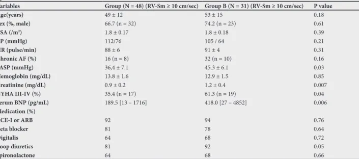
The results revealed no significant difference between the two groups regarding age, gender, body surface area, blood pressure, pulse rate, chronic atrial fibrillation, hemoglobin
level, and use of medication (P > 0.05). However, the patients in group B who had RV systolic dysfunction had significantly higher levels of serum BNP (P = 0.006), increased rate of poor functional status (NYHA III-IV) (P = 0.04), and higher levels of serum creatinine (P = 0.007). The clinical and demographic features of the study population according to RV-Sm values have been presented in Table 1.
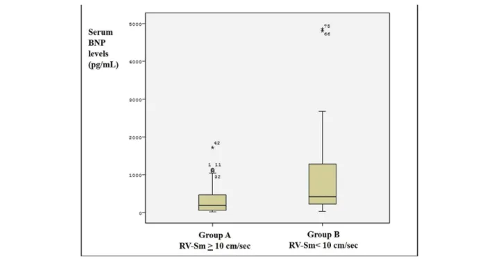
Furthermore, TAPSE and RV-Sm values were compared using Pearson’s correlation analysis whose results revealed correlation coefficient (r) = 0.623 and P < 0.001 (Figure 1). Thus, these two echocardiographic variables had a significant correlation, and RV-Sm values were used as a measure of RV systolic dysfunction in this study. According to Figure 2, the
Table 1. Baseline Demographic, Clinical, and Laboratory Parameters of the Study Population According to Right Ventricular
Systolic Function *
Variables Group (N = 48) (RV-Sm ≥ 10 cm/sec) Group B (N = 31) (RV-Sm ≥ 10 cm/sec) P value
Age(years) 49 ± 12 53 ± 15 0.18 Sex (%, male) 66.7 (n = 32) 74.2 (n = 23) 0.61 BSA (/m2) 1.8 ± 0.17 1.8 ± 0.18 0.39 BP (mmHg) 112/76 105 / 64 0.21 HR (pulse/min) 88 ± 6 91 ± 4 0.31 Chronic AF (%) 16 (n = 8) 32 (n = 10) 0.16 PASP (mmHg) 36,4 ± 7.1 45.3 ± 6.1 0.03 Hemoglobin (mg/dL) 13.8 ± 1.6 12.9 ± 1.5 0.85 Creatinine (mg/dL) 0.9 ± 0.2 1.2 ± 0.4 0.007 NYHA III-IV (%) 35.4 (n = 17) 61.3 (n = 19) 0.04 Serum BNP (pg/mL) 189.5 [13 – 1716] 418.0 [27 – 4852] 0.006 Medication (%) ACE-I or ARB 92 94 0.76 Beta blocker 81 78 0.64 Digitalis 64 68 0.72 Loop diuretics 81 92 0.05 Spironolactone 64 68 0.66
Abbreviations: BSA, body surface area; BP, blood pressure; HR, heart rate; AF, atrial fibrillation; PASP, pulmonary artery systolic pressure; BNP, B type natriuretic peptide; ACEI, angiotensin converting enzyme inhibitor; ARB, angiotensin receptor blocker
* Mann-Whitney U Test.
Figure 1. Pearson’s Correlation Graphic for the Two Parameters of Right Ventricular Systolic Dysfunction: TAPSE and RV-Sm (r = 0.623, P < 0.001)
patients in group B had higher serum BNP levels compared to group A.
The results demonstrated no significant difference between the two groups concerning LVESV, LVEDV, EF, SI, E/A ratio of the mitral inflow, left ventricular E/Em, and E/A ratio of the tricuspid inflow (P > 0.05). The group B patients who had RV systolic dysfunction were found to have statistically higher LAVI (51 ± 21 cm3/m2 vs. 43 ± 20
cm3/m2, P = 0.008), worse LV diastolic function (LV-E/Em
values: 20.2 ± 11.1 vs. 16.5 ± 7.4, P = 0.08), worse degree of FMR (severe FMR: 67% vs. 25%, P < 0.001), higher pulmonary artery pressure (40.7 ± 15.3 mmHg vs. 30.8 ± 19.2 mmHg, P = 0.01), and worse RV diastolic function (RV-E/Em values: 7 ± 2.6 vs. 5 ± 1.9, P = 0.001) compared to those in group A. The results of univariate analysis of the echocardiographic variables when the study population was divided into 2 groups according to RV systolic function
have been shown in Table 2.
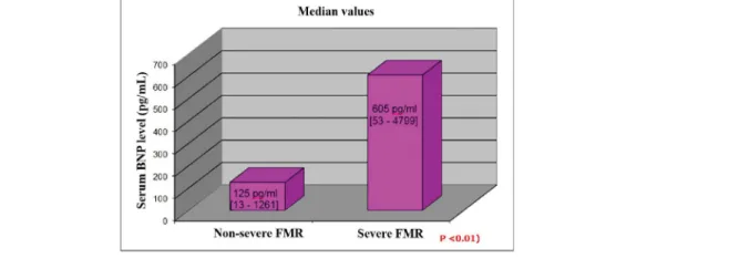
In order to determine the independent predictors of RV systolic dysfunction, the statistically significant variables predicting RV systolic dysfunction in univariate analysis (LAVI, LV-E/Em, severe FMR ratio, PASP, and RV-E/ Em) were evaluated through multivariate analysis. The results showed severe FMR (P = 0.006) and RV-E/Em (P = 0.016) as the independent predictors (Hosmer and Lemeshow test, P = 0.24; Nagelskerke R Square = 0.314) as presented in Table 3. Moreover, both severe FMR and RV-E/Em were found to have significant correlations with worsening functional status and high BNP levels (Figures 3 and 4, respectively).
5. Discussion
Prognostic factors related to NICMP are most commonly based on the echocardiographic parameters
Figure 2. Comparison of Serum BNP Levels between the Patients with (RV-Sm < 10 cm/sec, group B, N = 31) and without (RV-Sm > 10 cm/sec, group A, N = 48) Right Ventricular Systolic Dysfunction
Table 2. Analysis of The Study Population’s Echocardiographic Parameters according to Right Ventricular Systolic Function *
Variables Group (N = 48) (RV-Sm ≥ 10 cm/sec) Group B (N = 31) (RV-Sm ≥ 10 cm/sec) P value
EF (%) 32 ± 4 30 ± 5 0.29 LVESV (cm3) 126 ± 39 162 ± 55 0.45 LVEDV (cm3) 178 ± 43 211 ± 69 0.52 Sphericity 0.73 ± 0.08 0.71 ± 0.09 0.40 LV - Sm (cm/sec) 5.4 ± 1.3 5.0 ± 1.1 0.21 Mitral E/A 1.55 ± 0.8 1.7 ± 0.8 0.52 LV - E/Em 16.5 ± 7.4 20.2 ± 11.1 0.08 LAVI (cm3/m²) 43 ± 20 51 ± 21 0.008 FMR (severe, %) 25 (n = 12) 67 (n = 21) < 0.001 TAPSE (cm) 2.2 ± 0.3 1.6 ± 0.4 < 0.001 PASP (mmHg) 30.8 ± 19.2 40.7 ± 15.3 0.01 Tricuspid E/A 1.2 ± 0.3 1.1 ± 0.4 0.06 RV - E/Em 5 ± 1.9 7 ± 2.6 0.001
Abbreviations: EF, ejection fraction; LVESV, left ventricular end-systolic volume; LVEDV, left ventricular end-diastolic volume; LV-Sm, mitral annular systolic tissue Doppler velocity; E/A, pulse wave early diastolic velocity/late diastolic velocity; E/E’, pulse wave early diastolic velocity/tissue Doppler early diastolic velocity; LAVI, left atrial volume index; TAPSE, tricuspid annular plane systolic excursion; PASP, pulmonary artery systolic pressure
that reflect disease progression and hemodynamic deterioration (2). The present study aimed to determine the echocardiographic parameters that predict RV dysfunction and to assess the relationship between these parameters and functional capacity and serum BNP levels. The study results revealed severe FMR (EROA ≥ 0.2 cm2) as the
most important parameter predicting RV dysfunction. This can be explained by the increased LA pressure caused by chronic LA volume overload, leading to secondary pulmonary venous hypertension. Although right ventricle is initially normal, increased RV pressures caused by long-standing pulmonary hypertension lead to progressive RV systolic dysfunction. Echocardiographic detection of FMR in NICMP plays a critical role in preventing irreversible RV failure by optimizing medical therapy as well as by timing for possible interventions, such as surgical or percutaneous mitral repair (14-20).
Echocardiographic studies in NICMP have traditionally focused on the left ventricle and the associated mitral valve
and the left atrium as the primary affected structures. Recently, several studies have shown that RV dysfunction is strongly correlated to worsening of clinical and functional status along with a decreased survival rate (21, 22). Therefore, echocardiographic evaluation of RV anatomy and function has increasingly gained attention and several echocardiographic parameters were found to be related to RV dysfunction secondary to pulmonary hypertension (9, 23, 24).
A previous study from Dini et al. revealed that using TAPSE as an index of RV systolic dysfunction was significantly correlated to mortality and serum BNP levels in NICMP (25). A similar study by Bistola et al. also showed that combined assessment of RV systolic and diastolic dysfunction and serum BNP levels was a strong predictor of prognosis in heart failure population (26). In our study, RV systolic dysfunction was assessed with RV-Sm velocity, while tricuspid inflow diastolic pulsed-wave velocities (E/A ratio) were used for evaluating RV diastolic dysfunction.
Table 3. Multivariate Regression Analysis * Af the Echocardiographic Predictors of Right Ventricular Systolic Dysfunction
Variables Beta P value Odds ratio Confidence Interval
Severe FMR 1.47 0.006 4.38 1.5 - 12.6
RV-E/Em 0.29 0.016 1.33 1.5 – 1.69
Abbreviations: FMR, functional mitral regurgitation; RV-E/Em, pulse wave tricuspid inflow early diastolic velocity/right ventricular lateral annulus early diastolic tissue Doppler velocity
* Hosmer and Lemeshow test, P = 0.24, Nagelskerke R2 = 0.314
Figure 3. Comparison of the Functional Status of the Study Population When Grouped according to Their Functional Mitral Regurgitation Severity
Figure 4. Comparison of Serum BNP Levels of the Study Population When Grouped according to Their Functional Mitral Regurgitation Severity
The unique features of our study were investigating pure non-ischemic heart failure population and using multiple echocardiographic parameters (TAPSE, RV-Sm, RV-E/A, and RV-E/Em) to assess RV systolic and diastolic function. The study results indicated FMR and RV-E/Em as the independent predictors of RV systolic dysfunction.
It is well known that impairment of ventricular diastolic function, which is an energy-dependent active process, precedes ventricular systolic dysfunction (27). Systolic functions initially remain unaffected until impaired relaxation and increased filling pressures result in overt systolic dysfunction in coronary artery disease 28-29. Furthermore, in NICMP patients with similar EF, deterioration of diastolic functions has been found to be associated with worsening symptoms and increased mortality (27-30). According to the current study results, one of the most important echocardiographic parameters that reflected RV systolic dysfunction was RV diastolic dysfunction represented by RV-E/Em. Accordingly, analysis of RV diastolic functions with a simple echocardiographic parameter would be an important diagnostic tool for early detection of RV systolic dysfunction, enabling possible treatment options for prognosis.
Acknowledgements
There is no acknoeledgement.
Authors’ Contribution
Oguz Karaca: Study concept and design and acquisition of data. Anil Avci: Analysis and interpretation of data. Gamze Babur Guler: Analysis and interpretation of data. Onur Omaygenc: Drafting of the manuscript. Beytullah Cakal: Statistical analysis. Haci Murat Gunes: Critical revision of the manuscript for important intellectual content. Elnur Alizade: Analysis and interpretation of data. Ali Metin Esen: Statistical analysis. Irfan Barutcu: Administrative, technical, and material support. Bilal Boztosun: Study supervision.
Financial disclosure
There is no financial disclosure.
Funding/Support
There is no funding/support.
References
1. La Vecchia L, Paccanaro M, Bonanno C, Varotto L, Ometto R, Vincenzi M. Left ventricular versus biventricular dysfunction in idiopathic dilated cardiomyopathy. Am J Cardiol. 1999;83(1):120-2. A9.
2. Thomas DE, Wheeler R, Yousef ZR, Masani ND. The role of echocardiography in guiding management in dilated cardiomyopathy.
Eur J Echocardiogr. 2009;10(8):iii15-21.
3. Haddad F, Doyle R, Murphy DJ, Hunt SA. Right ventricular function in cardiovascular disease, part II: pathophysiology, clinical importance, and management of right ventricular failure.
Circulation. 2008;117(13):1717-31.
4. Barnard D, Alpert JS. Right ventricular function in health and disease. Curr Probl Cardiol. 1987;12(7):417-49.
5. Farb A, Burke AP, Virmani R. Anatomy and pathology of the right ventricle (including acquired tricuspid and pulmonic valve disease).
Cardiol Clin. 1992;10(1):1-21.
6. Stephanazzi J, Guidon-Attali C, Escarment J. [Right ventricular function: physiological and physiopathological features]. Ann Fr
Anesth Reanim. 1997;16(2):165-86.
7. Dickstein K, Cohen-Solal A, Filippatos G, McMurray JJ, Ponikowski P, Poole-Wilson PA, et al. ESC guidelines for the diagnosis and treatment of acute and chronic heart failure 2008: the Task Force for the diagnosis and treatment of acute and chronic heart failure 2008 of the European Society of Cardiology. Developed in collaboration with the Heart Failure Association of the ESC (HFA) and endorsed by the European Society of Intensive Care Medicine (ESICM). Eur
J Heart Fail. 2008;10(10):933-89.
8. Zoghbi WA, Enriquez-Sarano M, Foster E, Grayburn PA, Kraft CD, Levine RA, et al. Recommendations for evaluation of the severity of native valvular regurgitation with two-dimensional and Doppler echocardiography. J Am Soc Echocardiogr. 2003;16(7):777-802. 9. Miller D, Farah MG, Liner A, Fox K, Schluchter M, Hoit BD. The
relation between quantitative right ventricular ejection fraction and indices of tricuspid annular motion and myocardial performance. J
Am Soc Echocardiogr. 2004;17(5):443-7.
10. Meluzin J, Spinarova L, Bakala J, Toman J, Krejci J, Hude P, et al. Pulsed Doppler tissue imaging of the velocity of tricuspid annular systolic motion; a new, rapid, and non-invasive method of evaluating right ventricular systolic function. Eur Heart J. 2001;22(4):340-8. 11. Appleton CP, Jensen JL, Hatle LK, Oh JK. Doppler evaluation of
left and right ventricular diastolic function: a technical guide for obtaining optimal flow velocity recordings. J Am Soc Echocardiogr. 1997;10(3):271-92.
12. Barros MV, Machado FS, Ribeiro AL, Da Costa Rocha MO. Detection of early right ventricular dysfunction in Chagas’ disease using Doppler tissue imaging. J Am Soc Echocardiogr. 2002;15(10 Pt 2):1197-201.
13. Habib GB, Zoghbi WA. Doppler assessment of right ventricular filling dynamics in systemic hypertension: comparison with left ventricular filling. Am Heart J. 1992;124(5):1313-20.
14. Agricola E, Oppizzi M, Maisano F, De Bonis M, Schinkel AF, Torracca L, et al. Echocardiographic classification of chronic ischemic mitral regurgitation caused by restricted motion according to tethering pattern. Eur J Echocardiogr. 2004;5(5):326-34. 15. Bhamra-Ariza P, Muller DW. The MitraClip experience and
future percutaneous mitral valve therapies. Heart Lung Circ. 2014;23(11):1009-19.
16. Glower DD, Kar S, Trento A, Lim DS, Bajwa T, Quesada R, et al. Percutaneous mitral valve repair for mitral regurgitation in high-risk patients: results of the EVEREST II study. J Am Coll Cardiol. 2014;64(2):172-81.
17. Inoue T, Fujii K, Yugami S, Kitayama H, Saga T. Symmetrical papillary muscle approximation for functional mitral regurgitation with idiopathic dilated cardiomyopathy. J Heart Valve Dis. 2013;22(4):556-9.
18. Otsuji Y, Handschumacher MD, Schwammenthal E, Jiang L, Song JK, Guerrero JL, et al. Insights from three-dimensional echocardiography into the mechanism of functional mitral regurgitation: direct in vivo demonstration of altered leaflet tethering geometry. Circulation. 1997;96(6):1999-2008.
19. Perloff JK, Roberts WC. The mitral apparatus. Functional anatomy of mitral regurgitation. Circulation. 1972;46(2):227-39.
20. Punnoose L, Burkhoff D, Cunningham L, Horn EM. Functional mitral regurgitation: therapeutic strategies for a ventricular disease.
J Card Fail. 2014;20(4):252-67.
21. Jiang L, Levine RA, Weyman AE. Echocardiographic Assessment of Right Ventricular Volume and Function. Echocardiography. 1997;14(2):189-206.
22. Panidis IP, Ren JF, Kotler MN, Mintz G, Iskandrian A, Ross J, et al. Two-dimensional echocardiographic estimation of right ventricular ejection fraction in patients with coronary artery disease. J Am Coll
Cardiol. 1983;2(5):911-8.
23. Maslow AD, Regan MM, Panzica P, Heindel S, Mashikian J, Comunale ME. Precardiopulmonary bypass right ventricular function is associated with poor outcome after coronary artery bypass grafting in patients with severe left ventricular systolic dysfunction. Anesth Analg. 2002;95(6):1507-18. table of contents. 24. Zornoff LA, Skali H, Pfeffer MA, St John Sutton M, Rouleau JL,
Lamas GA, et al. Right ventricular dysfunction and risk of heart failure and mortality after myocardial infarction. J Am Coll Cardiol. 2002;39(9):1450-5.
25. Dini FL, Fontanive P, Panicucci E, Andreini D, Chella P, De Tommasi SM. Prognostic significance of tricuspid annular motion and plasma
NT-proBNP in patients with heart failure and moderate-to-severe functional mitral regurgitation. Eur J Heart Fail. 2008;10(6):573-80. 26. Bistola V, Parissis JT, Paraskevaidis I, Panou F, Nikolaou M,
Ikonomidis I, et al. Prognostic value of tissue Doppler right ventricular systolic and diastolic function indexes combined with plasma B-type natriuretic Peptide in patients with advanced heart failure secondary to ischemic or idiopathic dilated cardiomyopathy.
Am J Cardiol. 2010;105(2):249-54.
27. Section VI. Heart Failure and Transplantation In: Topol E, editor.
Textbook of Cardiovascular Medicine. Lippincott Williams and
Wilkins; 1998.
28. Braunwald E. The Denolin lecture. Congestive heart failure: a half century perspective. Eur Heart J. 2001;22(10):825-36.
29. Braunwald E. Congestive heart failure: a half century perspective.
European heart journal. 2001;22(10):825-36.
30. Topol EJ. Manual of Cardiovascular Medicine. second edition Heart Failure and Transplantation ed. Lippincot Williams and Wilkins; 2004. pp. 101–75.


