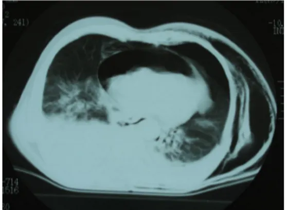
Dicle Tıp Dergisi, 2008 OLGU SUNUMU Cilt: 35, Sayı: 4 , (277-279)
Yazışma Adresi: Dr.Murat Orak, Dicle Üniversitesi Tıp Fak. 21280 D.BAKIR. Tel: 0412 248 80 01
Geliş Tarihi : 26.06.2007 Yayına Kabul Tarihi : 11.12.2007
74 Year-Old-Man With Pneumopericardium Due to Blunt Chest Trauma:
Case Report
Murat Orak1 , Mehmet Üstündağ2, Serdar Onat3 , Halil Doğan4 , Alper Avcı5
1,2,4Department of Emergency, Dicle University, Faculty of Medicine, Diyarbakır, Turkey 3,5Department of Thoracic Surgery, Dicle University, Faculty of Medicine, Diyarbakır, Turkey
SUMMARY
Pneumopericardium is the presence of air in the pericardial space. It may be seen in the context with severe blunt chest trauma. Usually, pneumopericardium is self-limiting and requiring no specific therapy. However, a continuous monitoring of the electrocardiography and the blood pressure is necessary at an intermediate care unit. We report a 74 year-old-man had been presented with Pneumopericardium, after a car striking. He had bilateral pneumohematothoraces, pneumomediastinum and bilateral multiple rib fractures. Pneumopericardium was diasappeared without surgical management like written in english literature. Frequent cardiac and vital signs monitoring and general support treatment were successful in our treatment.
Key Words: pneumopericardium, blunt chest trauma, intensive care unit
74 yaşındaki hastada künt göğüs travmasına bağlı pnömoperikardium: Olgu Sunumu ÖZET
Pnömoperikardium, pericardial boşlukta hava bulunmasıdır. Ciddi, künt toraks travmalarında oluşabilir. Pnömoperikardium, genellikle kendi kendini sınırlar ve özel bir tedaviye ihtiyaç duyulmaz. Bununla beraber, elektrokardiyografi ve kan basıncının sürekli olarak monitörize edildiği yoğun bakım ünitesinde takibi gerekmektedir. 74 yaş erkek hastada; araç dışı trafik kazası sonrasında oluşan, bilateral hemopnömotoraks, bilateral seri kot fraktürleri, pnömomediastinum’un eşlik ettiği pnömoperikardium vakasını sunmaktayız. Literatür bilgilerine paralel olarak, cerrahi bir girişime gerek kalmadan hastanın pnömoperikardiumu kaybolmuştur. Tedavi protokolü olarak; yakın vital bulgu ve kardiak monitörizasyon takipi altında genel destek tedavisi uygulanmış ve yeterli olmuştur.
Anahtar Kelimeler : Pnömoperikardium, künt göğüs travması, yoğun bakım.
INTRODUCTION
Pneumopericardium in adults is a rare disorder and may be seen most commonly after blunt chest trauma in combination with
pneumothorax or pneumomediastinum.
Motor vehicle accidents (72.2%) and falls (17.1%) are reported to be the most frequent causes of these blunt chest injuries in
polytraumatized patients1. Concomitant
injuries and complications of
pneumopericardium such as tension
pneumopericardium are the major causes of morbidity and mortality. The aim of us with this report is to emphasize importance of rapid diagnosing, immediate cardiac and vital monitoring, and being aware for complications which might be needed surgical management. We reported a 74 year-old-man had been presented with Pneumopericardium, after a car striking. He
had bilateral pneumohematothoraces,
pneumomedias-tinum and bilateral multiple rib fractures. He was diagnosed 5 hours after
M.Orak, M.Üstündağ, S.Onal, H. Doğan, A. Avcı Dicle Tıp Dergisi, 2008
278
accident and bilateral tube thoracostomies were performed in emergency department. Then he was hospitalized in the Thoracic Surgery Department intensive care unit.
CASE
A 74-year-old man was admitted to our hospital with complaining of increasing thoracic pain, aggravating dyspnea, reduced breathing sounds over the left hemithorax and subcutaneous emphysema at the 5th hour of the high-speed car striking while he had been walking. There were blood pressure of 110/80 mm Hg, and spontaneous breathing of 20/min. In electrocardiography (ECG); T wave negativeness in D2, D3, aVF, and low voltage were determined in all derivations. The antero-posterior chest radiography showed bilateral multiple rib fractures(2-3-4-5-6 of the left side and 1-2-3 of the right side),left pneumothorax, bilateral pleural effusion, pneumomediastinum and
pneumopericardium (Figure 1). The
subsequent axial computed tomography (CT) scan demonstrated bilateral multiple rib fractures, right pulmonary contusion, left pneumothorax, bilateral pleural effusion,
pneumomediastinum, nontension
pneumopericardium with a maximum diameter of 28mm (Figure 2). Injury Severity Score was 24 points. Bilateral chest tubes were inserted. The patient was
transferred to the Thoracic surgery
department intensive care unit for clinical
observation and breathing training.
Continuous monitoring of the blood pressure, central venous pressure, fever, and periodic chest radiography were performed. Thoracic tube drainage was 100cc from the left, and 50cc from the right at the first day.
No pneumothorax line, and no
pneumopericardium were seen in the chest anteroposterior radiography at the 3rd day,
and no drainage from right tube therefore right chest tube was ended at the 3rd day. The
left chest tube also ended at the 5th day. ECG showed normal myocardial function at the 6th day. The bronchoscopic aspiration was performed to patient due to obstructive atelectasis related to secretion at the 8th day.
After 2 weeks of follow up, he was stable. He was discharged at the 14th day. He was
healthy at the 3rd month of follow up control.
DISCUSSION
Pneumopericardium secondary to blunt chest trauma is generally due to 1 of 3 mechanisms: 1) penetration along pulmonary venous perivascular sheaths from ruptured alveoli to the pericardium, 2) pneumothorax with pleuropericardial tear, or 3) direct
tracheobronchial-pericardial communication2
The diagnosis of a
pneumopericardium is made by a plain radiography of the chest. The CT examination usually allows a more accurate assessment of the pneumopericardium and concomitant injuries of the chest. This evaluation is 4 to 5 times more sensitive than supine chest radiography at detecting intrathoracic injuries3,4. Also in our patient, plain chest graphies and CT examination showed concomitant thoracic wall and
intrathoracic injuries. Usually, the
pneumopericardium is self-limiting and requiring no specific therapy. A nontension pneumopericardium may initially appear as asymptomatic but can rapidly and suddenly progress into a life-threatening tension pneumopericardium. Therefore we followed up our patient with frequent ECG, continuous blood pressure, and arteriel blood
oxygen saturation monitoring. Initially, every pneumopericardium is a
nontension pneumopericardium. Gorecki et
al3 concluded that nontension
pneumopericardium in a stable patient, especially associated with lung and pleural injury, may rapidly progress to tension pneumopericardium. Because of intubation and an increase in airway pressure are probable precipitating factors. Especially,
patients with concomitant pulmonary
contusion and the need of intubation are at
high risk to develop a tension
pneumopericardium3,5. Our case was under
high risk for morbidity and mortality such as
M.Orak, M.Üstündağ, S.Onal, H. Doğan, A. Avcı Dicle Tıp Dergisi, 2008
279
intensive care unit follow-up period was longer than other blunt chest trauma patients. In cases of cardiac tamponade caused by tension pneumopericardium, a needle aspiration is required immediately, and a pericardial window with pericardial drainage is to be performed in the operation room1,5.
Intensive care unit patient follow-up, provides immediate surgical operation for life saving when needed.
CONCLUSION
Pneumopericardium is a rare
disorder after a blunt thoracic trauma. In most cases, the pneumopericardium is self-limiting and requiring no additional therapy. Concomitant injuries of the lung, heart, trachea, and bronchus have to be primarily excluded by CT scan, bronchoscopy, and ECG. Concomitant pneumothorax and hematothorax have to be treated by chest tube inserting. Because of the risk of cardiac tamponade, heart failure, or arrhythmias, all trauma patients with pneumopericardium
should be admitted to the intensive care unit with continuous ECG, blood pressure monitoring, and periodic chest radiography.
REFERENCES
1. Ladurner R, Qvick LM, Hohenbleicher F., et al. Pneumopericardium in blunt chest trauma after high-speed motor vehicle accidents. Am J Emerg Med 2005; 23: 83-86.
2. Spodick DH. Pericardial diseases. In: Braunwald E, Zipes DP, Libby P. (Editors). Heart disease: a textbook of cardiovascular medicine. 6th ed. Vol 2. Philadelphia: WB Saunders; 2001. p. 1823-1826.
3. Gorecki PJ, Andrei VE, Scehein M. Tension pneumopericardium in chest trauma. J Trauma 1999; 46: 954-956.
4. Ordog GJ, Balasubramamium S, Wasserberger J. Outpatient management of 357 gunshot wounds to the chest. J Trauma 1983; 23: 832-835.
5. Capizzi PJ, Martin M, Bannon MP. Tension pneumopericardium following blunt chest injury. J Trauma 1995; 39: 775-780.