Case
Reports
Acute Aortic Valve
Regurgitation Secondary
to Blunt Chest Trauma
Blunt injury to the cardiac valves leads to progressive acute ventricular failure, which often requires urgent surgical management. In this case report, we describe an acute aortic valve rupture caused by air-bag inflation during an automobile accident. Laceration of an aortic valve cusp was treated successfully with urgent aortic valve replacement. A concomitant orthopedic injury was treated electively 15 days after cardiac surgery.
Acute aortic valve rupture is a very rare complication of blunt chest trauma. We dis-cuss how to diagnose and manage this potentially catastrophic event. (Tex Heart Inst J 2001;28:312-4)
ardiac trauma is a common clinical problem in patients with multiple injuries. Almost 30% of all trauma patients have some form of cardiotho-racic injury.1Several intracardiac structures can be affected by nonpene-trating chest trauma, but serious aortic valve insufficiency due to rupture of aortic valve cusps is a rare complication. To our knowledge, the earliest published report of traumatic rupture of the aortic valve is that of Penderleath,2which appeared in 1830.
We present a case of laceration of the right coronary cusp of the aortic valve caused by the opening of an air bag during an automobile accident. The aortic valve was replaced successfully with a bioprosthesis. Survival after chest trauma depends upon early identification of the lesion and surgical repair. We discuss the principles of diagnosis and management of these unusual cases.
Case Report
In January of 2000, a 58-year-old man was admitted to the emergency room, after involvement in an automobile accident. He was awake and his vital signs were at first stable. He was suffering pain on the both sides of the sternum, caused by fracture, and pain at the right hip joint, caused by fracture and dislocation.
Physical examination revealed a widened pulse pressure (140/40 mmHg, for ex-ample) and a regular pulse rate of 120 beats/min. The abdomen was normal upon examination. Auscultation revealed an early diastolic murmur, with a grade 3/4 intensity. Chest radiography revealed a higher than normal cardiothoracic index and a widened mediastinum. Thoracoabdominal computed tomography (CT) showed cardiomegaly with a normal ascending aorta and multiple sternal frac-tures. The cranial CT was normal.
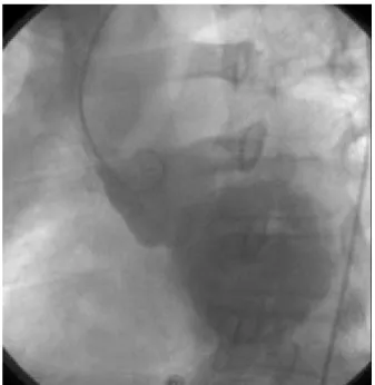
The patient was admitted to our intensive care unit for close monitoring. Transsternal echocardiography showed 4+ aortic regurgitation. Upon introduc-tion of a Swan-Ganz catheter, the pulmonary artery pressure measured 60 mmHg in systole. Cardiac catheterization showed normal coronary arteries and severe aortic regurgitation (Fig. 1). The patient was taken to the operating room, where transesophageal echocardiography (TEE), performed on the table, also confirmed 4+ aortic regurgitation.
We approached via a conventional median sternotomy, although the fracture made retraction difficult. There were no bone fragments. We found the heart to be distended. After initiation of cardiopulmonary bypass, we induced and main-tained cardiac arrest with a tepid solution of blood and potassium. Upon aortoto-my, we found avulsion of the right coronary cusp, which caused it to be flail (Fig. 2). Having decided that the aortic valve was irreparable, we resected it and
Mehmet Unal, MD Ergun Demirsoy, MD Abdullah Gogus, MD Harun Arbatli, MD Azmi Hamzaoglu, MD Bingur Sonmez, MD
Key words: Accidents,
traffic; air bags; aortic valve/surgery; heart valves/injuries; wounds, nonpenetrating From:The departments of Cardiovascular Surgery (Drs. Arbatli, Demirsoy, Sonmez, and Unal) and Orthopedics (Drs. Gogus and Hamzaoglu), Kadir Has University, Florence Nightingale Hospital, Istanbul 80220, Turkey
Address for reprints:
Mehmet Unal, MD, Istanbul Memorial Hospital, Piyale Pasa Bulvari 80270, Okmeydani, Istanbul, Turkey © 2001 by the Texas Heart®
Institute, Houston
Volume 28, Number 4, 2001 312 Aortic Valve Regurgitation Secondary to Blunt Trauma
implanted a 21-mm Medtronic-Hancock II® bio-prosthesis (Medtronic, Inc.; Minneapolis, Minn), which we chose because the patient would subse-quently undergo orthopedic surgery. The patient was weaned from bypass easily, and the new bioprosthesis was seen to be competent on TEE. The fractured ster-num was fixed with figure-eight wires.
The patient was extubated on the 1st postoperative day and discharged from the intensive care unit on the 5th day. His orthopedic procedure was done on the 15th day, and he was discharged from the hos-pital without any complication. Chest radiography performed at 3-month follow-up demonstrated that the cardiothoracic index had diminished. The patient underwent multiple orthopedic surgical procedures without complication. Fourteen months after his aortic valve replacement, he remained free of symp-toms.
Discussion
Aortic valve rupture resulting in acute aortic insuffi-ciency is an uncommon complication of blunt chest trauma. The evolution of sophisticated automotive safety systems is playing an important role in saving lives during crashes. Although 1 such device, the air bag, affords valuable protection against crushing in-juries of the head and body, it can cause other, less apparent, blunt trauma, including serious cardiotho-racic injuries.
The most common injury after blunt chest trauma is myocardial contusion, which itself is probably un-der diagnosed.3 Septal and free-wall ruptures of all 4 chambers have been well described in postmortem series; these studies suggest that the right ventricle is the most commonly ruptured chamber and that coro-nary artery disruption is rare.4Further, postmortem series and the clinical literature suggest that acute val-vular dysfunction after chest trauma is very rare.5,6
The greatest strain in the thoracic aorta is at the isthmus where the mobile thoracic aorta joins the more firmly fixed aortic arch; this is where most aor-tic ruptures are found. Indeed, rupture of the thoracic aorta accounts for 15% to 20% of deaths among vic-tims of automotive accidents.7 In contrast, cardiac valve injuries are seen very rarely. The mechanism of rupture of the aortic valve is thought to be a sudden increase in intrathoracic pressure during diastole, when the pressure gradient across the aortic valve is maximal and the valve is closed.8
Because of rapid deterioration of signs associated with heart failure due to acute aortic regurgitation, diagnosis of acute aortic valve rupture must occur early. Chest pain and dyspnea in association with a new diastolic murmur are the most common symp-toms and should create a high index of suspicion. Texas Heart Institute Journal Aortic Valve Regurgitation Secondary to Blunt Trauma 313
Fig. 1 Cardiac catheterization shows severe aortic regurgitation.
Fig. 2 Avulsion of the right coronary cusp was considered
irreparable.
In the setting of severe blunt chest trauma, echocar-diography is the noninvasive test of choice. For our patient, we chose transesophageal echocardiography following transthoracic echocardiography (TTE). If the patient is stable, TEE achieves better visualization of the valvular anatomy and myocardial function.9 Cardiac catheterization remains the gold standard for providing information concerning aortic insufficiency and abnormalities associated with the aortic root and coronary vessels, as well as information on ventricular function and pulmonary blood pressure. Our patient was stable enough, hemodynamically, to undergo car-diac catheterization.
Medical management is very important for multiple-injury patients in intensive care units. For example, the severity of aortic regurgitation is increased by the elevations in peripheral vascular resistance that are as-sociated with hemorrhage in patients who have sus-tained multiple injuries. In such a patient, peripheral vasodilators such as nitroglycerin remain the primary agents, provided that the patient’s blood pressure per-mits their use; another benefit is that these agents may decrease the pulmonary pressure as well.
The timing of surgical intervention remains an im-portant and controversial issue in patients with val-vular injuries. Deterioration of ventricular function, acute pulmonary edema, and a systemic diastolic blood pressure of less than 50 mmHg are indications for im-mediate surgery.10
Patients with chronic aortic insufficiency might tol-erate a new rupture better. However, even in these patients, delayed intervention can increase operative risks, because such patients might not develop symp-toms until myocardial dysfunction has become irre-versible.11
Nearly all authors recommend that surgical inter-vention be done as soon as possible. Although Hal-stead and coworkers12presented 3 valvular injury cases treated only with repair of the native aortic valve, we believe that valve replacement is usually necessary, because aortic valvuloplasty is associated in the long term with a high rate of failure, which requires reop-eration.12-14
References
1. Symbas PJ, Horsley WS, Symbas PN. Rupture of the as-cending aorta caused by blunt trauma. Ann Thorac Surg 1998;66:113-7.
2. Penderleath D. Case of death from rupture of one of the semilunar valves of the aorta. London Medical Gazette 1830; 7:109.
3. Westaby S, Brayley N. ABC of major trauma. Thoracic trauma—I. BMJ 1990;300:1639-43.
4. Parmley LF, Manion WC, Mattingly TW. Nonpenetrating traumatic injury of the heart. Circulation 1958;18:371-96. 5. Warburg E. Myocardial and pericardial lesions due to
non-penetrating injury. Br Heart J 1940;2:271-80.
6. Parmley LF, Mattingly TW, Manion WC, Jahnke EJ Jr. Nonpenetrating traumatic injury of the aorta. Circulation 1958;17:1086-101.
7. Sturm JT, McGee MB, Luxenberg MG. An analysis of risk factors for death at the scene following traumatic aortic rup-ture. J Trauma 1988;28:1578-80.
8. Munshi IA, Barie PS, Hawes AS, Lang SJ, Fischer E. Diag-nosis and management of acute aortic valvular disruption secondary to rapid-deceleration trauma. J Trauma 1996;41: 1047-50.
9. Weiss RL, Brier JA, O’Conner W, Ross S, Brathwaite CM. The usefulness of transesophageal echocardiography in diag-nosing cardiac contusions. Chest 1996;109:73-7.
10. Crosby IK, Muller WH Jr. Acquired disease of the aortic valve. In: Sabiston DC Jr, Spencer FC, editors. Gibbon’s Surgery of the chest. 4th ed. Vol 2. Philadelphia: WB Saun-ders; 1983. p. 1280-305.
11. Rosengart TK, Lang SJ. Valvular heart disease. In: Barie PS, Shires GT, editors. Surgical intensive care. Boston: Little, Brown and Co.; 1993. p. 577-612.
12. Halstead J, Hosseinpour AR, Wells FC. Conservative surgi-cal treatment of valvular injury after blunt chest trauma. Ann Thorac Surg 2000;69:766-8.
13. Pretre R, Faidutti B. Surgical management of aortic valve in-jury after nonpenetrating trauma. Ann Thorac Surg 1993; 56:1426-31.
14. Aris A, Delgado LJ, Montiel J, Subirana MT. Multiple in-tracardiac lesions after blunt chest trauma. Ann Thorac Surg 2000;70:1692-4.
Volume 28, Number 4, 2001 314 Aortic Valve Regurgitation Secondary to Blunt Trauma