METAL ION RELEASE AND SIGNALING IN
MOLECULAR LOGIC GATE DESIGN
A DISSERTATION SUBMITTED TO
THE GRADUATE SCHOOL OF ENGINEERING AND SCIENCE OF BILKENT UNIVERSITY
IN PARTIAL FULFILLMENT OF THE REQUIREMENTS FOR THE DEGREE OF
DOCTOR OF PHILOSOPHY IN
MATERIALS SCIENCE AND NANOTECHNOLOGY
By
TAHA BİLAL UYAR
October, 2016
ii
METAL ION RELEASE AND SIGNALING IN MOLECULAR LOGIC GATE DESIGN
By Taha Bilal Uyar October, 2016
We certify that we have read this dissertation and that in our opinion it is fully adequate, in scope and in quality, as a thesis of the degree of Doctor of Philosophy.
Engin Umut Akkaya (Principal Advisor)
Dönüş Tuncel
Salih Özçubukçu
Bilge Baytekin
Emrullah Görkem Günbaş
Approved for the Graduate School of Engineering and Science:
Ezhan Karaşan Director of the Graduate School
iii
ABSTRACT
METAL ION RELEASE AND SIGNALING IN MOLECULAR LOGIC
GATE DESIGN
Taha Bilal Uyar
Ph.D. in Materials Science and Nanotechnology Supervisor: Engin Umut Akkaya
October, 2016
Mimicking the biological structures is one of the main goals of the natural sciences, because processes are well-organized in nature despite its amazing complexity. On account of this, releasing metal ions at molecular level is a crucial topic owing to similar process in living organisms. In this thesis, we designed novel cage compounds for this purpose. Our novel cage molecule is activated with near-IR light while almost all of the cage compounds in the previous literature function in the UV region. Consequently, it is appropriate for using in biological systems. Release of zinc ions that has critical roles in human body successfully achieved by our novel compound in Part 2 of the thesis work.
Molecular logic concept is one of the promising areas of chemistry. Today, electronic devices consists of silicon based circuits that process information by using binary logic. Molecular logic offers alternative for silicon based devices. Using molecules to process data is a promising idea in the fields from electronics to biotechnology. In part 3, we proposed AND gate whose inputs are pH and glutathione while the response is singlet oxygen which can be used to destroy cancer cells. It is possible to process much complicated information by combining more than one logic gate. In first part of the thesis study, we designed modular molecular logic gates by cascading of three logic gates via metal ion signals.
In the last part, a fluorescent chemosensor was proposed for dopamine molecule, which is crucial in a number of biological processes at the human body.
Keywords: molecular logic gate, cage compounds, fluorescence, glutathione, photosensitizer, metal ion release, BODIPY, dopamine.
iv
ÖZET
METAL İYONU SALIMI ve MOLEKÜLER MANTIK
DEVRELERİNDE SİNYALİZASYON
Taha Bilal Uyar
Malzeme Bilimi ve Nanoteknoloji, Doktora Tez Danışmanı: Engin Umut Akkaya
Ekim, 2016
Biyolojik yapıların taklit edilmesi doğa bilimlerinin temel hedeflerinden bir tanesidir çünkü doğanın inanılmaz karmaşık yapısına rağmen çok düzenli bir işleyişi vardır. Bundan dolayı, canlı sistemlerde de gerçekleşen metal iyonu salımının moleküler seviyede gerçekleştirilmesi çok önemli bir konudur. Bu tezde, biz de bu amaca yönelik yeni kafes bileşikler tasarladık. Literatürdeki hemen hemen tüm kafes bileşikleri UV bölgesinde çalışıyorken, bizim yeni tasarladığımız kafes molekülü yakın IR ışığıyla aktif hale geliyor. Bu sebeple de biyolojik sistemlerde kullanımı uygundur. 2. Bölümde, insan vücudunda kritik rollerde bulunan çinko iyonunun salımı bizim yeni molekülümüzle başarılı bir şekilde gerçekleştirildi.
Moleküler mantık devreleri kimyanın gelecek vadeden alanlarından bir tanesidir. Günümüzde, elektronik cihazlar ikili sisteme göre işlem yapan silikon bazlı devrelerden oluşurlar. Moleküler mantık kapıları, fiziksel ve teknolojik bariyerlerden dolayı gelişiminin neredeyse sonuna gelmiş olan silikon bazlı cihazlara bir alternatif sunmaktadır. Elektronik alanından biyoteknolojiye kadar, bilgiyi işlemek için moleküllerin kullanılması ilham verici bir fikirdir. 3. Bölümde, pH ve glutatyon girdileriyle kanser hücrelerini yok etmek için kullanılan singlet oksijen üreten bir VE mantık kapısı tasarladık. Birden fazla mantık kapısını birleştirerek çok daha karmaşık işlemler yapmak da mümkündür. Tezin ilk kısmında, üç tane mantık kapısını metal iyonu vasıtasıyla birleştirerek modüler bir moleküler mantık kapsısı tasarladık. Son kısımda ise insan vücudundaki birçok biyolojik olayda çok kritik öneme sahip olan dopamin molekülü için bir floresans kemosensör tasarlandı.
Anahtar kelimeler: moleküler mantık kapısı, kafes bileşikler, floresans, glutatyon, ışık duyarlandırıcı, metal iyonu salımı, BODIPY, dopamin.
v
ACKNOWLEDGEMENT
I would like to express my hearty gratitude to my supervisor Engin Umut Akkaya for his deep insight, intense knowledge and support throughout my graduate studies. I am also grateful to him for teaching us how to become a good scientist. I will never forget his support throughout my life.
I am sincerely grateful to Ruslan Guliyev, Ahmet Atılgan, Sündüs Erbaş-Çakmak, Tuğba Özdemir-Kütük Yusuf Çakmak, Safacan Kölemen, Bilal Kılıç and Nisa Yeşilgül for their patience, great friendship and all their valuable contributions to the projects that we worked on together.
I would like to thank former and present group members of the Akkaya group Özlem Seven, Abdurrahman Türksoy, Seylan Ayan, Deniz Yıldız, Dilek Işık-Taşgın, Cansu Kaya, Serdal Kaya, Ceren Çamur, Darika Okeeva, Esra Tanrıverdi, Fazlı Sözmen, İlke Şimşek-Turan, José Luis Bila, Hale Atılgan, Tuğçe Karataş, Murat Işık, Ziya Köstereli, Onur Büyükçakır and rest of our group for their valuable support and friendship.
I would like to gratefully acknowledge my Thesis Committee Members, Assoc. Prof. Dr. Dönüş Tuncel and Assist. Prof. Dr. Salih Özçubukçu for their encouraging and fruitful discussions and advices for four years. Also, I would like to thank Assist. Prof. Dr. Bilge Baytekin and Assist. Prof. Görkem Günbaş for participating the dissertation committee.
I would like to thank to TÜBİTAK (The Scientific and Technological Research Council of Turkey) for financial support.
I also would like to thank all members of UNAM for facilities and multidisciplinary research atmospher.
Most importantly, I would like to thank my family for their love, support, and understanding.
vi
LIST OF ABBREVIATIONS
BODIPY : Boradiazaindacene
AcOH : Acetic Acid
CHCl3 : Chloroform
DDQ : Dichlorodicyanoquinone
DMF : Dimethylformamide
TFA : Trifluoroacetic Acid
THF : Tetrahydrofuran
Et3N : Triethylamine
TLC : Thin Layer Chromotography
ICT : Internal Charge Transfer
PET : Photoinduced Electron Transfer
ET : Energy Transfer
FRET : Förster Resonance Energy Transfer
HOMO : Highest Occupied Molecular Orbital
LUMO : Lowest Unoccupied Molecular Orbital
HRMS : High Resolution Mass Spectroscopy
vii
TABLE OF CONTENTS
CHAPTER 1: Introduction ... 1
1.1. What is Supramolecular Chemistry? ... 1
1.1.1. Basic Supramolecular Interactions ... 3
Electrostatic Interactions ... 3
Hydrogen Bonding ... 4
Van der Waals Forces ... 5
Π Interactions ... 5
Hydrophobic Effect ... 6
1.2. Luminescence ... 6
1.3. Fluorescent Dyes... 9
1.4. Molecular Sensors ... 10
1.5. Photophysical Aspect of Fluorescent Chemosensors ... 11
1.5.1. Photoinduced Electron Transfer (PET) ... 12
1.5.2. Internal Charge Transfer (ICT) ... 15
1.5.3. Energy Transfer (ET) ... 17
Dexter Type Energy Transfer... 18
Förster Type Energy Transfer ... 20
1.6. Sensing of Particular Ions or Compounds ... 23
1.6.1. Metal Ions ... 23
Zinc (II) Ion Sensors ... 24
1.6.2. Biological Thiols ... 25
Detection of Thiols ... 26
Disulfide Bond Cleavage ... 28
1.7. BODIPY Dyes ... 29
viii
1.8. Photodynamic Therapy (PDT) ... 36
1.8.1. Photophysical Background of PDT ... 36
1.8.2. Photosensitizers and Importance of Light ... 37
1.8.3. Reaction of Singlet Oxygen with Olefins ... 39
1.9. Caged Compounds ... 40
1.10. Molecular Logic Gates ... 44
1.10.1. Boolean Algebra... 44
1.10.2. Molecular Logic Gates ... 46
1.10.3. Higher Functions with Molecular Logic ... 48
CHAPTER 2: Modular Logic Gates: Cascading Independent Logic Gates via Metal Ion Signals ... 50
2.1. Objective ... 51
2.2. Introduction ... 51
2.3. Results and Discussion ... 52
2.4. Conclusion ... 58
2.5. Experimental Details ... 59
2.5.1. Additional Information... 69
CHAPTER 3: Near IR Triggered, Remote Controlled Release of Metal Ions: A Novel Strategy for Caged Ions ... 72
3.1. Objective ... 73
3.2. Introduction ... 73
3.3. Results and Discussion ... 74
3.4. Conclusion ... 82
ix
3.5.1. Additional Information... 95
CHAPTER 4: Selective Photosensitization through AND Logic Response: Optimization of pH and Glutathione Response of Activatable ... 101
4.1. Objective ... 102
4.2. Introduction ... 102
4.3. Results and Discussion ... 104
4.4. Conclusion ... 114
4.5. Experimental Details ... 115
4.5.1. Additional Information... 141
CHAPTER 5: BODIPY Assisted Dopamine Recognition ... 143
5.1. Objective ... 144
5.2. Introduction ... 144
5.3. Results and Discussion ... 145
5.4. Conclusion ... 148
5.5. Experimental Details ... 149
EPILOGUE ... 153
BIBLIOGRAPHY ... 155
x
LIST OF FIGURES
Figure 1. Conformity of host and guest in supramolecular chemistry. ... 1
Figure 2. Lock and key model for enzyme-substrate conformity. ... 2
Figure 3. Examples for different types of electrostatic interactions. ... 4
Figure 4. Hydrogen bonding in DNA. ... 4
Figure 5. Examples for π- π interactions types. ... 5
Figure 6. Energy releasing processes of an excited molecule. ... 7
Figure 7. The Perrin-Jablonski diagram. ... 7
Figure 8. Stokes’ shift. ... 8
Figure 9. Common fluorescent dyes in visible region. ... 9
Figure 10. Bifunctional sensor for both cation and anion. ... 11
Figure 11. Schematic representations of fluorescent chemosensors. ... 12
Figure 12. Schematic representation and mechanism of PET... 13
Figure 13. Example compounds for PET based chemosensors. ... 13
Figure 14. Schematic representation and mechanism of reverse PET. ... 14
Figure 15. An example molecular sensor for reverse PET. ... 14
Figure 16. Red and blue shifts according to energy gap between HOMO and LUMO in ICT based chemosensors. ... 15
xi
Figure 18. The fluorescent sensors that have similar structures show different responds
in the presence of H+. ... 17
Figure 19. Schematic representation of Förster and Dexter energy transfers. ... 18
Figure 20. Schematic illustration of Dexter electron exchange mechanism. ... 19
Figure 21. Examples for Dexter type energy transfer. ... 19
Figure 22. Another example for Dexter type energy transfer. ... 20
Figure 23. Schematic illustration of Förster electron exchange mechanism. ... 21
Figure 24. A literature example for BODIPY based Föster type energy transfer. ... 22
Figure 25. Another example for Föster type energy transfer. ... 22
Figure 26. Literature examples for metal ion sensors. ... 24
Figure 27. Some literature examples for Zn (II) ion sensors. ... 25
Figure 28. The chemical structure of Cysteine (Cys), homocysteine (Hcy) and glutathione (GSH). ... 25
Figure 29. Example for thiol sensor based on Michael addition... 26
Figure 30. Thiol probe based on PET mechanism. ... 27
Figure 31. Thiol sensor based on cyclization of aldehydes. ... 27
Figure 32. Thiol probe based on metal ions. ... 28
Figure 33. Thiol sensor based on disulfide bond cleavage. ... 28
Figure 34. Disulfide bond cleavage based FRET... 29
Figure 35. First synthesis of BODIPY dye. ... 30
xii
Figure 37. Quantum yields of BODIPY dyes that one them has methyl group at 1, 7
positions. ... 31
Figure 38. Free rotation of phenyl group that is attached to meso position of BODIPY. ... 31
Figure 39. Structures of the tetra-styryl BODIPY derivatives. ... 32
Figure 40. Schematic representation of 1, 3, 5, 7-tetrastyryl BODIPY derivatives. .. 33
Figure 41. Applications of BODIPY. ... 33
Figure 42. Examples for BODIPY based chemosensors. ... 34
Figure 43. BODIPY based photosensitizers for photodynamic therapy. ... 35
Figure 44. Example for BODIPY based photosensitizer that used in solar cells. ... 36
Figure 45. Photophysical processes in PDT. ... 37
Figure 46. Common photosensitizers in PDT. ... 37
Figure 47. Literature examples for BODIPY based photosensitizers. ... 38
Figure 48. The reaction of singlet oxygen with unsaturated organic compounds. .... 39
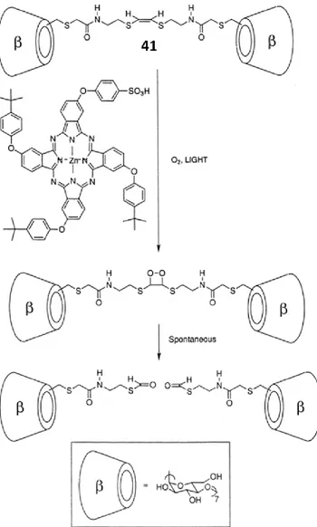
Figure 49. The reaction of singlet oxygen with unsaturated organic dithioethenyl bond. ... 39
Figure 50. Cleavage of dithioethenyl bond results formation of monomeric structures from dimers. ... 40
Figure 51. Photolysis reaction of nitrobenzyl-Caged ATP. ... 41
Figure 52. Commercial caged Ca(II) probes: NP-EGTA (left), DMNP-EDTA (right). ... 42
xiii
Figure 54. Photolysis and release of caged species from the typical
3’,5’-dialkoxybenzoin structure.. ... 43
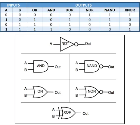
Figure 55. Schematic drawings and truth tables for common logic gates. ... 45
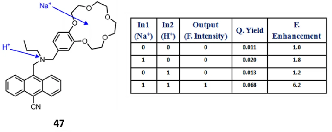
Figure 56. Literature example for AND molecular logic gate. ... 47
Figure 57. The example of AND logic gate by de Silva et al. ... 47
Figure 58. An example for XNOR and XOR molecular logic gates. ... 48
Figure 59. Half-adder logic gate and its truth table. ... 48
Figure 60. Literature example for half-adder molecular logic gate. ... 49
Figure 61. Independent INH and AND logic gates. ... 53
Figure 62. Fluorescence response of compound 2. ... 53
Figure 63. Cascading of the two independent gates INH and AND logic gates. ... 55
Figure 64. Absorbance spectra of compound 3. ... 56
Figure 65. Cascaded logic modules. ... 57
Figure 66. Spectral response of the cascaded INH-AND-AND logic modules. ... 58
Figure 67. Fluorescence response of compound 16 (Bodipy dye 2) upon uncaging of cage compound 15 (Caged Zn compound 1). ... 69
Figure 68. Fluorescence response of compound 16 (Bodipy dye 2) upon uncaging of 2 equivalents of compound 15 (Caged Zn compound 1). ... 69
Figure 69. Fluorescence response of compound 16 (Bodipy dye 2) upon uncaging of 2 equivalents of compound 15 (Caged Zn compound 1) in the presence of EDTA (1 equiv)... 70
xiv
Figure 71. Emission spectra of Compound 17. ... 71 Figure 72. Structures of proposed caged Zn (II) compounds (1 and 2) and the reporter molecule (3). ... 75 Figure 73. Modular design of the proposed caged compounds. ... 76 Figure 74. ITC titration curve of compound 13. ... 77 Figure 75. Working principle of the caged Zn(II) compound which can be activated by light of any chosen spectral region... 78 Figure 76. Fluorescence response of compound 3 (DPA-BOD) upon uncaging of 1+Zn(II). ... 79 Figure 77. Fluorescence response of compound 3 (DPA-BOD) upon uncaging of 2+Zn(II). ... 80 Figure 78. Fluorescence response of compound 3 upon uncaging of one equivalent o-nitrobenzyl Zn(II)-cage. ... 82 Figure 79. Decrease in absorbance spectrum of trap molecule DBPF in the presence of compound 8. ... 96 Figure 80. Absorbance decrease of DPBF at 414 nm with time in dichloromethane in the presence of compound 8. ... 97 Figure 81. Decrease in absorbance spectrum of trap molecule DBPF in the presence of methylene blue. ... 97 Figure 82. Absorbance decrease of DPBF at 414 nm with time in dichloromethane in the presence of methylene blue (reference). ... 98 Figure 83. Fluorescence response of compound DPA-BOD upon uncaging of Zn complex 1. ... 98
xv
Figure 84. Fluorescence response of compound DPA-BOD upon uncaging of Zn complex 2. ... 99 Figure 85. Fluorescence response of compound DPA-BOD upon uncaging of 1 equivalent Zn(II)-cage. ... 99 Figure 86. Control experiment of DPA-BOD. ... 100 Figure 87. Schematic representation of PS activation by acid and GSH. ... 103 Figure 88. Structures of distyryl-BODIPYs bearing different pH-sensitive groups with polyethylene glycol (PEG). ... 104 Figure 89. Chemical structure of AND logic construct of photosensitizer BOD 1 with GSH (red) and pH (blue) responsive moieties. ... 105 Figure 90. Normalized electronic absorption spectra of compounds 1 (black), 2 (red), 3 (blue) and 4 (green) in their neutral (solid) and protonated (dash) forms. ... 106 Figure 91. Chemical Structures of PS and Quencher (Q) modules... 107 Figure 92. Electronic absorption (top) and emission (bottom) spectra of 7.50 M BOD 1 (black), Quencher module (red) and PS module (blue) in THF. ... 108 Figure 93. Normalized electronic absorption spectra of neutral (solid) and deprotonated (dash) forms of compounds 5 (red), and micellar form of PS module of BOD 1 (black) in 40% THF/water and water respectively. ... 108 Figure 94. Comparison of normalized emission of PS module (black, solid) and absorption of Quencher module (red, dash) in THF depicting an excellent overlap for electronic energy transfer. ... 109 Figure 95. Electronic absorption spectra of neutral (black) and deprotonated (red) forms of micellar BOD 1 in water (a), and comparison of fluorescence spectra of equally absorbing micellar PS (red, dash) and BOD 1 (black, solid) in water (b, excited at 625 nm). ... 110
xvi
Figure 96. The cleavage of quencher from the photosensitizer in BOD 1 after
incubation with GSH for 12 h as analyzed by HRMS. ... 111
Figure 97. Emission spectra of micellar BOD 1 at the time of addition of 2.5 equivalents of GSH (black, solid) and after 12 h incubation with glutathione (red, dash) in water. ... 112
Figure 98. Control experiment with the solution containing the trap molecule only in acidic (black) and basic (red) acqueous conditions... 113
Figure 99. Comparison of 1O2 generation of micellar forms of molecular AND logic construct (7.50 M) in the presence of different combinations of inputs as followed by the decrease in 1O2 trap absorbance at 378 nm in water. ... 113
Figure 100. Comparison of initial 1O2 generation rate of BOD 1 as measured by the percent decrease in absorbance of trap molecule within 5 min (pink) or 1 h (bordeaux) of 625 nm light irradiation. ... 114
Figure 101. Mechanism of dopamine sensing with target molecule. ... 144
Figure 102. Structure-quantum yield relationship in Bodipy derivatives. ... 145
Figure 103. Structure of target and control molecules. ... 146
Figure 104. Fluorescence spectra of the compound 3 (1 μM) upon increasing dopamine concentrations. ... 146
Figure 105. Fluorescence spectra of the compound 2 (1 μM) upon increasing dopamine concentrations. ... 147
Figure 106. Fluorescence spectra of the compound 5 (1 μM) upon increasing dopamine concentrations. ... 148
xvii
LIST OF TABLES
Table 1. Summary of protonation dependent absorbance change of compounds 1-5 and BOD1 and their experimental pKa values.a ... 106 Table 2. Photophysical characterization of BOD 1, PS and Quencher.a ... 109
1
CHAPTER 1
1. Introduction
1.1. What is Supramolecular Chemistry?
Jean-Marie Lehn won the Nobel Prize in 1987 for his work in young research area, which is called “supramolecular chemistry” [1]. He defined this area as the “chemistry of molecular assemblies and of the intermolecular bond” [2]. In addition, there are some other expressions such as “chemistry beyond the molecule”, “the chemistry of the non-covalent bond” and “non-molecular chemistry”. In the supramolecular chemistry, there are host and guest molecules that stand together by non-covalent interactions that is illustrated in the figure 1 below.
Figure 1. Conformity of host and guest in supramolecular chemistry.
The supramolecular chemistry is relatively young discipline and it has been studied since the late 1960s. However, it began with Emil Fischer almost a hundred years ago. He defined the term “lock and key principle” that explain structural fit of enzyme-substrate long time ago [3]. Then, this ‘structural fit’ has been expressed with this model for long time. This concept is based on molecular recognition that an enzyme interact with only particular substrate. Almost in the same time with Fischer, Alfred Werner conducted his research in coordination chemistry, which stands on also non-covalent interactions [4]. Then in 1937, Wolf described the “übermolecül” term for self-associated carboxylic acid structures by hydrogen bonding [5]. Watson and Crick
2
solved the double helix structure of DNA in 1953 [6]. In 1967, crown ethers were discovered incidentally by Charles Pederson [7]. First “supramolecular” term is used by Jean-Marie Lehn in the late 1970s: “Just as there is a field of molecular chemistry based on the covalent bond, there is a field of supramolecular chemistry, the chemistry of molecular assemblies and of the intermolecular bond”.
Figure 2. Lock and key model for enzyme-substrate conformity.
Supramolecular chemistry mainly includes two categories, which are host-guest chemistry and self-assembly. The type of structure is determined according to size and shape. In the host-guest chemistry, host molecules are remarkably bigger than guest molecules. In these model, guest molecules are surrounded by host molecules. In the self-assembly model, size of two molecules relatively close to each other [8]. In the nature, there are several examples for both host-guest and self-assembly models. For instance, enzymes and their substrates are example of host-guest complexes. A substrate binds to binding site of an enzyme. A binding site is a region that has proper size, geometry and chemical environment to interact to some specific molecule or ion. It is important concept in the especially in biochemistry. Another example for host-guest interaction is the coordination chemistry. Metal ion is host-guest for the large ligands that especially macrocyclic compounds. The self-assembly model also has several examples from the nature. Non-covalent interaction, which is hydrogen bond between the chains of DNA is one of the most known examples of self-assembly structure. In the self-assembly structures, there is an equilibrium between two or more species that are building blocks of the main structure. The self-assembly process is usually spontaneous and reversible.
3
Supramolecular chemistry is used in wide range. Its application areas are catalysis [9], molecular recognition [10], molecular devices [11], light harvesting systems [12], solar cells [13], and molecular logic gates [14]. It is still growing and popularity of supramolecular chemistry is increasing rapidly every day.
1.1.1. Basic Supramolecular Interactions
Supramolecular chemistry is built by non-covalent interactions. Supramolecular structures are hold by these interactions, which are weaker than chemical bonds. The energy in a single bond of these interactions is between 2 kj mol-1 to 200 kj mol-1 but they are uncommonly has the energy greater than 100 kj mol-1. One of the most significant features of these weak interactions is being reversible, which plays a vital role in the existing of life. These supramolecular interactions are electrostatic interactions, hydrogen bonding, Van der Waals forces, π interactions and hydrophobic effect.
Electrostatic Interactions
Electrostatic interactions based on attraction forces between positive and negative charges such as metal ions, polar molecules, etc. Electrostatic interactions split into three categories, which are (i) ion-ion interactions, (ii) ion-dipole interactions, and (iii) dipole-dipole interactions. Ion-ion interaction is the strongest one and it is called ionic bond, which is the one of the chemical bonds. It is non-directional interaction different from the other two electrostatic interactions. Ion-dipole interaction is stronger than dipole-dipole because ion has more charge than polar molecule, which has partial charge only. For example, NaCl solution has ion-dipole interaction that is between polar water molecule and sodium or chloride ions. Dipole-dipole interaction is found between two polar molecules and relatively weak interaction. The example for the dipole-dipole is interaction between acetone molecules.
4
Figure 3. Examples for different types of electrostatic interactions.
Hydrogen Bonding
When hydrogen atom binds to electronegative atom with covalent bond, electron of hydrogen withdraw by this atom and hydrogen atom has strong partial positive charge anymore. In this situation, hydrogen atom which is positively charged interacts with other electron rich atoms and that is called hydrogen bonding. For hydrogen bonding, hydrogen atom must covalently connect to most electronegative atoms, which are fluorine, oxygen, and nitrogen. Hydrogen bonding is the strongest intermolecular interaction according to experimental results. For example, HF molecule has higher boiling point than HI molecule, although HI is much heavier. Hydrogen bonding is specific form of dipole-dipole interaction, but it is considerably stronger than others because positively charged hydrogen atom is so small that results much stronger interaction. In the nature, there are many examples for hydrogen bonding in vital part of biological systems. The interaction between the chains of DNA is the one of the most known example. There are two hydrogen bonds between adenine and thymine and three between guanine and cytosine and these hydrogen bonds hold the chains together.
5 Van der Waals Forces
Van der Waals forces are the common name of all of the intermolecular interactions that includes dipole-dipole, dipole-induced dipole, and induced dipole-induced dipole. A molecule which has even no permanent dipole can have instant dipole because of the non-homogeneous distribution of electrons of molecule. That instant dipole causes polarization of neighboring atoms or molecules and that is named London dispersion forces which is weakest intermolecular interaction. All of atoms or molecules have London dispersion forces which is also known as induced dipole-induced dipole interactions. Almost all of hydrocarbons are good example of the molecules which have no polarity so majority of hydrocarbon molecules have only London dispersion forces.
Π Interactions
π interactions consist of three main group π- π interaction, cation- π interaction and anion- π interaction. The most known is π- π stacking interaction which is because of the interaction between p orbitals. π- π interaction divided into two types which are face-to-face and edge-to-face interactions. π stacking interactions of nucleobases contribute to stabilizing of DNA double helix structure. Another example is that organic compounds which have aromatic ring are dissolved better in benzene on account of π stacking interactions. Cation- π interactions are very strong as hydrogen bonding as, so it is used for sensing of some cations. Na ion-benzene interaction is example for this interaction.
6 Hydrophobic Effect
Like dissolves like is simple rule in the general chemistry. Polar solvents dissolve in polar compounds and non-polar dissolves non-polar. Non-polar molecules aggregate in polar solvents owing to the desire of decreasing surface of touching area. Hydrophobic effect splits into two type with respect to energy; enthalpic and entropic hydrophobic effects. Hydrophobic effect is used in biochemistry to study some biological facts such as protein folding.
1.2. Luminescence
Mostly, particles are in their ground states at room temperatures. These particles absorb the energy when they are exposed to a radiation. A particle, which is absorbed photon is called excited. Emission of ultraviolet, visible or infrared photons from this excited particle is defined “luminescence”. In Latin, lumen means light and ‘luminescenz’ was used for the first time by Eilhardt Wiedemann in 1888. There are many types of luminescence such as photoluminescence, electroluminescence, chemiluminescence, etc. Photoluminescence is a kind of luminescence and excitation occurs with light. Photoluminescence is composed of fluorescence and phosphorescence which are particular type of luminescence. There are many other de-excitation (returning ground state) pathways for an excited molecule which are internal conversion, energy transfer, excimer formation, intersystem crossing, etc. (Figure 6) [15]. These processes are explained in following paragraph.
7
Figure 6. Energy releasing processes of an excited molecule.
The Perrin-Jablonski diagram [16] uses to show absorption and de-excitation processes in simple way. S and T letters represent the singlet and triplet energy states. The difference between them is spin of the electron. Energy levels which are between the singlet or triplet energy states are vibrational levels.
Figure 7. The Perrin-Jablonski diagram.
The first process is absorption, which is the fastest process in the all of them. After the absorption process, the molecule is excited and there are several de-excitation ways to
8
ground state. Internal conversion is non-radiative vibrational relaxation between vibrational levels, which have same spin multiplicity. It is not efficient way for transition from S1 to S0 or T1 to T0, so probably fluorescence or phosphorescence can
be observed in this situation. Fluorescence is radiative process which electron passes away S1 to S0. Actually emission is fast process as much as absorption but some cases
that staying in S1 or other non-radiative processes can be reason for delay. Another
way for de-excitation is intersystem crossing which is the transition singlet to triplet energy state. Intersystem crossing is non-radiative process and takes place between the vibrational levels, which have same energy. Although transition between states that have different multiplicity is forbidden, large spin-orbit coupling makes possible this replacement. Phosphorescence, which is radiative way for relaxation is slow transition T1 to S0. De-excitation non-radiative relaxation is usually preponderant to
phosphorescence due to T1 to S0 transition is forbidden. Phosphorescence can be
observed mostly in low temperatures.
Figure 8. Stokes’ shift.
In most cases, absorption and fluorescence wavelengths are different from each other and fluorescence wavelength is the longer one. The reason of this situation is that rapid vibrational relaxation of excited electron to lower energy levels in non-radiative way. The difference between absorption and fluorescence wavelength is named as Stokes’ shift [17]. Fluorescence wavelength is independent from the absorption wavelength
9
because of the reason mentioned above. This cases is called Kasha’s Rule [18]. Today, it is known that there are exceptions for Kasha’s Rule.
1.3. Fluorescent Dyes
Working principle of fluorescent dyes are completely different from the traditional ones. In traditional dyes, dye absorbs the particular wavelengths of white light then the complementary color left behind is seen. For example, an orange dye means that blue light is absorbed and remaining orange color is observed. Process of fluorescent dyes are explained before. Fluorescent organic dyes, which have emission in ultraviolet (UV), visible (VIS), infrared (IR) and near-infrared (NIR) regions have many application areas from medicine to nanotechnology. The figure shows that dyes that are commonly used in the applications [19].
Figure 9. Common fluorescent dyes in visible region.
Almost all of the visible region is covered by the organic dyes, which have some advantages in different aspects. Naphthalene, pyrene and coumarin based dyes are
10
usually found in UV region. On the other hand, IR dyes are consist of fluorescein, rhodamine, bodipy, and cyanine derivatives. All of these dyes are different from each other according to their properties, which are photochemical features, structures, spectral features and chemical characteristics. All of the dye families have both advantages and disadvantages in many aspects. For instance, fluorescein is one the most used organic dyes in biological applications because of good water solubility, high quantum yield and molar coefficient. However, it has also some disadvantages in the aspects of self-quenching, photobleaching and pH sensitivity [20]–[22].
1.4. Molecular Sensors
In common definition, a sensor is a matter that detect changes in a particular place or medium and supply an output. An ideal chemosensor should be selective for a particular ion, molecule or particle in addition to determine quantity of analyte. They have very wide application areas from medicine to environment. A couple of examples for using areas of chemosensors are that detection of a toxic compound in body and measurement of heavy metal amount in drinking water [23].
Importance of molecular sensor is increasing in recent days because of high costs and low detection limits of other sensing methods. Molecular sensors are highly interdisciplinary area and they many types in respect of analyte type, sensing mechanism, etc.
Optical sensors is one of the most known and used types of molecular sensors. Their working principle is based on interaction with light. Fluorescent sensors are a common type of optical sensors on account of properties that high sensitivity, low cost and fast response. The difference between wavelength of absorbed and emitted lights provides an advantage for fluorescent molecular sensors. Fluorescence is different from absorbance in aspect of the ratio between signal intensity and concentration. Fluorescence signal intensity can be increased by incident beam power on the other absorbance signal is proportional to concentration of sample essentially. Therefore, fluorescent sensors can detect picomolar levels while micromolar levels can be
11
measured with absorbance. Moreover, fluorescent sensors are reliable since there is only a stable molecule and light interaction [15].
Today, fluorescent sensors are widely used by scientist in many application areas from biological studies to nantechnological applications. For example, the fluorescent sensor, which is according to Lee et. al. is capable to sense both cation and anion (Figure 10). Binding of Pb (II) causes conformational changes that quench excimer fluorescence. There is also F- anion moiety, which is triazacrown by hydrogen bonding. Interaction with F- anion results in PET which is quenching of fluorescence [24].
Figure 10. Bifunctional sensor for both cation and anion.
1.5. Photophysical Aspect of Fluorescent Chemosensors
A fluorescent probes consist of two main group, which are receptor and fluorophore parts. These parts are recognition and signaling moieties. Receptor part is very important in terms of sensitivity and selectivity [25]. Receptor should have good and selective affinity for target particle and not influenced from environmental effects such as temperature, pH, etc. The fluorophore that gives optical output is as significant as receptor part. Purpose of fluorophore is that converting chemical inputs to optical output such as fluorescence emission. Fluorescent probe can be designed in two types. In first type, receptor is attached directly to fluorophore and it is part of conjugation
12
system of fluorophore. Other type of fluorescent senor have also spacer part which can be aryl group to separate receptor from π conjugation system of fluorescent dye [26].
Figure 11. Schematic representations of fluorescent chemosensors.
1.5.1. Photoinduced Electron Transfer (PET)
Photoinduced electron transfer (PET) is observed in the fluorescent probe whose structure is fluorophore-spacer-receptor type. In this type of fluorescent sensor, receptor is not part of conjugation because of the spacer, however still they are close enough for electronic interaction [27].
Figure 12 shows that working mechanism of PET. At the beginning, fluorophore absorbs a photon which excites electron from highest occupied molecular orbital (HOMO) to lowest unoccupied molecular orbital (LUMO). Receptor’s donor atom that is commonly nitrogen atom has HOMO whose energy level is between the molecular orbitals mentioned before. After excitation, an electron of donor atom’s HOMO moves to empty positon in fluorophore’s HOMO. That transfer of electron prevents the returning of excited electrons. All of these events result in quenching of emission.
13
Figure 12. Schematic representation and mechanism of PET.
There are many examples for PET based fluorescent chemosensors in the literature [28]–[30]. The figure 13 shows that different PET based molecular sensors for various metal ions. Compound 2 [31] is very simple example for aza-crown ether based fluorescent probe whose fluorophore is anthracene. In other examples, cryptand [32] and podand [33] is attached as receptor. In this fluorescent sensors, binding of some alkali or transition metal ions prevents the quenching of emission.
Figure 13. Example compounds for PET based chemosensors.
On the other hand, there are reverse PET based fluorescent chemosensors too. In this probes, binding of analyte causes quenching of emission. The figure shows mechanism
14
of reverse PET. Binding of analyte decrease the LUMO of donor atom and then excited electron moves the vacancy on the LUMO, so emission is quenched.
Figure 14. Schematic representation and mechanism of reverse PET.
An example for the reverse PET is compound 5, which is designed and synthesized by Akkaya group. Fluorophore is BODIPY that will be explained at next section. Binding Zn (II) ion to bipyridine receptor quench the emission by oxidative PET [34].
15 1.5.2. Internal Charge Transfer (ICT)
When receptor is attached to fluorophore directly, it becomes a part of conjugation too. In this type of fluorescent probes, binding of analyte changes in dipole moment that causes Stokes shift by intramolecular charge transfer from donor to acceptor. The positive or negative effect to excited state dipole results in changes of not only emission spectrum but also absorption spectrum [35].
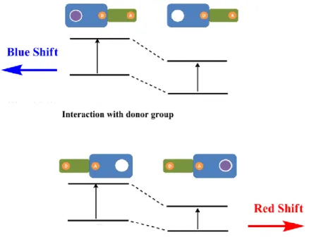
ICT based fluorescent sensors split into two type. First one is that an electron donor group, which can be amino group is attached to fluorophore. Binding of cation to receptor decreases electron density of electron donor group that means less conjugation. As a result, blues shift in absorbance and fluorescence spectrums is observed. That can be explained also charge-dipole interaction. In the excited state, electron donor group is positively charged, therefore binding of a cation results in interaction between two positively charged particles. In this case, destabilization of excited state is higher than ground state, so energy gap between HOMO and LUMO increases which means blue shift [36].
Figure 16. Red and blue shifts according to energy gap between HOMO and LUMO in ICT based
16
On the contrary, second type of ICT based probes indicates red shift in the fluorescence. Electron with drawing group like carbonyl is connected to fluorophore changes the result completely. Interaction between the cation and electron withdrawing group improve the electron withdrawing ability of this group. Stabilization and broader conjugation occurs with gaining of more electron for fluorophore by binding of analyte. A cation stabilize the electron withdrawing group, which is negatively charged in the excited states. Therefore, energy of excited states reduce more than ground states that results in decreasing the energy gap between HOMO and LUMO. In conclusion, red shift is observed in both absorbance and fluorescence spectrums [37].
There are many examples for ICT based fluorescent sensors. When metal ion is bound to compound 6 [38] and 7 [39], blue shift is observed. Receptor moiety is crown in these probes.
Figure 17. Examples for ICT based sensor which has crown ether as receptor.
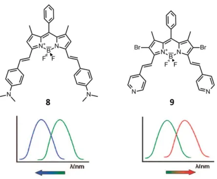
Although they have similar structures, the shift in their absorbance and fluorescence spectrums is completely different for compounds which are shown in figure 18 [40]. Because their receptor parts are different that one of them electron donating aniline group the other one is electron withdrawing pyridine. As a result, these two compounds exhibit opposite spectral shift upon proton binding.
17
Figure 18. The fluorescent sensors that have similar structures show different responds in the
presence of H+.
1.5.3. Energy Transfer (ET)
Light-harvesting antenna system is used many phenomena in nature [41]. For example, green plants have light-harvesting mechanism to gather more sunlight from different wavelengths in photosynthesis. A wide scale of sunlight is collected by only one photosynthetic reaction center alone by the help of light-harvesting complexes. Inspired by these examples from the nature, many scientists try light-harvesting mimicking applications [42]. In supramolecular chemistry, designing and synthesis of artificial light-harvesting systems is the research area, whose popularity increases rapidly in recent years.
Donor and acceptor fluorophores are required to make energy transfer system. It can be explained simply that donor fluorophore is excited with light then the emission from acceptor part is observed [43]. There are two types of energy transfer mechanism that Förster and Dexter energy transfers (Figure 19).
18
Figure 19. Schematic representation of Förster and Dexter energy transfers.
Characterization of energy transferred can be performed by tracking of parameters, which are lifetime, quantum yields, decreasing of donor emission and increasing of acceptor emission. Moreover, it is related to rate of deactivation of excited system. The requiring time of energy transfer must be less than lifetime of excited donor to carry out energy transfer successfully [44].
Dexter Type Energy Transfer
In Dexter type, energy is transferred by electron exchange. Orbital overlapping is required for electron transfer, so fluorophores are connected to each other by conjugated bridge. Because of that reason it is also named through-bond energy transfer. In this type of energy transfer, electron moves from HOMO of donor to LUMO of acceptor [45].
19
Figure 20. Schematic illustration of Dexter electron exchange mechanism.
There is an equation which shows rate constant for Dexter type energy transfer.
K
dexter= K J exp(-2R
DA/ L)
In this equation K symbolizes orbital interaction, J is normalized integral of spectral overlapping, RDA is space between donor and acceptor, and L is Van der Waals radii
[44].
According to equation, Dexter energy transfer is much dependent on the distance between donor and acceptor. Energy transfer yield increases exponentially with increasing distance. Therefore, they have to very close to each other for efficient energy transfer. Dexter energy transfer is also named as ‘short range energy transfer’.
Figure 21. Examples for Dexter type energy transfer.
Figure 21 [46] shows that examples for through bond energy transfer. Anthracene-BODIPY cassettes are designed to understand effect of distance in Dexter type energy transfer that is published by Burgress et al. Results are interesting because compound 11 has more efficient energy transfer unlike compound 10. The reason is that steric
20
hindrance in compound 10 disturbs the conjugation, so energy transfer efficiency decreases.
Figure 22. Another example for Dexter type energy transfer.
Another example for Dexter type energy transfer is shown in figure 22 [47]. It consists of three BODIPY units two of them are donors and the other one is acceptor that they are linked to each other with conjugated bridge. It is clarified that through bond energy transfer decreases with distance.
Förster Type Energy Transfer
In Dexter energy transfer, orbital interaction is required, on the other hand Fluorescence (Förster) energy transfer (FRET) becomes in case of spectral overlapping. Energy is transferred from donor fluorophore to acceptor non-radiatively. It was introduced by Förster first time in 1948 [48] and today it has many applications in the fields from biotechnology to electrochemistry. The scientists need FRET since in some cases Stokes’ shift of a fluorophore is not enough to distinguish the difference. Förster energy transfer depends on several factors. It is similar to Dexter type, FRET is also much dependent on distance between donor and acceptor fluorophores. Energy transfer efficiency is inversely proportional to sixth power of distance. The other
21
significant factor for efficiency is spectral overlap of donor’s emission and acceptor’s absorbance [49].
Figure 23. Schematic illustration of Förster electron exchange mechanism.
The electronical mechanism of FRET is shown in figure x. After the excitation of donor, excited donor transfer its energy to acceptor in non-radiative way which is called resonance. Then excited electron of acceptor moves to ground state by emitting light.
The rate equation of FRET is given below:
Where D represents excited state lifetime of donor in the lack of acceptor, d is the
distance between donor and acceptor and Rc is critical radius which is the distance when kET equals to real decay rate of donor. The equation of Rc is given below:
Where D symbolizes the emission quantum yield in the lack of acceptor, K is
orientation factor and n represents refractive index of solvent, N is Avogadro’s number and J is the integral of spectral overlap [50].
22
Figure 24. A literature example for BODIPY based Föster type energy transfer.
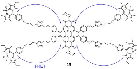
In the figure 24 [51], one of the most known example is shown. In this example, BODIPY fluorophores as donors are attached to perylene based acceptor unit. Click chemistry is used to bind donors to acceptor in this study. FRET efficiency is calculated as 99% that is satisfying result.
Figure 25. Another example for Föster type energy transfer.
Another example is compound 14 [52] that three BODIPY units are covalently attached each other via click reaction. Excitation wavelengths are 501 nm and 572 nm
23
and emission wavelength is 662 nm that there is huge difference between them. Energy transfer yield is 99% for both donors.
1.6. Sensing of Particular Ions or Compounds
1.6.1. Metal Ions
Metal ions participate in several important biological processes from muscle contraction to nerve impulses. Moreover, catalytic amount of cations are involved in binding site of many enzymes. Not only lack of some metal ions but also overdose of them are the reasons of various diseases directly or indirectly. There are many examples for relation between ion levels and some illnesses. For example, control of lithium is considerable in the treatment of manic depression and also potassium amount in high blood pressure [53]–[56].
Transition metals have critical roles in environmental pollution and biological metabolism [57]. Therefore, observing and tracking of their concentration is important for understanding of their toxic and pollutant effects. These metal ions are especially Pb (II), Cd (II), Hg (II), etc. that have many negative effects on the balance of nature. Selectivity is still challenge to design a good receptor, which has affinity for only one cation relatively. Because of that reason, many types of receptor synthesized and this field is very comprehensive. For example, majorly oxygen atoms containing crown ethers derivatives can be used to sense alkali or alkali earth metals that are hard metals relatively. According hard and soft acid and bases (HSAB) theory, hard acids interact with hard bases better and soft prefers soft also because of charge to size ratio. Another example is that usually Hg (II) receptors consist of Sulphur containing crown ethers due to HSAB again. Besides HSAB, size also important itself only that size compatibility between ion and moiety is required. In the figure 26, there are examples for different metal chemosensors. Compound 15 is sodium selective fluoroionophore [58]. Compounds 16,17 and 18 are capable to sense Hg(II) ions [59]–[61].
24
Figure 26. Literature examples for metal ion sensors.
Zinc (II) Ion Sensors
Zinc (II) ion is the second most abundant transition metal ion in the brain and also it participates as main component in many biological processes. Major amount of zinc presents as tightly bound to proteins, on the other hand minority is in the mobile form as intracellular zinc ions which are existing in the tissues, brain, intestine, pancreas, and retina, however the exact function is still obscure in the many biological processes. Moreover zinc ions, which are coming from intracellular metalloproteins have a role in programmed cell death called ‘apoptosis’. Zinc ion is very critical in the several disorders such as Alzheimer’s disease, epilepsy, Parkinson’s disease, ischemic stroke and infantile diarrhea. In spite of intensive research on role of zinc ion in these processes, a lot uncertain points about its function should be clarify. Therefore, it is still challenge to design new receptors to monitor quantity of zinc ions [62]–[64].
25
Figure 27. Some literature examples for Zn (II) ion sensors.
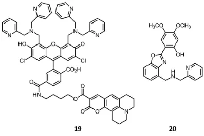
There are many examples of fluorescent sensors for zinc ions. Compound 19 [65], which is shown in the figure 27 is the example of ratiometric sensor for intracellular zinc ion. There is ester linkage between coumazin group and fluorescein and that is hydrolyzed by esterase enzyme in the cell. After leaving of coumazin, fluorescein that emits at 534 nm is sensitive to zinc concentration. Compound 20 [66] is another example that is developed O`Halloran et al. It is water soluble and very selective to zinc ion. Intracellular zinc ion can be monitored by emission of this molecule.
1.6.2. Biological Thiols
Cysteine (Cys), homocysteine (Hcy) and glutathione (GSH) are biological thiols, which are very important roles in enzyme functionality and preservation of redox states because of the good nucleophilic properties. The change in amount of biological thiols is related with many diseases such as occlusive vascular, premature arteriosclerosis, leukemia, diabetes, etc. [67]–[69].
26
Glutathione (GSH) is tripeptide, which consists of glutamate, glycine and cysteine. In the synthesis of GSH, the enzymes that γ-glutamylcysteine synthetase and glutathione synthetase participate as catalyzer. In the tumor cells GSH concentration is more than normal cells 1000-fold. GSH takes part in antioxidant response, cell proliferation, regulation of gene expression, etc. Lack of GSH causes the diseases, which are Parkinson’s disease, AIDS, Alzheimer’s disease, cancer and heart attack [70], [71].
Detection of Thiols
Detection of the thiols that are Cysteine (Cys), homocysteine (Hcy) and especially glutathione (GSH) is certainly significant because of their properties mentioned before [72], [73]. Two characteristic of thiols are helpful for sensing of them. These two properties that they are good nucleophiles and they have affinity to metal ions are very beneficial to design fluorescent-based thiol probes. Many reactions and methods, which are Michael addition, cyclization, metal complex formation, redox reactions and disulfide bond cleavage are used in sensing of thiols. pH of medium is also important in sensing because of the acidity of thiol derivatives [74], [75].
Figure 29. Example for thiol sensor based on Michael addition.
Compound 21 [76] is example for Michael addition based fluorescent probes. The coumarin that includes α, β-unsaturated ketone derivative has no emission. After binding of thiol derivative, it starts to give emission. This PET based thiol sensor is shown in the figure 29.
27
Figure 30. Thiol probe based on PET mechanism.
In the figure 30, there is another thiol sensor example. This example based on cleavage of sulfonamide and sulfonate. Thiol derivative attacks to electron deficient 2,4-dinitrophenyl sulfonyl part of compound 22 and that results in turning on the emission of fluorescein because of de-sulfonylation [77].
Figure 31. Thiol sensor based on cyclization of aldehydes.
Compound 23 [78] is a successful example for sensing of thiols by cyclization of aldehydes. The reaction between bisaldehyde and cysteine or homocysteine causes the shift of absorption maximum to longer wavelength. Monoaldehyde derivative of compound 23 is more selective for Cys. The reason is related with formation five-membered ring at end of reaction.
28
Figure 32. Thiol probe based on metal ions.
Another method to sense thiols is using their high affinity to metal ions. Kim et. al. were used iminocoumarin which includes Cu (II) ion for sensing thiols at pH 7.4. Adding of thiol to compound 24 [79] results in coumarinaldehyde, which has high emission as distinct from the initial non-emissive metal complex. Compound 24 is another example for PET based turn-on fluorescent sensor for thiols.
Disulfide Bond Cleavage
Thiols are strong reducing agents and by this way they can break the disulfide bonds. First example is compound 25 [80] which consists of coumarin and porphyrin derivatives that are connected with disulfide bond. Coumarin is donor and porphyrin is acceptor part of the structure and there is fluorescence resonance energy transfer (FRET) between them. In the presence of thiols, disulfide bond is cleaved, therefore FRET is over at this point. Emission shifts from red to blue color. By this way, thiols can be sensed easily and detected by naked eye.
29
Another example for disulfide bond cleavage is compound 26 [81] as shown in figure 34. There are three main parts in this structure, which are rhodamine, BODIPY and folate that is folic acid receptor. Aim of folate is targeting of folic acid that is found more in some type of cancer cells. In this structure, BODIPY and rhodamine fluorophores are donor and acceptor respectively. There is FRET in lack of GSH. Concentration of GSH in tumor cells is more than normal cells almost 1000 times. In the presence of GSH, disulfide bond is broken and emission wavelength is changed from 595 nm to 520 nm.
Figure 34. Disulfide bond cleavage based FRET.
1.7. BODIPY Dyes
4,4-difluoro-4- borata-3a, 4a-diaza-s-indacene which is abbreviated as BODIPY was first discovered accidentally by Treibs and Kreuzer in 1968 [82]. They used acetic anhydride to acylate the 2,4-dimethylpyrrole in the presence of BF3·OEt2. Dipyrrin
30
compound was synthesized by acid catalyzed then dipyrrin formed complex with borontrifluoride to yield BODIPY (Figure 35). Almost 20 years later, Haugland and Kang explored the fluorescence properties of BODIPY in 1988 [83], [84]. After the recognition of BODIPY as a fluorophore, it has been used in many applications in the areas from molecular sensors to biomedical materials. There are many reaction sites on BODIPY which can be modified by nucleophilic and electrophilic substations.
Figure 35. First synthesis of BODIPY dye.
BODIPY has many excellent properties against the other fluorescent dyes. For instance, BODIPY dyes absorbs and emits strongly in the fields from visible to near-IR. Not only quantum yields but also absorption coefficients of BODIPY dyes are quite high [85]. Furthermore, they have thermal and photostability in different types of mediums and solubility in organic solvents is very good. They are usually neutral molecules, therefore insensitive to pH and polarity of solvents [86].
Figure 36. Structure and numbering of BODIPY core.
The most significant characteristic of BODIPY dyes is probably easy chemistry. It possible to functionalize almost all positions on BODIPY and the yields of reactions are satisfactory. BODIPY family gains new members with each of this modification that every new member has different chemical and photophysical features [87].
31
Figure 37. Quantum yields of BODIPY dyes that one them has methyl group at 1, 7 positions.
Every positon effects different property of BODIPY dye that is mentioned before [88]– [91]. For instance, quantum yield of BODIPY is influenced remarkably with aromatic substation on meso positon. In the figure 37, the difference between fluorescence quantum efficiencies of two BODIPY dyes are very large while their structures are very similar. In phenyl-BODIPY (without methyl groups), possible conjugation between phenyl group and BODIPY core causes quenching of emission. Presence of methyl groups at 1 and 7 positions keeps the phenyl group at perpendicular to BODIPY core. Methyl groups prevent the free rotation because of steric hindrance. As a result of that structural change, the fluorescent quantum yield increases enormously.
Figure 38. Free rotation of phenyl group that is attached to meso position of BODIPY.
Modification of meso position is realized with acid-catalyzed in the synthesis part. Water-soluble groups, receptors for particular ions or molecules and donor-acceptor moieties can be attached to meso position.
32
2 and 6 positon of BODIPY is less positive, so electrophilic reactions are preferred instead nucleophilic. Sulfonation, nitration, halogenation and formylation of these positions can be possible because of the electron rich characteristic. Binding of heavy atoms like bromine and iodine causes intersystem crossing, by this way fluorescence of BODIPY decreases. This substation makes possible producing singlet oxygen, which is very important for photodynamic therapy.
Modification of 1, 3, 5 and 7 positions allows several additional functions to BODIPY [86], [92]. The most used reaction to functionalize especially 3 and 5 positions is Knoevenagel condensation [93]. In this reaction, Dean-Stark apparatus [94] is used for to get rid of water, which forms as product. By using this reaction, several types of electron donor and withdrawing aldehydes can be attached to these positons. They cause red shift in absorption and emission spectrum [95] so that it can be proper for biological applications.
Figure 39. Structures of the tetra-styryl BODIPY derivatives.
Akkaya group stated that 1 and 7 positions are also reactive as 3 and 5 positions [96]. Tetra-styryl BODIPY dyes were synthesized by Akkya group in 2009 (Figure 39). Then, Ziessel et al reported the tetra-styryl BODIPY whose absorption wavelength is 720 nm and emission wavelength is 800 nm (Figure 40) [97]. Also both of two groups, which have many study on BODIPY show that 3 and 5 positions are more acidic than 1 and 7 positions. Modification of BODIPY with some groups such as dimethyl amino
33
and pyridine makes possible pH sensitive applications in the area from sensors to drug delivery.
Figure 40. Schematic representation of 1, 3, 5, 7-tetrastyryl BODIPY derivatives.
1.7.1. Applications of BODIPY
BODIPY dyes has many significant properties which are mentioned before. Because of that adorable features, it has wide application area from sensing to solar cells. The figure shows the application areas of BODIPY.
Figure 41. Applications of BODIPY.
There are many examples in the literature for BODIPY based chemosensors because of the properties such as easy modification, photostability and thermal stability, high
34
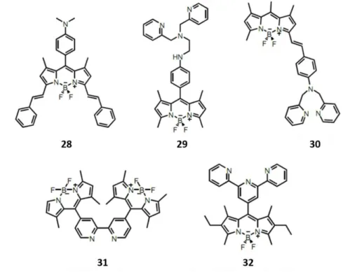
fluorescence quantum efficiency, etc. Daub and Rurack synthesized BODIPY based chemosensor first time in 1997 [98]. In the figure 42, some examples for PET and ICT based BODIPY sensors for particular ions are given. In compound 28 [92], aniline group is attached to meso postion and that is for sensing of proton. Compound 29 [99] and 30 [100], which are dipicolylamine substituted BODIPYs sense in order of zinc and cadmium ions. In compound 31 [34] and 32 [101], receptor moieties are bipyridine and terpyridine groups respectively.
Figure 42. Examples for BODIPY based chemosensors.
Another application area of BODIPY dyes is photodynamic therapy (PDT) which will be explained in detailed later. PDT is cancer treatment method that photosensitizer in the tissue produce singlet oxygen in the case irradiation with light. Singlet oxygen, which is produced by photosensitizer destroys the cancer cells. IR and Near-IR photosensitizers are more proper for PDT because penetration of these lights to human skin is better. PDT is used especially in the treatment of skin cancer. BODIPY based dyes, which has often heavy atoms on 2 and 6 positons are used as photosensitizers. There are examples for BODIPY based PDT agents in the figure 43. For example,
35
water soluble groups attached to compound 33 [102] to increase solubility in water because these molecules must work in aqueous media to destroy cancer cells. Also substitution on 3 and 5 positions increases conjugation that ensures longer wavelengths. Compound 34 [89] has very low fluorescence quantum yield that means better intersystem crossing. It is designed by Nagano et al and works in organic solvents. Compound 35 [103] and 36 [104] are another examples for water soluble BODIPY based photosensitizers by the help of sulfonate and carboxylic groups.
Figure 43. BODIPY based photosensitizers for photodynamic therapy.
Popularity of dye-sensitized solar cells (DSSCs) increases rapidly similar to other alternative energy sources nowadays. Many scientist conduct research on DSSCs to increase efficiency. BODIPY dyes are also used in this field and Nagono is leading scientist which used BODIPY in DSSCs first time. Usually carboxylic group (-COOH) is used for attaching dye to TiO2 surface. In the figure 44, compound 37 [105] was
designed by Akkaya et al. for DSSCs. Conversion efficiency of this BODIPY based photosensitizer is reported as 1,66%.
36
Figure 44. Example for BODIPY based photosensitizer that used in solar cells.
1.8. Photodynamic Therapy (PDT)
Photodynamic therapy (PDT) is very young cancer treatment method whose importance is increasing rapidly in recent days [106]–[109]. PDT consists of three main parts, which are photosensitizer, light and oxygen molecule. In the existence of O2, photosensitizer is irradiated with light and by this way triplet oxygen molecule is
excited indirectly. Unlike chemotherapy and radiotherapy, PDT is non-toxic and submissive treatment method. There are plenty of benefits against to radiotherapy and chemotherapy. First, side effects are very low because of the local application and it can be performed many times to same location due to it has almost no harmful effect and toxic properties. Another advantage is that functionalization of photosensitizer is possible with respect to types of cell, cancer, treatment and light. Disadvantage of PDT is difficulty in penetration of light, which can be furthest 5-10 mm. PDT is applicable usually skin cancer because of the penetration problem [110].
1.8.1. Photophysical Background of PDT
Photosensitizers are usually the organic dyes that have conjugated systems. In photosensitizer, the electron at ground state is excited with light to excited singlet state. The electron at excited singlet state is short-lived and it has different relaxation pathways such as fluorescence, interval conversion, intersystem crossing, etc. the spin
37
of excited electron changes with intersystem crossing from singlet to triplet state which has longer life time. The reason of long life time is that triplet-singlet transition called phosphorescence is forbidden transition. There is also another possible transition which is spin allowed between excited triplet photosensitizer and triplet oxygen that in at ground state. By this transition, highly reactive singlet oxygen species form [111]. The process is shown in the figure 45 [112].
Figure 45. Photophysical processes in PDT.
1.8.2. Photosensitizers and Importance of Light
Photosensitizers that absorb visible and near-IR light have wide range of application fields. The distinctive feature of a PS is that it not only absorbs light in the traditional sense, but it can also transfer this resulting energy to the molecular oxygen to generate highly reactive singlet oxygen molecule [113]. The most common PSs are shown in Figure 46.
38
Porphyrins and Roze Bengal are common singlet oxygen generating compounds, however do not have strong absorption in the therapeutic window of the body. Phthalocyanine related sensitizers are more promising in terms of absorption profiles. Although the Boradiazaindacene (BODIPY) derivative depicted in figure 46 has an absorption maxima at 540 nm, its near-IR absorbing derivatives was easily synthesized and reported in the literature. Below, in figure 10, three different BODIPY based near-IR absorbing PSs are illustrated. All, absorb light in the 650-700 nm range, which is compatible with the therapeutic window of the body. While compound 38 [114] was introduced by Killoran et al., photosensitizers 39 [102] and 40 [115] were reported by the Akkaya group.
Figure 47. Literature examples for BODIPY based photosensitizers.
In PDT, wavelength of the light is important as characteristic of photosensitizers. Light must penetrate deeply to tissue to excite photosensitizer. Some compounds and structures in tissue such as macromolecules, organelles, cell layer, hemoglobin, and water interact with light in aspect of not only scattering but also absorption. The best penetration into tissue is possible between IR and near IR wavelengths [116]. For example, penetration of light whose wavelength is 700-850 nm is almost two times of 600 nm whose penetration depth is 1-3 mm [117].