Minireview
MRI-guided gene therapy
Xiaoming Yang
a, Ergin Atalar
a,b,*aDepartment of Radiology, Johns Hopkins University, United States
bDepartment of Electrical and Electronics Engineering, Bilkent University, EA 212, Bilkent Ankara 06800, Turkey
Received 4 April 2006; accepted 8 April 2006 Available online 19 April 2006
Edited by Horst Feldmann
Abstract MRI has the ability to generate high-contrast and high-resolution images, to obtain multiple diagnostic evaluations of organ function and morphology, and to provide multiple image planes with no risk of ionizing radiation. Recent efforts have fo-cused on using MR technology to monitor gene delivery, to en-hance gene transfection/transduction, and to track gene expression. This review summarizes the current status of MRI-guided gene therapy.
Ó 2006 Published by Elsevier B.V. on behalf of the Federation of European Biochemical Societies.
Keywords: Magnetic resonance imaging; MRI-guided therapy; Gene therapy
1. Introduction
Gene therapy is an exciting frontier in medicine today. It is designed to target the causes rather than the symptoms of dis-ease. Our current knowledge about the biodistribution and pharmacokinetics of gene therapy relies mainly on the results of in vitro laboratory examinations of tissues obtained at biopsy or autopsy. Thus, it is essential to develop non-invasive methods to monitor and guide gene therapy.
To date, different imaging modalities, such as magnetic res-onance imaging (MRI), ultrasound, nuclear medicine, and optical imaging, have been applied to the monitoring of gene therapy[1]. MRI has several prominent advantages compared to other imaging techniques, including the ability to generate high-contrast and high-resolution images, to obtain multiple diagnostic evaluations of organ function and morphology, and to provide multiple image planes with no risk of ionizing radiation. Recent efforts have focused on using MR technol-ogy to monitor gene delivery, to enhance gene transfection/ transduction, and to track gene expression [2]. This review summarizes the current status of MRI-guided gene therapy.
2. MRI-monitored gene delivery
The efficient delivery of genes to the target is the first key step in successful gene therapy. During the primary gene deliv-ery procedure, one needs to see how the delivered genes
distrib-ute within the target and whether the delivered genes localize at a high concentration at the target. An initial study at-tempted to validate the feasibility of using MRI to monitor gene delivery to the vessel walls[3]. The authors mixed a mar-ker gene, green fluorescent protein (GFP) gene carried by a lentiviral vector, with a T1 MR contrast agent, gadolinium (Gd). The GFP–Gd mixture was then locally transferred into swine artery walls via a catheter-based approach, while the delivery process was monitored under real-time MRI. Cross-sectional magnetic resonance images showed an enhanced bright ring as the GFP–Gd mixture was transferred into the ar-tery wall, which correlated with GFP-positive cells of the arte-rial tissues according to subsequent histological staining. Another attempt was to evaluate the possibility of using MRI to monitor agent delivery to the prostate under MRI guidance[4,5]. After percutaneously positioning an MR-com-patible biopsy needle into the prostate, the authors used MRI to visualize the distribution of a Gd–trypan blue mixture that was injected into the prostate via a biopsy needle. The results of this study show an accurate correlation between MRI and histology with regard to the intra-prostate distribution of Gd–blue mixture (Fig. 1).
Intraluminal MRI may greatly facilitate the development of MRI-guided gene therapy in other organs and systems. For example, recent studies have demonstrated the feasibility of generating intraluminal MRI of non-cardiovascular systems, such as the esophagus[6]and hepatobiliary system[7]. These intraluminal MRI techniques enable us to (a) observe the lumi-nal walls at high-resolution scales; and (b) combine the intra-luminally placed MR devices with different existing interventional procedures, including gene therapy. These advantages of intraluminal MRI should provide further opportunities to use MRI to guide and monitor gene therapies in various organs and systems of the body.
3. MRI-enhanced gene expression
One of the current limitations of gene therapy is its low capability to sufficiently transfect/transduce genes/vectors into the targets. For example, the in vivo transfection/transduction of genes in vasculatures is very low, e.g., approximately only 1% for non-viral gene-carrying vectors and less than 5% for viral gene-carrying vectors [8,9]. To overcome this problem, imaging scientists have begun to explore the potential of com-bining MR techniques with other techniques, such as radiofre-quency (RF) and focused-ultrasound, to enhance gene transfer and gene expression.
*Corresponding author. Fax: +90 312 442 6482.
E-mail address:[email protected](E. Atalar).
0014-5793/$32.00 Ó 2006 Published by Elsevier B.V. on behalf of the Federation of European Biochemical Societies. doi:10.1016/j.febslet.2006.04.027
The principle of RF-enhanced gene transfection/transduc-tion and expression is based on the fact that RF can create lo-cal heat. A few in vitro studies have demonstrated that controlled heating can enhance gene transfection, with the pro-posed mechanisms of (a) fracturing tissues by heat; (b) increas-ing the permeability of the plasma membrane and cell metabolism; and (c) increasing of the activity of heat-sensitive heat shock proteins[10–12]. Recent efforts have focused on the combination of MRI and RF-enhanced vascular gene expres-sion. Using a loopless MR antenna as an intravascular heat delivery vehicle, investigators first designed an intravascular MR/RF system[13,14], which enabled generation of not only local RF heat but also MR thermal mapping to monitor and control the RF heat distribution at the targeted vessels[13]. In the subsequent in vitro and in vivo studies, the authors have further evaluated the feasibility of using the intravascular MR/ RF system to enhance vascular gene expression[15], and to en-hance vascular endothelial growth factor (VEGF) gene ther-apy of in-stent neointimal hyperplasia (Fig. 2)[16].
The principle of low-frequency ultrasound (US)-enhanced gene transfection/transducion and expression is based on the fact that ultrasound may induce cell-membrane porosity, re-duce the thickness of the unstirred layer at the cell surface, and facilitate the passage of genes/vectors across cell mem-branes [17,18]. However, low-frequency ultrasound does not offer high-resolution imaging of the gene-targeted vessel wall and may be technically feasible only when treating superficial organs due to difficulties in precisely focusing the ultrasound power in deep structures. To deal with this problem, investiga-tors have developed a method that uses MRI to guide the direction of focused-ultrasound energy to the targets, and thus, enhance gene expression at the targets.
A non-invasive, physical approach to the combination of MRI-guided focused ultrasound (MRI-FUS) with a heat-sen-sitive promoter (hsp) was reported to help the enhancement of gene expression [10,19]. In one study with natural hsp70 induction in rat leg by MRI-FUS, investigators demonstrated increased expression of the hsp70 mRNA at the MRI-FUS-treated region[10]. In another study, a stable modified C6 gli-oma cell line was used to carry a fused gene coding for thymi-dine kinase (TK) and green fluorescent protein (GFP) under control of the human hsp 70 promoter. In vivo tumors were subjected to a 3-min temperature elevation using MRI-FUS with a constant temperature, and were analyzed 24 h after the heat shock for GFP fluorescence. Preliminary results showed the elevated expression of TK-GFP[10,19]. In addi-tion, some authors have reported that the blood–brain barrier can be consistently opened with focused ultrasound exposures in the presence of an ultrasound contrast agent. Indeed, MR imaging signal intensity changes may be useful in the detection of blood–brain barrier opening during sonication [20]. The interesting point here is that therapeutic genes may be carried by liposome- or polymer-based ultrasound contrast, which may then permit high doses of genes to pass through the blood–brain barrier and enhance gene therapy in the neurolog-ical system under MRI guidance.
4. MRI tracking of gene expression
After therapeutic genes are successfully transferred into the targets, gene expression must be tracked, basically to discover (a) at what level genes express, and (b) for how long genes function at the targets. Currently, it is not possible to directly Fig. 1. Human injection trials: Histology and magnetic resonance monitoring of distributions show close agreement. First column: A 0.018-in. diameter nitinol injection needle was placed by hand into the prostate, and T1-weighted gradient echo images were acquired. Second column: Following injection of 0.3 ml of 30 mM Gd–DTPA, mixed with trypanblue tissue dye (over 30 s), a second set of T1-weighted gradient echo images was acquired. Enhancement due to the gadolinium solution is clearly visible. Third column: Blue stained tissue, seen in gross tissue sections of the prostate, corresponds well with the enhancement seen in MR images[5].
visualize any genes/vectors themselves with any clinical imag-ing modality. As molecular imagimag-ing rapidly emerges as a bio-medical research discipline, imaging scientists have promoted the concept of using MRI as a non-invasive imaging tool to monitor, in vivo, specific molecular and cellular processes, for example, gene expression. MRI tracking of gene expression is primarily based on the ability of MRI to detect gene-ex-pressed downstream products, such as proteins and enzymes.
The initial investigations of MRI tracking of gene expression were focused on imaging of introduced genes, which express MR substrate-attractable receptors or enzymes, into the tar-gets. The receptors or enzymes can bind or metabolize para-magnetic substrates to the target at high concentrations, which then generate MR signals at the targets[21]. For exam-ple, the human transferrin receptor (TfR) genes express, on the cell surface, high-level TfRs on a variety of tumor cell types, which then can be imaged using transferrin (Tf)-bound super-magnetic contrast agents[22]. Tyrosinase, an enzyme normally involved in melanogenesis at the target, can remarkably bind metals, such as iron, which thus produces high signal intensity on T1-weighted images [23]. The principle of tyrosinase-in-duced MRI is primarily based on the mechanism of tyrosinase in catalyzing the conversion of low-relaxivity substrates into high-relaxivity products.
Recent progress in nanotechnology has opened up new avenues for extending the usefulness of MRI to track gene expression. Micro/nanoparticle-based MR imaging of gene
expression depends primarily on the production of smart con-trast agents to target specific biochemical pathways, such as a receptors or enzymes that are expressed or activated by the transferred genes at the targets. A functional smart contrast agent requires the conjugation of a ligand with MR-detectable nanoparticles or MR contrast agent molecules. Thus, once transgenes express the downstream receptors, such as cell-sur-face receptors, the systemically delivered MR smart contrast agents can concentrate specifically on these expressed receptors via the receptor–ligand interaction (pair), which has been re-ferred to as ‘‘cell-surface receptor imaging’’ [24]. A recent study also reported the synthesis of two novel gadolinium-bisamides, which were designed as enzyme-activated contrast agents for MRI, with a net increase in longitudinal (R1) relax-ivity[25].
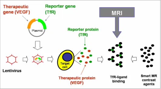
The gene-expressed receptors or enzymes mentioned above are actually considered MRI reporters or markers for the gen-eration of MR imaging. Our final goal in developing the tech-nology of MRI for gene therapy is to detect the expression of a given therapeutic gene rather than that of a marker gene. To this end, it is essential to build dual gene-carrying vectors, which carry and express simultaneously two genes, a reporter gene (such as TfR gene), and a therapeutic gene (such as VEGF gene). Thus, the indirect assessment of functional VEGF gene expression can be achieved by the direct detection of TfR gene expression since two genes are simultaneously ex-pressed from the same vector (Fig. 3).
Fig. 2. Microscopy examination of stented arteries with VEGF gene therapy only (A) and with intravascular MR/RF-enhanced VEGF gene therapy (B). In-stent neointimal hyperplasia (open arrow) is thinner in VEGF/RF-treated artery (B) than in VEGF-only-treated artery (A). H&E staining, 68X.
Fig. 3. Dual gene-carrying plasmid is constructed within a lentiviral vector, which thus expresses simultaneously an MR imaging reporter, the (TfR), and a therapeutic protein, the VEGF, at the target cell. Transferrin-conjugated smart MR contrast agents bind TfRs via the receptor–ligand binding, which is then imaged by MRI. Thus, the success of directly detecting the TfR by MRI indicates indirectly the functionality of the VEGF.
5. MRI of gene therapy follow-up
One of the prominent advantages of MRI is its ability to generate not only anatomic imaging for observation of organ morphology, but also functional imaging for assessment of or-gan perfusion, and metabolic and mechanical functions. This unique advantage makes MRI a powerful tool for the fol-low-up of a given gene therapy. For example, after gene ther-apy of cardiac ischemia/infarction, one may use perfusion MRI to evaluate the recovery of ischemic tissues, MR tagging to assess the mechanical properties of the gene-treated cardiac region, and spectroscopic imaging to examine the metabolic status of the gene-treated heart. Targets for vascular gene ther-apy currently include limiting restenosis after balloon angio-plasty and stent placement, inhibiting vein bypass graft intimal hyperplasia/stenosis, therapeutic angiogenesis for car-diac and lower-limb ischemia, and prevention of thrombus for-mation[26]. While catheter angiography is still the standard method by which to follow vascular gene transfer, advanced MR techniques, such as MR angiography and perfusion MR imaging, are becoming useful non-invasive methods by which to assess the success of gene therapy in vascular diseases.
6. Conclusion
Gene therapy is a promising approach to the treatment of many forms of disease, including cancer. MR technology is an important imaging tool in this era. The development of intraluminal MR techniques provides a great opportunity to combine MRI with exciting interventional techniques, which permits precise monitoring of the biodistribution of the deliv-ered genes/vectors to the targets under MRI guidance. The MR thermomapping technique should play a critical role in the control of the RF- and focused ultrasound-mediated enhancement of gene expression. Target-specific molecular MR imaging associated with nanotechnology has already of-fered basic medical science an extremely useful in vivo evalua-tion tool for the non-invasive or minimally invasive imaging of gene therapy. Functional MR imaging obtained pre- and post-gene therapy should become an ideal approach to assess the success of gene therapy. While there are still many challenges that face MR imaging, the growing number of MRI applica-tions in the field of gene therapy represents a bright future for MR imaging in modern medicine.
References
[1] Weissleder, R. and Mahmood, U. (2001) Molecular imaging. Radiology 219, 316–333.
[2] Yang, X. (2003) Imaging of vascular gene therapy. Radiology 228, 36–49.
[3] Yang, X., Atalar, E., Li, D., Serfaty, J., Wang, D. and Kumar, A., et al. (2001) Magnetic resonance imaging permits in vivo mon-itoring of catheter-based vascular gene delivery. Circulation 104 (14), 1588–1590.
[4] Susil, R.C., Krieger, A., Derbyshire, J.A., Tanacs, A., Whitcomb, L.L. and Fichtinger, G., et al. (2003) System for MR image-guided prostate interventions: canine study. Radiology 228 (3), 886–894.
[5] Chowning, S.L., Susil, R.C., Krieger, A., Fichtinger, G., Whit-comb, L.L. and Atalar, E. (2006) A preliminary analysis and model of prostate injection distributions. Prostate 66 (4), 344–357. [6] Shunk, K.A., Lima, J.A.C., Heldman, A.W. and Atalar, E. (1999) Transesophageal magnetic resonance imaging. Magn. Reson. Med. 41 (4), 722–726.
[7] Arepally, A., Georgiades, C., Hofmann, L., Choti, M., Thuluv-ath, P. and Bluemke, D. (2004) Hilar cholangiocarcinoma: staging with intrabiliary MRI. AJR 183, 1071–1074.
[8] Nabel, E. and Leiden, J. (1999) Gene transfer approaches for cardiovascular disease in: Molecular Basis of Cardiovascular Disease (Chien, K., Ed.), pp. 86–112, W.B. Saunders, Philadelphia. [9] Yla-Herttuala, S. and Martin, J. (2000) Cardiovascular gene
therapy. Lancet 355 (9199), 213–222.
[10] Madio, D., van-Gelderen, P., DesPres, D., Olson, A., de-Zwart, J. and Fawcett, T., et al. (1998) On the feasibility of MRI-guided focused ultrasound for local induction of gene expression. JMRI 8, 101–104.
[11] Tang, M., Redemann, C. and Szoka, F. (1996) In vitro gene delivery by degraded polyamidosmine dentrimers. Bioconjug. Chem. 7 (6), 703–714.
[12] Doukas, A. and Flotte, T. (1996) Physical characteristics and biological effects of laser-induced stress waves. Ultrasound Med. Biol. 22 (2), 151–164.
[13] Qiu, B., El-Sharkawy, A.M., Paliwal, V., Gao, F., Karmarkar, P. and Atalar, E., et al. (2004) Simultaneous RF-heating and temperature-monitoring at the vessel wall using intravasuclar MR-imaging/RF-heating system. Magn. Reson. Med. 54, 226– 230.
[14] Qiu, B., Yeung, C., Du, X., Atalar, E. and Yang, X. (2002) Development of an intravascular heating source using an MR imaging-guidewire. JMRI 16, 716–720.
[15] Du, X., Qiu, B., Zhan, X., Kolmakova, A., Gao, F. and Hofmann, L., et al. (2005) Intravascular MR/radiofrequency-enhanced vascular gene transduction/expression: feasibility study in pigs. Radiology 236, 939–944.
[16] Gao, F., Qiu, B., Kar, S., Zhan, X., Hofmann, L. and Yang, X.M. (2006) Intravascular magnetic resonance/radiofrequency may enhance gene therapy of atherosclerotic in-stent stenosis. Acad. Radiol. 13, 526–530.
[17] Lawrie, A., Brisken, A., Francis, S., Tayler, D., Chamberlain, J. and Crossman, D., et al. (1999) Ultrasound enhances reporter gene expression after transfection of vascular cells in vitro. Circulation 99, 2617–2620.
[18] Unger, E., McCreery, T. and Sweitzer, R. (1997) Ultrasonic enhances gene expression of liposomal transfection. Invest. Radiol. 32 (12), 723–727.
[19] Guilhon, E., Quesson, B., Moraud-Gaudry, F., de Verneuil, H., Canioni, P. and Salomir, R., et al. (2003) Image-guided control of transgene expression based on local hyperthermia. Mol. Imaging 2 (1), 11–17.
[20] Hynynen, K., McDannold, N., Vykhodtseva, N. and Jolesz, F.A. (2001) Noninvasive MR imaging-guided focal opening of the blood–brain barrier in rabbits. Radiology 220 (3), 640– 646.
[21] Allport, J. and Weissleder, R. (2001) In vivo imaging of gene and cell therapies. Exp. Hematol. 29, 1237–1246.
[22] Moore, A., Josephson, L., Bhorade, R., Basilion, J. and Weiss-leder, R. (2001) Human transferrin receptor gene as a marker gene for MR imaging. Radiology 221, 244–250.
[23] Enochs, W., Petherick, P., Bogdanova, A., Mohr, U. and Weissleder, R. (1997) Paramagnetic metal scavenging by melanin: MR imaging. Radiology 204, 417–423.
[24] Winnard, P. and Raman, V. (2003) Real time non-invasive imaging of receptor–ligand interactions in vivo. J. Cell. Biochem. 90, 454–463.
[25] Querol, M., Chen, J.W., Weissleder, P. and Bogdanov Jr, A. (2005) DTPA-bisamide-based MR sensor agents for peroxidase imaging. Org. Lett. 28, 1719–1722.
[26] Manninen, H. and Yang, X. (2005) Imaging after vascular gene therapy. Eur. J. Radiol. 56, 165–170.