JTu5A.77.pdf CLEO 2017 © OSA 2017
Infrared Absorption Spectroscopy of Monolayers with Thin
Film Interference Coatings
Sencer Ayas[*a,b], Gokhan Bakan[a,c], Erol Ozgur[a], Kemal Celebi[a] and and Aykutlu Dana[a]
a
UNAM, Institute of Materials Science and Nanotechnology, Bilkent University, Ankara, 06800, Turkey.
b
Department of Radiology, Canary Center at Stanford for Cancer Early Detection, Stanford University School of Medicine
c
Department of Electrical and Electronics Engineering, Atilim University, Ankara, 06830, Turkey *[email protected]
Abstract: We report high performance Infrared spectroscopy platforms based on interference
coatings on metal using CaF2 dielectric films and Ge2Sb2Te5 (GST) phase-change films. IR vibrational bands of proteins and organic monolayers are also detected.
OCIS codes: (300.0300) Spectroscopy; (240.0240) Optics at surfaces
1. Introduction
Infrared absorption spectroscopy is a powerful method to directly probe the vibrational signatures of molecules, enabling label-free biochemical analysis[1], [2]. However, because of the poor interaction between the infrared field and nanoscale molecules, conventional infrared absorption methods require large numbers of molecules to collect significant information, making them insufficient to detect thin films or monolayers, which is crucial for
biomolecular sensing. Plasmonic field enhancement by surface nanostructuring has been widely studied in recent years, enabling the technique called Surface Enhanced Infrared Absorption Spectroscopy (SEIRA) [3]–[6]. However, as other plasmonic enhancement methods, SEIRA also requires patterned plasmonic structures that provide optical resonances close to the vibrational modes of the molecules, narrowing down the detection bandwidth. Hence, the resonance wavelength of the plasmonic structure has to be tuned [5], [7]. To overcome the need for tuning by increasing the bandwidth, spatially multiplexed designs with multispectral responses have also been demonstrated[4]. Here, we propose thin film structures for IR sensing and demonstrate the advantages of such structures over onventional plasmonic structures (SEIRA), in terms of fabrication, signal intensity, bandwidth and spatial detection range. We also present IR-based detection of self-assembled monolayers and proteins on these platforms[8], [9].
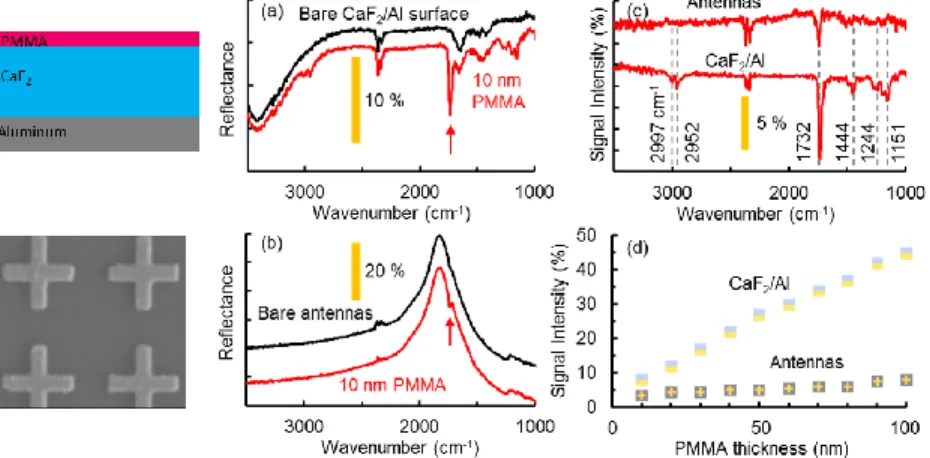
Fig 1. Experimental comparison of the CaF2/Al surface to the plasmonic antenna-array, for IR spectroscopy. Reflection spectra of bare and 10 nm
PMMA coated CaF2/Al surface (a), and plasmonic antenna surface (b). (c) Background subtracted signal intensities. (d) The thickness dependent
comparison of CaF2/Al and plasmonic antenna-array. 2. Results
Figure 1 shows the infrared absorption spectroscopy performances of the thin film surfaces and plasmonic
structures. We have selected PMMA as the probe material, Al as the reflector, CaF2 as the dielectric and Ag to form the plasmonic antennas that are defined by e-beam lithography on silicon. The thickness of CaF2 is chosen as 900 nm, to have a field enhancement above unity at the IR range of λ: 3-12 µm. Thus, the reflection spectrum without PMMA is mainly flat above ~3200 cm-1, as expected by the simulations, except the dips at 3400 and 1640 cm-1, which can be attributed to the O-H modes (Fig.1a). This broad bandwidth enables easy identification of all the PMMA bands ranging from 3000 to 1000 cm-1, even when there is a very thin layer of PMMA on CaF2 (Fig.1a).
JTu5A.77.pdf CLEO 2017 © OSA 2017
The reflection from the 10-nm-thick PMMA coated antennas, however, only shows the major PMMA peak at 1732 cm-1 (Fig.1b) with lower signal intensity (3.5% vs. 7.7%) as shown in the background-subtracted data in (Fig.1c). Besides the bandwidth and signal intensity, the spatial extent of the field enhancement by the CaF2 surfaces is much larger. Thus, when a thicker (100 nm) layer of PMMA is coated on both surfaces, the dip at 1732 cm-1 reaches 45% of the total reflection for the CaF2 surface, while the same dip is a mere 8% for the antenna surface (Fig.1d).
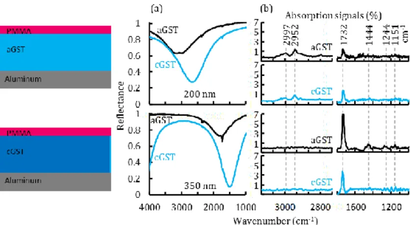
Fig 2. Infrared absorption spectroscopy of 10 nm PMMA films on GST/Al surfaces. (a) Reflection spectra of 10 nm PMMA coated aGST/Al and cGST/Al surfaces. 200 and 350 nm GST thicknesses are used to address PMMA’s lower and higher wavelength absorption bands. (b) Absorption of the PMMA layers extracted from the reflection spectra in (a). The major PMMA absorption bands are highlighted with dashed lines.. In Fig 2, we have demonstrated a thermally tunable IR spectroscopy platform based on GST phase-change films on Al mirror. The infrared absorption sensing performance of the fabricated surfaces are tested using 10 nm thick PMMA layers. The GST thicknesses are chosen as 200 and 350 nm targeting the PMMA vibrational bands around 3000 and 1500 cm-1, respectively. The PMMA absorption bands are observed as narrow dips on the reflection spectra (Fig. 2a). aGST (200 nm)/Al surface can sense the PMMA absorption bands at 2997, 2952 and 1732 cm-1. Crystallization of the GST layer, prior to coating the PMMA layer, lowers the absorption at 2997, 2952 cm-1 and enhances the absorption at 1732 and 1444 cm-1 (Fig. 2b). The major PMMA band at 1732 cm-1 is observed with a signal intensity of ~7% on aGST (350 nm)/Al surface. This surface is particularly good at sensing all the vibrational bands between 1732 and 1151 cm-1. On cGST(350 nm)/Al surface, although the signal intensity for 1732 cm-1 band drops to 3.5%, the higher wavelength absorption bands (1192-754 cm-1) appear as clear peaks.
3. Conclusion
In conclusion, we have demonstrated broadband (CaF2/Al) and tunable IR spectroscopy platform using interference coatings on metal mirrors. The proposed surfaces outperform plasmonic counterparts in terms of signal
enhancement, bandwidth and spatial extend of field.
4. References
[1] A. Hartstein, J. R. Kirtley, and J. C. Tsang, “Enhancement of the Infrared Absorption from Molecular Monolayers with Thin Metal Overlayers,” Phys. Rev. Lett., vol. 45, no. 3, pp. 201–204, Jul. 1980.
[2] C. Kendall et al., “Vibrational spectroscopy: a clinical tool for cancer diagnostics.,” Analyst, vol. 134, no. 6, pp. 1029–45, Jun. 2009. [3] R. Adato et al., “Ultra-sensitive vibrational spectroscopy of protein monolayers with plasmonic nanoantenna arrays.,” Proc. Natl. Acad.
Sci. U. S. A., vol. 106, no. 46, pp. 19227–32, Nov. 2009.
[4] H. Aouani et al., “Plasmonic Nanoantennas for Multispectral Surface-Enhanced Spectroscopies,” J. Phys. Chem. C, vol. 117, no. 36, pp. 18620–18626, Sep. 2013.
[5] L. V Brown, K. Zhao, N. King, H. Sobhani, P. Nordlander, and N. J. Halas, “Surface-enhanced infrared absorption using individual cross antennas tailored to chemical moieties.,” J. Am. Chem. Soc., vol. 135, no. 9, pp. 3688–95, Mar. 2013.
[6] R. Adato and H. Altug, “In-situ ultra-sensitive infrared absorption spectroscopy of biomolecule interactions in real time with plasmonic nanoantennas.,” Nat. Commun., vol. 4, p. 2154, Jan. 2013.
[7] D. Rodrigo et al., “Mid-infrared plasmonic biosensing with graphene,” Science (80-. )., vol. 349, no. 6244, pp. 165–168, Jul. 2015. [8] S. Ayas, G. Bakan, E. Ozgur, K. Celebi, and A. Dana, “Universal Infrared Absorption Spectroscopy Using Uniform Electromagnetic
Enhancement,” ACS Photonics, vol. 3, no. 3, pp. 337–342, Mar. 2016.
[9] G. Bakan, S. Ayas, E. Ozgur, K. Celebi, and A. Dana, “Thermally Tunable Ultrasensitive Infrared Absorption Spectroscopy Platforms Based on Thin Phase-Change Films,” ACS Sensors, Nov. 2016.