• Sonuç bulunamadı

Therapeutic bronchoscopic interventions for malignant airway obstruction: A retrospective study from experience on 547 patients.

N/A
N/A
Protected

Academic year: 2021

Share "Therapeutic bronchoscopic interventions for malignant airway obstruction: A retrospective study from experience on 547 patients."

Copied!
6
0
0

Yükleniyor.... (view fulltext now)

Tam metin

(1)

Therapeutic bronchoscopic interventions for

malignant airway obstruction

A retrospective study from experience on 547 patients

Levent Dalar (MD)

a,∗

, Cengiz Özdemir (MD)

b

, Yasin Abul (MD)

c

, Levent Karasulu (MD)

b

,

Sinem Nedime Sökücü (MD)

b

, Aysegül Akbas (MD)

d

, Sedat Alt

ın (MD)

b

Abstract

There is no definitive consensus about the factors affecting the choice of interventional bronchoscopy in the management of malignant airway obstruction. The present study defines the choice of the interventional bronchoscopic modality and analyzes the factors influencing survival in patients with malignant central airway obstruction. Totally, over 7 years, 802 interventional rigid bronchoscopic procedures were applied in 547 patients having malignant airway obstruction. There was a significant association between the type of stent and the site of the lesion in the present study. Patients with tracheal involvement and/or involvement of the main bronchi had the worst prognosis. The sites of the lesion and endobronchial treatment modality were independent predictors of survival in the present study. The selection of different types of airway stents can be considered on the base of site of the lesion. Survival can be estimated based on the site of the lesion and endobronchial brochoscopic modality used.

Abbreviations:APC= Argon plasma coagulation, COPD = chronic obstructive pulmonary disease, ICU = intensive care unit.

Keywords:bronchoscopy, lung cancer, malignant airway stenosis, stent, survival

1. Introduction

Endobronchial treatment with or without stenting is commonly used as a tumor debulking method for malignant central airway obstruction.[1,2]

A clinically significant airway obstruction may be caused by growth of an intraluminal tumor or by compression from an extrinsic tumor. Bronchogenic tumors, malignant lymphomas, esophageal tumors, tumors of larynx or thyroid, or metastases from an extrathoracic tumor may involve the central airways.[3]

The clinical symptoms and signs of these advanced malignancies include stridor, dyspnea, and obstructive pneumonia.

To the best of our knowledge, there is no absolute consensus about the factors influencing the choice of interventional bronchoscopic modality in the management of malignant airway stenoses with or without stenting. The purposes of the present study were to define the clinical factors which have potential to influence the choice of the interventional modality with or

without stenting for patients having malignant central airway obstruction. This study includes a large number of clinical experiences from the literature with a favorable follow-up period.

2. Methods

2.1. Study design and selection of patients

The present study was designed as a retrospective cohort study in which all the available data were retrospectively reviewed for patients referred to the Interventional Pulmonology Unit for central airway obstruction between August 2005 and January 2013 and for patients who were diagnosed as having malign airway obstruction and who underwent endobronchial treatment. All the patients in the cohort were followed up till January 2014. In studies with a retrospective analysis of standard diagnostic data, no ethical approval is needed in Turkey.

The Dumon (Novatech, France) stents were inserted using appropriate equipment as described in the literature.[4]

A diode laser operating at a wavelength of 980 nm with 4 to 25 Watt, pulsed mode (Biolitec, Ceralas D 25; Jena, Germany) was applied for the endobronchial treatment. Argon plasma coagula-tion (40 Watt, blended mode-continuousflow) was applied using an instrument manufactured by ERBE Elektromedizine GBMH (Tubingen, Germany). Standardized protocols for appropriate power selections were used in accord with to the manufacturer’s recommendations.

Cryotherapy was performed using the ERBOKRYO system (Elektromedizine GBMH, Tübingen, Germany).

All the patients were intubated by a rigid bronchoscope (Efer Endoscopy, Paris, France) under general anesthesia using standardized techniques and mechanical debridement was applied when it was necessary. All the patients had symptomatic relief after endobronchial treatment and stenting.

2.2. Collection of data, statistical analysis, and measurements Clinical and radiological data were obtained by using the hospital electronic database, chart reviews, and radiological systems. The Editor: Jianfeng Li.

The authors have no funding and conflicts of interest to disclose.

a

Istanbul Bilim University, Faculty of Medicine, Department of Pulmonary Medicine, Istanbul,b

Yedikule Thoracic Diseases and Thoracic Surgery Hospital, Interventional Pulmonology Unit, Istanbul,c

Karadeniz Technical University Faculty of Medicine, Department of Pulmonary and Critical Care Medicine, Trabzon,

d

Ministry of Health Bucak State Hospital, Burdur, Turkey.

Correspondence: Levent Dalar, Sisli Florence Nightingale Hospital, Sisli Istanbul, Turkey (e-mail: leventdalar@gmail.com).

Copyright© 2016 Wolters Kluwer Health, Inc. All rights reserved. This is an open access article distributed under the Creative Commons Attribution-NonCommercial License, where it is permissible to download, share and reproduce the work in any medium, provided it is properly cited. The work cannot be used commercially.

Medicine (2016) 95:23(e3886)

Received: 3 January 2016 / Received infinal form: 14 April 2016 / Accepted: 16 May 2016

Published online 1 May 2016

http://dx.doi.org/10.1097/MD.0000000000003886

Observational Study

Medicine

(2)

primary outcomes of the present study were defined as the response to stenting and endobronchial treatment of the patients, the rate of complications of the stenting and endobronchial treatment. Multiple and recurrent applications of stenting and total procedures for endobronchial treatment were also recorded. The chi-square test was used to assess the significance of factors influencing the choice of interventional brochoscopic treatment modality. A Kaplan–Meier log-rank analysis was used to determine the factors for survival. Factors which were significant on the basis of a univariate analysis were then analyzed using the Cox regression model. A P value of <0.05 was accepted as statistically significant.

3. Results

3.1. Clinical features of the study population

Totally, 802 interventional rigid bronchoscopic procedures were applied in 547 patients having a malign airway obstruction. The male to female ratio was 432 to 115 and median age was 63 years. The median follow-up period was 5.3 months (range, 0–100 months). Demographical findings and clinical features of the study population were summarized in Table 1.

3.2. Interventional bronchoscopic features of study population

The total laser application was performed in 250 procedures for 178 patients, argon plasma coagulation was used in 373 procedures for 257 patients, cryotherapy was performed in 93 procedures for 54 patients, and stents were applied during 171 procedures in 147 patients. Overall, 94 Y-stents and 52 tube stents (Novatech, La Ciotat, France) were placed into the central airways of patients having malign airway obstruction. Total flexible bronchoscopy (Olympus Evis Exera, Tokyo, Japan) was used in 391 patients for follow up of endobronchial treatment and/or stenting. For follow up, aflexible bronchoscopy was used in 100 patients after they had stenting. A laser was most commonly used in lung malignancies and was combined with stenting in 36 patients in the present study. Argon plasma coagulation (APC) was most commonly used for lung malignancies and was combined with stenting in 65 patients in the present study. Cryotherapy was most commonly used in lung malignancies and was combined with stenting in 5 patients in the present study.

The diagnoses and types of the malignancies are summarized in Table 2. Bronchoscopic view of the lesions and stents were shown in Figs. 1 and 2. Bronchogenic lung cancers were the most common

type of malignancy causing central airway obstruction followed by lymphoma and metastases from extra extrathoracic sites. The placement of an endobronchial stent was most commonly used for primary lung cancers in the present study. The primary lesions were confined only to the trachea in 65(11.9 %) patients; confined to the trachea plus right main bronchus in 87 (15.9 %) patients; confined to the trachea plus left main bronchus in 20 (15.9 %) patients; confined to the trachea plus both main bronchi in 121 (22.1 %) patients, and the right bronchial system and as well as left bronchial system were involved in 9 (1.6%) patients.

There was a significant association between the type of stent used and the site of the lesion (P=0.013). Insertion of Y stent alone was most frequently used for the lesions confined to both the left and the right bronchial system combined with a coexisting tracheal involvement. Tube stents were more frequently placed for lesions located in only the trachea or in left main bronchus. All patients reported substantial improvement in their dyspnea after stenting. There was no significant association between the use of the laser alone and the site of the lesion (P>0.05). There was a significant association between the use of Argon plasma coagulation and the site of the lesion (P=0.029). The Argon plasma coagulation was more frequently used for the lesions involving both trachea plus a main bronchus.

4. Complications

The complications were seen in total in 10.8% (59 out of 547) patients. Complications following endobronchial treatment were observed in 59 patients. Arrhythmias during endobronchial treatment were seen in 4 patients, a hypertensive attack was seen

Table 1

Demographicalfindings and number of patients according to clinical features and type of the tumor in the study population. Small cell carcinoma Nonsmall cell carcinoma Lymphoma Thyroid cancer Malignant mesothelioma Neuroendocrine tumor Carcinoid Malignant mesenchymal tumor Other metastasis Age (years±SD) 61.25±10.25 62.50±10.30 65.2±17.04 60.25±10.84 64±11.31 68.33±10.35 51.90±16.73 49.67±18.00 53.29±17.87 M:F ratio 42:11 351:60 6:3 1:3 2:0 5:4 12:17 5:4 4:10 Smoking history 44 352 5 3 2 8 19 5 8 COPD 12 55 3 — — 1 4 2 2 ICU stay 16 71 1 — — 3 5 2 2 Dyspnea 40 274 8 1 1 5 10 4 13 Cough 17 178 6 1 — 4 5 2 4 Hemoptysis 2 89 — 2 2 1 2 2 — Sputum production 10 81 4 1 — 1 2 1 3

COPD, Chronic obstructive pulmonary disease; ICU, Intensive care unit.

Table 2

Causes of airway obstruction and distribution of patients. Underlying malignancy causing of

airway obstruction

Number of patients n=547

Squamous cell cancer 199

Small cell cancer 53

Adenocarcinoma 31

Nonsmall cell lung cancer undefined 181

Carcinoid tumor 29

Lymphoma 9

Thyroid cancer 4

Renal cell cancer 4

Malign mesothelioma 2

Neuroendocrine tumor 9

Malign mesenchymal tumor 9

(3)

in 9 patients, and oxygen desaturation was seen 24 patients. Restenosis due to the tumor progression during an acute follow-up period was seen in 20 patients.

The patency of the airway lumen, the control of bleeding, and reduction of tumor size were achieved in all of the cases. 4.1. Prognosis and survival analysis

A total of 151 patients were still alive at the end of follow-up period. The median follow-up time was 5.3 months (range 0–100 months). Factors significantly affecting survival using the Kaplan–Meier log-rank analysis were type of malignancy causing central airway obstruction (P<0.01) (Fig. 3), site of lesion (P< 0.01) (Fig. 4), and the type of endobronchial treatment modality (P=0.01) (Fig. 5). Multivariate analysis using a Cox regression model showed that site of lesion and endobronchial treatment

modality were independent predictors of survival (Fig. 6) (Table 3).

5. Discussion

The primary indication for endobronchial treatment is palliation of central airway obstruction.[2,5]The common indications for

airway stenting are presence of intrinsic lesions, extrinsic compression, complex strictures, or a central airway fistula.[3]

There are no definitive data about factors such as type of disease or site of the lesion which may affect the use of a single or combined modality or stenting in the treatment of endoluminal lesions. The location, shape, and length of stenosis is an important consideration to determine the choice of stent.[1] A

significant association between the type of stent and the site of the lesion was seen in the present study. A bifurcated Y stent alone

Figure 1. (A) A 57-year-old patient with squamous cell carcinoma type and lobulated mass lesion located in midportion of the trachea causing 65% of the lumen obstruction before the endobronchial treatment. (B) Tumor was coaugulated with diode laser and obstruction was relieved totally by rigid bronchoscopic intervention. Scabbard trachea was also seen and noticed easily after the endobronchial treatment.

Figure 2. (A) A 54-year-old patient with internal and external tracheal tumoral compression who presented with respiratory insufficiency and tracheal lumen was obstructed>90%. (B) Early findings seen after dilatation of the distal trachea by rigid bronchoscopy. (C) Late findings seen after dilatation of the distal trachea by rigid bronchoscopy showing decreased tracheal osbtruction. (D) Placement of Dumon Y stent that was located on carina.

(4)

was most frequently used for lesions confined to both the left and the right bronchial system, having a coexisting tracheal involvement in the present study and tube stents were more commonly used [employed] for the lesions located in only the trachea or in the left main bronchus in the present study. The Dumon Y-stent has been proved to be useful and has been proposed to be well-tolerated in the management of malignant disease involving the main carina.[6] All the stents were well

tolerated in the present study. Y stents are usually used when dealing with long stenoses and strictures involving the trachea and the main bronchi. The inner surface of Y stents is smoother and no tumor tissue can grow between the single stents. Prevention of migration is another advantage of Y stents as they are located on the carina. Tube stents or so called straight stents have been used for placement in the trachea or larger bronchi.[3]

Follow-upflexible bronchoscopy was used in 100 patients after that had stenting in the present study. A study of this issue proposed the conclusion that routine follow-up bronchoscopies are not justified.[7]However follow-up bronchoscopy may reveal

a complication or a side-effect of stenting. If granulation tissue is present during follow-up bronchoscopy it is not obligatory that it

be removed. If the patient is not symptomatic, no action may be required. However, in the case of growth of further granulation tissue, the patient can be symptomatic and postobstructive pneumonia may be revealed. Therefore, early follow-up bronchoscopy may prevent this complication.[3]

Endobronchial stents are most frequently used for re-establish-ing the patency of narrowed central airways in lung malignancies. Malignant lymphomas, esophageal tumors, tumors of larynx or thyroid, or metastases from extrathoracic organs can also involve the major airway in addition to primary lung malignancies.[3] Primary lung cancers were the most common indication for endobronchial treatment and stenting in the present study. Other malignancies included lymphoma, tumors of the larynx, and metastasies from extrathoracic organs in the present study.

The present study showed that all the patients had symptom-atic relief after endobronchial treatment and stenting. Airway stenting is a useful treatment modality besides the other interventional bronchoscopic procedures. It not only provides rapid relief of symptoms and an improved quality of life but also gives additional time for adjuvant chemoradiotherapy that might provide prolonged survival.[2,8,9]

Complications related to the placement of the stents include reobstruction of the lumen by tumor, granuloma formation at the

Figure 5. Type of endobronchial treatment modality as a factor affecting survival of the study population represented as the Kaplan–Meier curve (P= 0.01).

Figure 6. Cumulative survival of study cohort during 365 days (1 year) period. Figure 3. Type of malignancy causing central airway obstruction as a factor

affecting survival of the study population represented as the Kaplan–Meier curve (P<0.01).

Figure 4. Site of the lesion as a factor affecting survival of the study population represented as the Kaplan–Meier curve (P<0.01).

(5)

stent side, mucous plugging and migration of the stent, or with the use of a laser, both perforation of airway wall andfire.[2,10] The stent-related complications were observed in 20 patients in the present study.

Total laser applications were performed in 250 procedures for 178 patients and there was no significant association between the use of the laser alone and the site of the lesion in the present study. One of the main indications for the use of laser bronchoscopy includes obstructive lesions of the trachea, the left and right main bronchi, the bronchus intermedius, and the lobar orifices that compromise ventilation and the severely symptomatic patients having dyspnoea, stridor, an intractable cough, or hemoptysis. The most common indication is inoperable lung malignancies having endobronchial features. In most cases, laser bronchoscopy is used together with other treatment modalities such as stenting, external beam irradiation, and brachytherapy.[2] Lasers were most commonly used in lung malignancies and combined with stenting in the present study.

External compression of the airways is the only absolute contraindication for the use of a laser. Hypoxemia, bronchoe-sophageal fistula, and coagulopathy are other relative contra-indications for laser use. Hypoxemia, bleeding, perforation, fistula formation, and fire are laser-related complications. Inspired oxygen concentration should be kept<40% to prevent the risk offire.[2]Hypoxemia was observed in 7 patients who had

received laser application in the present study.

Argon plasma coagulation was used in 373 procedures for 257 patients as an interventional bronchoscopic modality. Argon plasma coagulation was more frequently used for lesions

involving both trachea plus a main bronchus. Argon plasma coagulation is used with an ionized argon gas jetflow to conduct electrons providing a noncontact mode of treatment.[11]

Indications for argon plasma coagulation include coagulation of hemorrhage from the tracheabronchial system, and treatment of granulomatous lesions in relation with airway stents. Immediate symptomatic relief is provided by using argon plasma coagulation for tumor coagulation followed by mechanical debulking; this has been the accepted consensus strategy.[12]

Cryotherapy was used in 93 procedures for 54 patients. Cryotherapy is the endobronchial treatmtent modality of extreme cold for local destruction of viable tissue. Cryotherapy has a delayed action compared to laser or electrosurgery. The action of cryotherapy occurs from a few hours to a few days after application. Cryotherapy has been used for paliation of endobronchial exophytic obstructive lesions.[12] Cryotherapy was most commonly used in lung malignancies and combined with stenting in 5 patients in the present study.

There is no consensus about the factors influencing the choice of interventional bronchoscopic modality in the management of malignant airway stenoses with or without stenting. This issue has been raised by Chhajed et al and reported that there was no significant year effect for the treatment modality (laser, stent, or both) and the underlying disease as well as the nature of the lesion. On the other hand, the use of laser alone was reported as significantly influenced by the presence of lesions below the main bronchi.[13]In the present study, argon plasma coagulation was more frequently used for lesions involving both trachea plus a main bronchus.

Table 3

Characteristics of the study population affecting survival after interventional bronchoscopic procedures for malignant central airway obstruction. Survival P Characteristics Mean months (%95 CI) 3 months % 6 months % Univariate Multivariate

Type of underlying malignancy 0.006 0.7

Squamous cell lung cancer 21.5 (16–27) 62 47

Small cell lung cancer 15.3 (6.3–24.4) 38 28

Adenocarcinoma 28.5 (18.9–38.1) 73 70

Non-small cell lung cancer undefined 21 (14.4–27.6) 90 82

Carcinoid tumor 84.8 (78.9–90.7) 96 96

Lymphoma 20.8 (1.8–39.8) 71 57

Thyroid cancer 10.7 (5.9–15.5) 75 75

Renal cell cancer 25.6 (15.8–35.5) 100 100

Malign mesothelioma 1 (0.3–1.6) — —

Neuroendocrine tumor 7.1 (2.3–11.9) 55 44

Malign mesenchymal tumor 33.8 (14.3–53.3) 55 55

Other metastasis 13.4 (2.7–24) 42 28

Site of the lesion <0.001 <0.001

Trachea 20.1 (12.1–28) 56 43

Right main bronchus 43.2 (34.6–51.8) 73 60

Left main bronchus 41.6 (33.8–49.4) 77 68

Trachea+right main bronchus 14.4 (8–20.8) 46 38

Trachea+left main bronchus 10.1 (4.2–16) 47 36

Trachea+right+left main bronhus 10 (6.4–13.6) 41 28

Right main+Left main bronchus 13.3 (0.1–26.8) 33 13

Endobronchial treatment 0.013 0.02

Laser only 22.4 (16–27.8) 71 57

Argon plasma coagulation only 29.7 (21.7–37.9) 58 49

Cryotherapy only 20.9 (3.9–37.8) 57 35

Stent only 10.7 (5.9–15.4) 38 26

Laser+stent 7.9 (1.8–14.1) 42 11

(6)

The patients who had involvement of the trachea or both main bronchi had a worse prognosis than having a malignancy restricted to 1 lung. The patients in whom only laser therapy or only argon plasma coagulation was performed as an endobron-chial treatment had a better survival compared to other modalities of endobronchial treatment.

There are some limitations of this study. All cases are initially Stage IIIB and stage IV patients, but unfortunately we do not have the data of final stages in this retrospective cohort. Survival analysis should be based on the final stages to reflect the association with endobronchial therapy.

In conclusion, there is no consensus about the factors influencing the choice of interventional bronchoscopic modality in the management of malignant airway stenoses with or without stenting. There was a significant association between the type of stent and the site of the lesion in the present study. Patients with tracheal involvement or with involvement of the main bronchi had the worst prognosis. The site of the lesion and the endobronchial treatment modality were independent predictors of survival in the present study. Further prospective randomized trials are needed to define the factors influencing the choice of interventional bronchoscopic modality in the management of malignant airway stenoses.

Acknowledgements

The authors thank Prof RW Guillery from Oxford University for the English correction of the manuscript.

References

[1] Lee P, Kupeli E, Mehta AC. Airway stents. Clin Chest Med 2010;31:141–50.

[2] Bolliger CT, Sutedja TG, Strausz J, et al. Therapeutic bronchoscopy with immediate effect: laser, electrocautery, argon plasma coagulation and stents. Eur Respir J 2006;27:1258–71.

[3] Freitag L. Airway stents. Eur Respir Mon 2010;48:190–217. [4] Dumon JF. A dedicated tracheobronchial stent. Chest 1990;97:328. [5] Ernst A. Airway obstruction and stenting: more complex than we

thought? Am J Respir Crit Care Med 2004;169:1081–2.

[6] Dutau H, Toutblanc B, Lamb C, et al. Use of the Dumon Y-stent in the management of malignant disease involving the carina: a retrospective review of 86 patients. Chest 2004;126:951–8.

[7] Matsuo T, Colt HG. Evidence against routine scheduling of surveillance bronchoscopy after stent insertion. Chest 2000;118:1455–9. [8] Cavaliere S, Venuta F, Foccoli P, et al. Endoscopic treatment of

malignant airway obstructions in 2,008 patients. Chest 1996;110: 1536–42.

[9] Colt HG, Harrell JH. Therapeutic rigid bronchoscopy allows level of care changes in patients with acute respiratory failure from central airways obstruction. Chest 1997;112:202–6.

[10] Ernst A, Silvestri GA, Johnstone D. Interventional pulmonary proce-dures: guidelines from the American College of Chest Physicians. Chest 2003;123:1693–717.

[11] Grund KE, Storek D, Farin G. Endoscopic argon plasma coagulation (APC)first clinical experiences in flexible endoscopy. Endosc Surg Allied Technol 1994;2:42–6.

[12] Bolliger CT, Mathur PN, Beamis JF, et al. ERS/ATS statement on interventional pulmonology. European Respiratory Society/American Thoracic Society. Eur Respir J 2002;19:356–73.

[13] Chhajed PN, Somandin S, Baty F, et al. Therapeutic bronchoscopy for malignant airway stenoses: choice of modality and survival. J Cancer Res Ther 2010;62:204–9.

Şekil

Figure 2. (A) A 54-year-old patient with internal and external tracheal tumoral compression who presented with respiratory insufficiency and tracheal lumen was obstructed &gt;90%
Figure 6. Cumulative survival of study cohort during 365 days (1 year) period.Figure 3

Referanslar

Benzer Belgeler

In this case report; we presented the first case of giant TAS presenting with both spontaneous bleeding and respiratory distress needing tracheotomy for upper airway obstruction

ABSTRACT Objective: To evaluate the location of rhabdomyomas in the heart, and the spontaneous regression, clinical and echocardio- graphic findings and association of rhabdomyomas

In our study, we aimed to evaluate tracheobronchial infectious agents in sputum culture, and the relationship between cautious microorganisms and airway obstruction in

年老、虛弱、手臂無力者,可協助在上臂加壓使血管擴張,或漸歇性使用止血 帶紮緊上臂,加速靜脈動脈化。 冰敷原則 冰敷時每次約

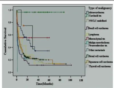
study 11 , which investigated the role of neoadjuvant chemotherapy in patients with resectable malignant pleural mesothelioma, similar survival data were obtained in patients who

In fact, a recent study showed that CMR performed dur- ing index hospitalization with MVO assessment provides bet- ter prognostic stratification of STEMI patients who underwent

All patients who were operated for antibradycardic pace- maker pulse generator and/or lead reimplantation in our institu- tion underwent ipsilateral venography through the ipsilateral

Conclusion:­The presence of deep vein thrombosis, idiopathic events, high D-dimer levels at the end of the first year and hereditary risk factors seem to be associated