Does lengthening after acute correction negatively
affect bone healing during distraction osteogenesis?
Correspondence: Özgür Karakoyun, MD. Namık Kemal Üniversitesi Tıp Fakültesi, Ortopedi ve Travmatoloji Anabilim Dalı, Tekirdağ, Turkey.
Tel: +90 282 – 250 50 00 e-mail: [email protected] Submitted: August 11, 2014 Accepted: December 12, 2014
©2015 Turkish Association of Orthopaedics and Traumatology
Available online at www.aott.org.tr doi: 10.3944/AOTT.2015.14.0275 QR (Quick Response) Code
doi: 10.3944/AOTT.2015.14.0275
Özgür KARAKOYUN1, Metin KÜÇÜKKAYA2, Mehmet Fatih EROL1
1Namık Kemal University Faculty of Medicine, Department of Orthopaedics and Traumatology, Tekirdağ, Turkey 2İstanbul Bilim University Faculty of Medicine, Department of Orthopaedics and Traumatology, İstanbul, Turkey
Leg lengthening with distraction osteogenesis can be performed safely using external fixators. The length-ening over nail (LON) and plating after lengthlength-ening (PAL) techniques have been developed to decrease complications and external fixator time. The distraction phase of these procedures is carried out with external
fixators. The consolidation phase is completed with an intramedullary nail or plate. Telescopic intramedullary nails, which enable internal lengthening, have been de-veloped.[1–3] In addition to decreasing complications
due to external fixators such as pin-tract infections or joint contractions due to muscle function limitations of
Objective: Lengthening after acute correction has a negative effect on bone healing during distraction
osteogenesis. In this study, we investigated whether correcting an acute deformity prior to lengthening resulted in a negative effect on bone healing.
Methods: Patients with shortened femora were assigned to 3 matched groups. Retrograde femoral
nailing after distal metaphyseal-diaphyseal osteotomy was used in all cases. Group 1 (9 femora) in-cluded cases of lengthening >4 cm using intramedullary distraction devices after acute correction. Group 2 (16 femora) included pure lengthening cases of ≥4 cm using intramedullary distraction de-vices. Group 3 (13 femora) included cases of lengthening ≥4 cm with lengthening and the retrograde nailing method (LORN) following acute correction.
Results: Healing indices and full weight-bearing times of patients were evaluated. Mean lengthening
values were 6.6 (range: 4–14 cm), 5.7 (range: 4–8 cm), and 5.2 cm (range: 4–6.5 cm) in Groups 1–3, respectively, and mean radiographic consolidation index and full weight-bearing times were 31.0±8.2, 30.2±5.5, and 39.0±5.0 day/cm in Groups 1–3, respectively. The consolidation index was significantly better in the Groups 1 and 2 compared to that in Group 3, but no difference was detected between Groups 1 and 2.
Conclusion: Acute correction had no negative effect on bone healing after distraction osteogenesis
us-ing new-generation intramedullary distraction devices. We suggest that the negative impact on healus-ing and the prolonged consolidation index in patients undergoing LORN may be due to impaired perios-teal blood supply due to fixator pins.
Keywords: Deformity correction before lengthening; distraction osteogenesis; fitbone; intramedullary
fixator pins with these nails, patients’ comfort increases during treatment.
Angular and/or rotational deformities are also typi-cally seen in shortened bone. These deformities can be corrected in an acute or gradual fashion during length-ening with external fixators according to basic principles of distraction osteogenesis.[4] Such deformities are
cor-rected acutely by lengthening, nailing, and LON tech-niques, as gradual correction is not possible.
Case series of lengthening after acute deformity cor-rection with external fixators have been published.[5,6]
These studies report that lengthening after acute correc-tion has no negative effect on bone healing. However, no report has investigated the effects of acute correction on bone healing prior to lengthening using LON or length-ening nail techniques.
In this study, we investigated whether acute defor-mity correction before lengthening resulted in a negative effect on bone healing.
Patients and methods
Thirty-four patients undergoing 38 femoral lengthening procedures >4 cm due to a leg length discrepancy or for cosmetic reasons between 2008 and 2012 were included in this study. The lengthening was performed with in-tramedullary distraction devices (Fitbone®, Wittenstein, Igersheim, Germany; Precice®, Ellipse Technology, Ir-vine, CA, USA) and the lengthening over retrograde nail (LORN) techniques. All osteotomies were performed to the distal metaphyso-diaphyseal area of the femur. Lengthening of ≥4 cm metaphyso-diaphyseal was ob-tained in all cases. Smokers and patients with metabolic bone diseases were excluded from this study.
Patients were categorized into 3 groups. Group 1 in-cluded patients with femoral deformities and limb length discrepancy who were undergoing lengthening with in-tramedullary distraction devices after acute correction of the deformity (Figure 1). Patients who required only lengthening of the femur were included in Group 2. Group 3 contained cases with a femoral deformity and limb length discrepancy that were lengthened with the LORN technique after acute correction.
Group 1 contained 9 patients with 6 post-traumat-ic deformities (5 females and 4 males; mean age: 25.2 years; range: 12–41 years). Poliomyelitis sequela was the etiology in 2 patients. One patient had an idiopathic an-gular deformity and a short femur. Three patients had valgus deformities, 2 had varus and external rotation deformities, 2 had varus and procurvatum, 1 had an ex-ternal rotation, and 1 had procurvatum. Mean femoral
lengthening was 6.6 cm (range: 4–14 cm). We observed the “running back” phenomenon in 2 cases, but the ex-pected lengthening values were achieved in those cases.
Sixteen femora of 13 patients with 6 cases of post-traumatic shortening were lengthened in Group 2 (9 fe-males and 4 fe-males; mean age: 25.6 years; range: 15–43 years). Other etiologies were fibular hemimelia in 1 pa-tient, achondroplasia in 1, and poliomyelitis in 1. Two patients presented idiopathic shortening of the femur, and 2 were undergoing lengthening for cosmetic reasons. No acute deformity was corrected in these patients. Mean femoral lengthening was 5.7 cm (range: 4–8 cm).
Thirteen femora of 12 patients who underwent femoral lengthening using the LORN technique after acute deformity correction were included in Group 3 (3 females and 9 males; mean age: 22.5 years; range: 16–35 years). Two idiopathic and 8 post-traumatic cases were included. The etiologies of the remaining patients were poliomyelitis in 1 and rickets in 1. Four patients had a valgus rotation, 1 an internal rotation, 3 an external ro-tation, 1 a valgus rotation and procurvatum deformity, 1 a valgus and external rotation, 1 a varus rotation, and 2 had a varus rotation and a procurvatum deformity. Three patients had additional tibial deformities. Mean femoral lengthening was 5.2 cm (range: 4–6.5 cm).
Joint range of motion (ROM), muscle strength, pres-ence of joint contracture, and rotational deformities were noted during the preoperative physical examination. De-formities were analyzed, and the shortened limbs were measured with standard long-leg radiograms. Medul-lary diameters were measured, and osteotomy sites were defined on actual-size direct radiograms of the bone segment to be lengthened. Radiological and laboratory studies confirmed no infection risk after intramedullary nailing. Nail insertion points and locations of polar screws to secure the bone in the correct position were defined on the radiograms in cases with additional femoral deformi-ties at the osteotomy level during preoperative planning.
The knee was flexed 45° while the patient was placed in the supine position. The 1st insertion guide wire was placed in the femoral intercondylar area through the pa-tellar tendon percutaneously under fluoroscopic control. One Schanz screw (DePuy Synthes, Zuchwil, Switzer-land) was placed at the proximal and distal ends of the femur before the osteotomy to prevent an iatrogenic ro-tational deformity. Following osteotomy, the medullary canal was reamed, and a nail was placed. Nail function was tested before wound closure. Cases with additional femoral deformities were corrected after osteotomy, and polar screws were placed according to the preoperative plan. LORN was carried out using the 3-Schanz pin
technique as described by Küçükkaya et al.[7]
Statistical analyses were carried out using the SPSS 18 software (SPSS Inc., Chicago, IL, USA). The Krus-kal-Wallis test was used to compare the 3 groups, since the data were not normally distributed. The Mann-Whitney U-test was used for dual comparisons between the groups. A p value <0.05 was considered to indicate statistical significance.
Results
The average follow-up period was 30.8 months (range: 16–45 months). Mean lengthening was 6.6 cm (range: 4–14 cm) in Group 1, 5.7 cm (range: 4–8 cm) in Group 2, and 5.2 cm (range: 4–6.5 cm) in Group 3. No differ-ence in lengthening was observed among the groups.
The mean angular deformity correction in Group 1 was 9° (range: 5–15°). Three patients had rotational de-formities in this group. Mean corrected rotational defor-mity was 25° (range: 20–30°). Mean corrected angular
deformity was 8° (range: 4-11°) in Group 3. Four cases of rotational deformities were corrected with a mean angle of 18.7° (range: 10–25°).
The mean bone healing index (BHI) was 31.0±8.2 days/cm in Group 1, 30.2±5.5 days/cm in Group 2, and 39.0±5.0 days/cm in Group 3. No difference in the con-solidation index was observed between Groups 1 and 2 (intramedullary distraction devices; p=0.83). How-ever, the consolidation index was significantly better in Groups 1 and 2 compared to that of Group 3 (p<0.05).
No knee or hip joint contracture was detected in any case. None of the cases had osteomyelitis or developed a superficial infection. In the LORN group, mild super-ficial pin-tract infections developed and were treated with proper antibiotherapy. This problem did not cause changes in treatment course in any cases.
Discussion
It is well documented that gradual deformity
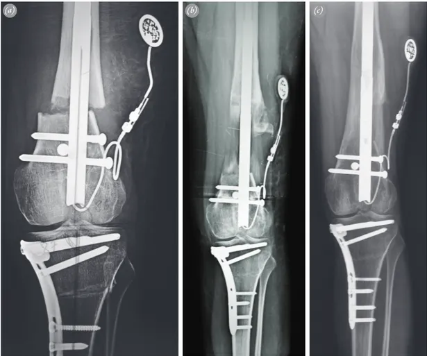
correc-Fig. 1. A 15-yr-old female with angular deformity and 8-cm shortening of the left femur accompanying a deformity of the
ipsilateral tibia. The tibial deformity was corrected acutely with osteotomy and fixed with plate and screws. (a) Left
femur; a Fitbone® intramedullary nail was applied after correcting the acute deformity. (b) Anteroposteror view 3
months after lengthening was completed. (c) One-year follow-up X-ray.
tion during lengthening with external fixators is a safe method. Considering studies on lengthening after acute angular correction, Noonan et al. lengthened 40 bone segments of 35 patients with external fixators follow-ing acute correction by open wedge osteotomy.[5] Mean
lengthening was 4.1 cm (range: 1.5–11 cm), mean angu-lar correction was 19° (range: 5–45°), and BHI was 52 days/cm. Donnan et al. reported results of lengthening following acute deformity correction in 11 tibial and 46 femoral segments using external fixators.[6] They found
that mean lengthening was 6.4 cm (range: 1.0–7.0 cm), mean angular correction was 23° (range: 7–45°), and the BHI was 55.5 day/cm. They used open wedge diaphyse-al osteotomy and grouped patients into 3 groups accord-ing to the degree of angular deformity. The groups in-cluded cases with angular deformities of 0–15°, 16–30°, and >30°. They found a significant difference in BHI in patients with an angular deformity >30°. Kamegaya et al. lengthened 14 tibiae and 2 femora of 11 patients fol-lowing acute angular correction using unilateral external fixators.[8] In their results, mean lengthening was 32.4
mm (range: 20–50 mm), mean angular correction was 23.8° (range: 14–40°), and BHI was 32.6 days/cm. They used open wedge osteotomy in cases with angular defor-mities <20° and dome osteotomy for angular deformi-ties >20°. They postulated that the area of bone contact between fragments increased by using these osteotomy techniques during the period before lengthening started. BHI values were equivalent in all 3 studies mentioned above. We used a metaphyseal drill osteotomy technique in all cases and prefer open wedge osteotomy to correct angular deformities. Mean lengthening in Group 1 was 6.6 cm (range: 4–14 cm), mean angular correction was 8.6° (range: 5–15°), and BHI was 31.0±8.2 days/cm. Mean lengthening in Group 2 was 5.7 cm (range: 4–8 cm), and BHI was 30.2±5.5 days/cm. Mean lengthen-ing in the LORN group was 5.2 cm (range: 4–6.5 cm), mean angular correction was 7.9° (range: 4–11°), and BHI was 39.0±5.0 days/cm. No case had an angular de-formity >30° in our series. Sufficient bone contact was obtained between fragments during the prelengthening period because we osteotomized the bone segment in the metaphyseal area.
Thaller et al. reported results of 6 femora and 4 tibiae lengthened in 11 patients using expandable nails. Mean BHI in their series was 27 days/cm.[1] They performed
acute deformity correction prior to lengthening in 3 cases without experiencing any complications. Krieg et al. used Fitbone® to lengthen 6 femora and 2 tibiae of 8 patients.[2] Six of their cases had angular deformities
with a mean mechanical axis deviation (MAD) of +22
mm (range: 10–35 mm). They corrected these deformi-ties acutely and reported a final MAD of +5 mm (range: 0–10 mm) before lengthening, with a mean BHI of 26 days/cm (range: 19–41 days/cm). Al-Sayyad length-ened 9 femora and 5 tibiae using Fitbone® in 10 patients, with a mean BHI of 24 days/cm (range: 20–39 days/ cm).[3] They did not report any challenge during
treat-ment of 3 patients who were acutely corrected prior to lengthening. BHI values before lengthening were similar during acute correction in our study to those reported by these studies. We found no significant differences in mean BHI values between intramedullary distraction devices.
BHI values of 26 and 24 days/cm have been reported in studies of lengthening with intramedullary distraction devices.[3,9] BHI values were 32.6, 52, and 55.5 days/cm
in lengthening studies that used external fixators and
LON.[5,6,8] In our study, BHI values were 30.2±5.5 and
39.0±5.0 days/cm in the 2 groups in which lengthening was performed with intramedullary distraction devices. These results reveal that BHI was greater in the exter-nal fixator group than in the intramedullary distraction device group. We believe that this result occurred due to damage to soft tissue and bone caused by the external fixator pins.
The limited number of cases and small degree of the corrected deformities were limitations of our study. However, our results are sufficient to comment on the effects of acute deformity correction before lengthening.
The acutely corrected angular deformities in our study were <30°. Therefore, we have no results on the negative effects of acute correction on BHI in patients with angular deformities >30°. We also cannot provide a neurovascular injury risk value for our patients. A short-ened extremity accompanying an angular deformity is a common clinical occurrence which is accepted as a lengthening limitation. However, our results reveal that lengthening can be safely carried out after acute correc-tion of angular deformities <30° by metaphyseal oste-otomy.
Conflics of Interest: No conflicts declared. References
1. Thaller PH, Fürmetz J, Wolf F, Eilers T, Mutschler W. Limb lengthening with fully implantable magnetically actuated mechanical nails (PHENIX(®))-preliminary re-sults. Injury 2014;45 Suppl 1:60–5.
2. Krieg AH, Speth BM, Foster BK. Leg lengthening with a motorized nail in adolescents : an alternative to external fixators? Clin Orthop Relat Res 2008;466:189–97.
3. Al-Sayyad MJ. Lower limb lengthening and deformity cor-rection using the Fitbone motorized nail system in the ado-lescent patient. J Pediatr Orthop B 2012;21:131–6. 4. Ilizarov GA. The transosseous osteosynthesis: theoretical
and clinical aspects of the regeneration and growth of tis-sue. Berlin: Springer-Verlag; 1992.
5. Noonan KJ, Price CT, Sproul JT, Bright RW. Acute cor-rection and distraction osteogenesis for the malaligned and shortened lower extremity. J Pediatr Orthop 1998;18:178– 86.
6. Donnan LT, Saleh M, Rigby AS. Acute correction of low-er limb deformity and simultaneous lengthening with a
monolateral fixator. J Bone Joint Surg Br 2003;85:254–60. 7. Kucukkaya M, Karakoyun O, Kuzgun U. Lengthening
over a retrograde nail using 3 Schanz pins. J Orthop Trau-ma 2013;27:13–7.
8. Kamegaya M, Shinohara Y, Shinada Y. Limb lengthen-ing and correction of angulation deformity: immediate correction by using a unilateral fixator. J Pediatr Orthop 1996;16:477–9.
9. Dinçyürek H, Kocaoğlu M, Eralp IL, Bilen FE, Dikmen G, Eren I. Functional results of lower extremity lengthening by motorized intramedullary nails. Acta Orthop Trauma-tol Turc 2012;46:42–9.