Dicle Tıp Dergisi, 2007 Cilt: 34, Sayı:4, (272-274)
* Dicle University, Faculty of Medicine, Department of Anatomy ** Diyarbakır State Hospital, Physical Medicine 272
The Variations of Torg Ratio with Gender in Patients with Neck Pain
Özlen Karabulut*, Zülfü Karabulut** SUMMARY
The Torg Ratio which may help to identify patients at risk for cervical spine cord injuries is an indicator of cervical canal stenosis determined by dividing the sagittal spinal canal diameter by the corresponding sagittal vertebral-body diameter. It was reported that male and female subjects showed differences in previous studies. The aim of this study was to investigate these differences. Our study that included the subjects 45 women and 45 men with neck pain was carried out at Diyarbakır State Hospital. The mean ages of male and female participants were 30 and 33. We measured both diaemeters on the lateral radiographs of the cervical spine in mm by using a Vernier Calipper.
Women had smaller sagittal spinal canal and vertebral body diameters at all levels. The narrowest sagittal spinal canal diameters measured were at C4 level in men and at C2 level in women. The mean Torg Ratio was 0.73 at C4 in men and 0.80 at C2 in women. We found larger Torg Ratios in females at all cervical vertebral levels except C2 level.
Key Words: Torg Ratio, Cervical Canal Stenosis
Boyun Ağrısı Olan Hastalarda Cinsiyete Göre Torg Oranı Farklılıkları
ÖZET
Torg Oranı, bir vertebrada spinal kanal çapının tekabül eden omur cismi sagital çapına bölünmesi ile elde edilen, servikal omurga yaralanması yönünden riskli hastaların teşhis edilmesine yardımcı olabilen bir göstergedir. Kadın ve erkeklerde bu oranın farklılıklar gösterdiği çalışmalarda kaydedilmiştir. Çalışma amacımız bu farkları araştırmaktı. Boyun ağrısı olan 45 kadın ve 45 erkek hastayı kapsayan çalışmamız Diyarbakır Devlet Hastanesinde gerçekleştirildi. Yaş ortalaması erkekler için 30 kadınlar için 33 saptandı. Her iki çap yan servikal grafide Vernier Calipper ölçüm aleti yardımı ile mm. cinsinden ölçüldü. Kadınlarda ölçülen sagital spinal kanal çapları ve omur cismi çapları her seviyede daha küçüktü. Ölçülen en dar sagital spinal kanal çapı erkeklerde C4 kadınlarda C2 seviyesindeydi. Ortalama Torg oranı erkeklerde C4 seviyesinde 0,73 ve kadınlarda C2 seviyesinde 0,80 bulundu. C2 dışında tüm servikal omurga düzeylerinde bu oran kadınlarda daha büyük bulundu.
Anahtar Kelimeler: Torg Oranı, Servikal Kanal Darlığı INTRODUCTION
The Torg Ratio is an indicator of cervical canal stenosis determined by dividing the sagittal spinal canal diameter (SDD) by the corresponding sagittal vertebral body diameter (VBD) (1). The results of the method may help to determine the presence of spinal stenosis
...
and to identify patients at risk for cervical spine cord injuries (2). The ratio of the sagittal diameter of the cervical canal to that of the vertebral body was first proposed by Torg et al. as an indicator of the degree of developmental canal narrowing (3). Using of this method may
Ö Karabulut ve Z. Karabulut Dicle Tıp Dergisi 2007
273 exclude magnification errors resulting from
nonstandardized film-tube distances (4). For these reasons the method is reliable and directly reflects the risk for spinal injuries. In a previous study a ratio of less than 0.80 has been shown to have a high sensitivity for transient cervical cord neurapraxia (CCN) in athletes (5). Another study showed that a ratio of less than 0.70 indicated significant spinal stenosis (4). In previous studies it was reported that male and female subjects showed differences in Torg Ratios when measured (6). This study aims to determine the variations of Torg Ratios between males and females in patients with neck pain and also aims to identify the narrowest vertebral level for each gender.
METHODS
This study was carried out at Diyarbakır State Hospital, during the period from September 2005 to May 2006; it included the subjects who were 45 women and 45 men with neck pain. The mean ages of male and female participants were 30 and 33. We measured the sagittal spinal canal diameter and vertebral body diameter on the lateral plain radiographs of the cervical spine. A plain lateral radiograph of the cervical spine was taken with the neck in a neutral position with the patient standing.
Lateral plain radiographs were taken with a 180 cm film to tube distance. We measured the sagittal spinal canal diameter and vertebral body diameter from C2 to C7 vertebrae. The measurement points were marked on the radiograph with a pencil. The distances between markings were measured. For these measurements we used a Vernier Calipper. We measured the both diameters in mm. We reported these values. We found the Torg Ratio for each patient. The Torg Ratio was determined by dividingthe sagittal spinal-canal diameter by the corresponding sagittal vertebral-body diameter. The following values were obtained:
Sagittal developmental diameter of the cervical canal (SDD): The distance between the cephalocaudal midpoint of the posterior aspect of the vertebral body to the nearest point on the corresponding spinal laminar line.
Anteroposterior diameter of the vertebral body (VB): The distance between the cephalocaudal midpoints of the anterior and posterior surfaces of the vertebral body.
Torg Ratio (SDD/VB): The ratio of the sagittal developmental diameter of the canal to the anteroposterior diameter of the vertebra .
We used students t tests for the statistical analysis.
RESULTS
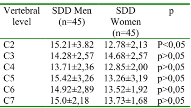
There isn’t a significant difference between two genders’ sagittal canal diameters except C2 level. But at C2 level women had significantly smaller sagittal canal diameters.
The narrowest sagittal spinal canal diameters were at C4 level (13.71 mm) in men and at C2 level (12.78 mm) in women (Table 1). Table 1. The mean SDD Values for men and women according to cervical vertebral levels Vertebral level SDD Men (n=45) SDD Women (n=45) p C2 15.21±3.82 12.78±2,13 P<0,05 C3 14.28±2,57 14.68±2,57 p>0,05 C4 13.71±2,36 12.85±2,00 p>0,05 C5 15.42±3,26 13.26±3,19 p>0,05 C6 14.92±2,89 13.52±1,92 p>0,05 C7 15.0±2,18 13.73±1,68 p>0,05
The vertebral body diameters were larger in men at all levels but the difference is not statically meaningful.
The vertebral body diameter was observed where the narrowest was C3 level (17.00 mm) in men and C7 level (15.96 mm) in women (Table 2).
Table 2. The mean VBD Values for men and women according to cervical vertebral levels Vertebral Level VBD Men (n=45) VBD Women (n=45) p C2 17.71±2,74 15.68±1,82 p>0,05 C3 17.92±3,11 16.00±2,01 p>0,05 C4 18.21±3,32 15.31±2,29 p>0,05 C5 18.00±3,12 16.68±3,30 p>0,05 C6 18.14±2,67 16.78±2,32 p>0,05 C7 18.14±2,56 17.05±2,27 p>0,05 - - - - -
Cilt: 34, Sayı:4, (272-274)
274
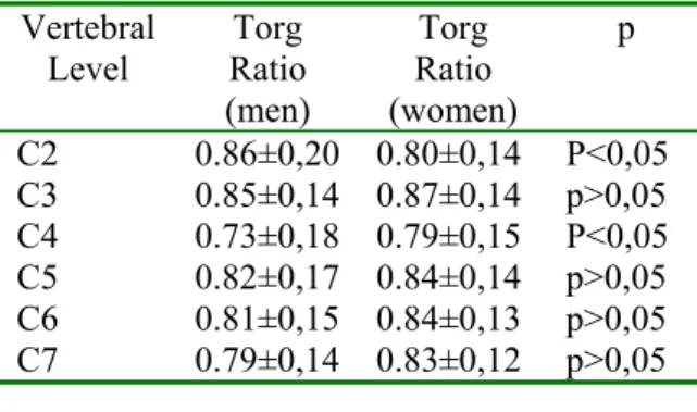
We found significant differences at two levels between the Torg Ratios of two genders in correlation. These were C2 and C4 vertebras. At only C2 level women had smaller Torg Ratios. At all other levels men had smaller ratios. At C4 men had the smallest ratios. (Table 3).
Table 3. Torg Ratios for men and women according to cervical levels
Vertebral Level Torg Ratio (men) Torg Ratio (women) p C2 0.86±0,20 0.80±0,14 P<0,05 C3 0.85±0,14 0.87±0,14 p>0,05 C4 0.73±0,18 0.79±0,15 P<0,05 C5 0.82±0,17 0.84±0,14 p>0,05 C6 0.81±0,15 0.84±0,13 p>0,05 C7 0.79±0,14 0.83±0,12 p>0,05 DISCUSSION
The Torg Ratio is an accurate indicator of spinal stenosis when radiographs are used. This is because the ratio avoids measurement differences caused by different target distances, object to film distance magnification errors common with radiographs. The spinal canal is compared with the vertebral body at the same spinal level (2,4). The vertebral body dimensions reported by Torg et al. were numerically similar to their corresponding canal diameters, resulting in ratios approaching unity throughout the cervical spine. However, the study of Herzog et al. demonstrated vertebral bodies were proportionately larger than the canal diameters at each level. This resulted in smaller sagittal diameter to vertebral body ratios of around 0.86 whereas the ratios reported by Torg et al. were significantly larger at around 1.00(5,7). In Lim’s study women had smaller SDDs at all levels of the cervical spine and female VBs were similar to their corresponding SDDs in size. Men had larger VBs when compared with corresponding SDDs. This resulted in small Torg Ratios in men (6).
In our study we found vertebral body dimensions proportionately larger than their corresponding canal diameters in both women and men. This resulted in small Torg Ratios both in women and in men. Women had the
smallest and significantly different SDDs at C2 level and the smallest Torg Ratio at the same level. Men had the smallest Torg Ratio at C4 level which was significantly different. There is no meaningful difference in SDD for women and men at that level but the largest VBD for men was at that level.
REFERENCES
1.Ryan T. Tierney, Catherine Maldjiant, Carl G, et al. Cervical Spine Stenosis Measures in Normal Subjects. Journal of Athletic Training by the National Athletic Trainers’ Association. 2002; 37: 190-193
2.Pavlov H, Torg JS, Robie B, et al. Cervical Spinal Stenosis: Determination with Vertebral Body Ratio Method. Radiology. 1987; 164: 771-775.
3.Torg JS, Pavlov H, Genvario SE, et al. Neurapraxia of the Cervical Spinal Cord with Transient Quadriplegia, J Bone Joint Surg. Am 1986; 68: 1354-1370
4.Castro FB Jr, Ricciardi J, Brunnet ME, et al. The Torg Ratio, and the Cervical Spine. Am J Sports Med. 1997; 25: 603-608.
5.Torg JS, Naranja RJJ, Pavlov H, et al. The Relationship of Developmental Narrowing of the Cervical Spinal Canal on Reversible and Irreversible Injury of the Cervical Spinal Cord in Football Players. J Bone Joint Surg Am 1996; 78-1308-1314
6.Jit-Kheng Lim, Hee-Kit Wong. Variations of the Cervical Spinal Torg Ratio with Gender and Ethnicity. The spine Journal 4 2004; 396-401.
7.Herzog RJ, Wiens JJ, Dillingham MF, et all. Normal Cervical Spine Morphometry and Cervical Spinal Stenosis in Asymptomatic Professional Football Players. Plain film Radiography, Multiplanar Computed Tomography and Magnetic Resonance Imaging. Spine 1991; 16: 178-186.
Corresponding Author Özlen KARABULUT
Dicle University, Faculty of Medicine Department of Anatomy, Diyarbakır E-mail: [email protected]