Biomedical Materials
In vitro biocompatibility of plasma-aided
surface-modified 316L stainless steel for intracoronary
stents
To cite this article: Cem Bayram et al 2010 Biomed. Mater. 5 055007
View the article online for updates and enhancements.
Related content
Microwave plasma activation of a polyvinylidene fluoride surface for protein immobilization
C Vasile, M C Baican, C M Tibirna et al.
-A plasma polymerization technique to overcome cerebrospinal fluid shunt infections
D Çökeliler, H Caner, J Zemek et al.
-Topical Review
Yan-Peng Jiao and Fu-Zhai Cui
-Recent citations
Surface modification of stainless steel for biomedical applications: Revisiting a century-old material
Aliya Bekmurzayeva et al
-Improving Corrosion Resistance of 316L Austenitic Stainless Steel Using ZrO2 Sol-Gel Coating in Nitric Acid Solution Mahdi Kazazi et al
-Electrodepositing salicylate modified PHEMA on stainless steel surface for hemocompatibility
B. Chen et al
Biomed. Mater. 5 (2010) 055007 (8pp) doi:10.1088/1748-6041/5/5/055007
In vitro biocompatibility of plasma-aided
surface-modified 316L stainless steel for
intracoronary stents
Cem Bayram
1,2, Alpay Koray Mizrak
3, Sel¸cuk Akt ¨urk
4,
Hurkan Kur¸saklio˘glu
5, Atila Iyisoy
5, Ahmet Ifran
6and
Emir Baki Denkba¸s
1,71Nanotechnology and Nanomedicine Division, The Institute For Graduate Studies in Science and
Engineering, Hacettepe University, 06800, Ankara, Turkey
2Biochemistry Division, Department of Chemistry, Aksaray University, 68100, Aksaray, Turkey 3Institute of Materials Science and Nanotechnology, Bilkent University, UNAM, 06800, Ankara, Turkey 4Department of Physics, Mugla University, 48000 K¨otekli, Mugla, Turkey
5Department of Cardiology, School of Medicine, Gulhane Military Medicine Academy, 06018, Ankara,
Turkey
6Department of Hematology, School of Medicine, Gulhane Military Medicine Academy, 06018, Ankara,
Turkey
E-mail:[email protected]
Received 27 April 2010
Accepted for publication 18 August 2010 Published 15 September 2010
Online atstacks.iop.org/BMM/5/055007
Abstract
316L-type stainless steel is a raw material mostly used for manufacturing metallic coronary stents. The purpose of this study was to examine the chemical, wettability, cytotoxic and haemocompatibility properties of 316L stainless steel stents which were modified by plasma polymerization. Six different polymeric compounds, polyethylene glycol, 2-hydroxyethyl methacrylate, ethylenediamine, acrylic acid, hexamethyldisilane and hexamethyldisiloxane, were used in a radio frequency glow discharge plasma polymerization system. As a model antiproliferative drug, mitomycin-C was chosen for covalent coupling onto the stent surface. Modified SS 316L stents were characterized by water contact angle measurements
(goniometer) and x-ray photoelectron spectroscopy. C1s binding energies showed a good correlation with the literature. Haemocompatibility tests of coated SS 316L stents showed significant latency (t-test, p < 0.05) with respect to SS 316L and control groups in each test. (Some figures in this article are in colour only in the electronic version)
Introduction
In today’s developing world, coronary artery disease (CAD) is the leading cause of cardiovascular mortality worldwide, with over 5 million deaths per year. The risk factors of CAD rise rapidly and continuously in developing countries [1]. CAD is conditioned with the deposition of fibro-fatty clusters—also known as atherosclerotic plaques—in the coronary arteries, preceded and accompanied by inflammation. Atherosclerosis is a chronic and widespread immunoinflammatory disease
7 Author to whom any correspondence should be addressed.
of large- and medium-sized arteries fuelled by atherogenic lipoproteins, in particular modified LDL [2,3]. The common non-surgical treatment is percutaneous transluminal coronary angioplasty (PTCA). The treatment of balloon angioplasty with intravenous metallic stents for cardiovascular system diseases has been the method of choice in the world with bypass surgery, but some complications exist that threaten human health and clash with the solution of metallic stents [4–9].
Early restenosis is still one of the major clinical problems of stent implantations with a 30% incidence in the first
Biomed. Mater. 5 (2010) 055007 C Bayram et al
three months following the operation [4, 5]. Secondary complications are coagulation on the metallic surface, inflammation of tissues and release of toxic metal ions and complexes by corrosion of the metal surface [6]. The implantation causes arterial wall injury. The steel stent itself has a thrombogenic nature. To minimize these complications and to provide maximum biocompatibility of metallic stents for human health has been a major goal since the discovery of the implantation method by biocompatible material sciences and medicine. The term biocompatibility is a phenomenon that occurs at the interface between the biomaterial and the host tissues. Although bulk properties dictate the mechanical properties of biomaterials, tissue–biomaterials interactions are surface phenomena and are governed by surface properties. These interactions have been hypothesized to occur within a narrow zone of less than 1 nm [10]. Biomaterials must possess bulk properties that meet other requirements, especially mechanical properties in order to have a proper function in its environment. To eliminate complications and increase biocompatibility, alternative and more effective surface modification techniques have been discovered [11–13]. Stainless steel is still the most widely used material for manufacturing intracoronary stents; however it is insufficient in haemocompatibility. There are numerous examples in the literature related to the disadvantages of metallic stents [14–17]. The modification of stent surfaces can be made with many techniques such as dipping, electrospraying, chemical vapour deposition, physical vapour deposition, plasma ion immersing and spraying. The stent modifications with methods mentioned above have many examples in the past two decades. Yoshioka et al reported that alginic acid coating onto SS 316L stents reduces platelet adhesion by up to 75% [18]. The stents which were coated with titanium nitric oxide by the physical vapour deposition method decreased vessel narrowing up to 45% compared with a negative control [19]. Spraying of active agents containing polymer solutions is also a useful approach in the surface treatment of stainless steel stents [20]. The thickness of casted polymer and adhesion forces play key roles in the effectiveness of surface modification. Coating by dipping and solvent-casting methods can result in a lack of uniformity, and also the thickness of the coating greater than the desired values makes the surface act as a bulky structure. Poor adhesion of a polymeric coating material onto the stent surface may cause it to delaminate under the dynamic conditions of bloodstream. Recently, surface modifications related to plasma processes have produced good and promising results. Radio frequency plasma treatment offers an advantageous mechanism to alter the surface properties of biomaterials and medical devices without affecting the mechanical properties of bulk structures. Such coatings are tightly adherent, conformal and easily applied. They also exhibit excellent biocompatibility qualities [21]. Saito et al reported that fluorinated diamond-like carbon films via the plasma-aided chemical vapour deposition technique could be a promising candidate for blood-contacting interventional devices with a dramatic decrease in the number of adhering platelets with respect to the untreated samples [22]. Plasma treatment of stainless steel stents before coating
with drug-eluting polymers incorporates functional groups that prevent the polymeric films from delaminating from the stent surface [23].
The surface treatment of 316L-type stainless steel stents was carried out with six different materials (polyethylene glycol (PEG), 2-hydroxyethyl methacrylate (HEMA), ethylenediamine (EDA), hexamethyldisilane (HMDS), hexamethyldisiloxane (HMDSO) and acrylic acid (AA)) by plasma-aided deposition. The polymeric precursors above were previously studied with blood-containing biomaterials, especially with the radio frequency glow discharge (RFGD) method [24–27]. The literature and commercial products have numerous examples on drug variety but lack data on covalently coupled active agent studies. Mitomycin C (MMC) is a potent DNA crosslinker and is used in several cancer therapies intravenously. Because of having antimitogenic ability, MMC was also previously used in in vivo vascular inflammation and neointimal hyperplasia studies [28,29].
Immobilization of the anticoagulant and antimitogenic agents on the stent surface using biodegradable ester or peptide bonds allows a sustained release of the agents [30]. The purpose of this study was to generate different surface characteristics and functional groups on the surface which can be useful for covalent coupling of bioactive agents subsequently.
Experimental details
Surface pre-cleaning of SS 316L stents
To avoid any interfering ion or radical that would come from the impurities of the surface during the plasma surface modification, the surfaces were pre-cleaned with piranha solution consisting of sulfuric acid (98% w/w) and hydrogen peroxide (30% w/w) in a 3:1 ratio for 5 min [20]. The pre-cleaned stents were then rinsed with distilled water two times for 10 min in a water sonication bath. The pre-cleaned SS 316L stents were dried in a vacuum oven and kept in closed glassware.
Plasma modification of SS 316L stents
The surface of the SS 316L stents were modified with the RFGD plasma deposition technique. A plasma modification system (Vacuum Praha, Czech Republic) was equiped with a 13.56 MHz radio frequency generator. The plasma reactor was attached with a vacuum pump for the evacuation of reactor gas. The reactor was fed with a monomer tank and argon gas during the process.
In the surface modification method six different monomers and prepolymers, AA (Acros, Belgium), PEG (Acros, Belgium, Mw= 300), HEMA (Aldrich, USA), EDA (Fluka, USA), HMDS and HMDSO (Merck, Germany), were used at two different power levels (25 and 50 W) and for a 10 min timeline.
The stents were placed perpendicularly onto a stereofoam support deployed in the middle of the electrodes with 1 cm spaces between each of the species. Argon gas was passed through the reactor at 0.1 mbar pressure to sweep away any
reactive species like oxygen and nitrogen. Subsequently, the reactor was fed with a coating polymer and the glow discharge initiated at two power levels (25 and 50 W). The plasma process lasted for 10 min and argon gas was passed through the chamber again to sweep away any gaseous residue. The stents were kept in vacuum for 10 min for the stabilization of the coating.
Mitomycin-C immobilization
The 316L stents containing –COOH groups, the surfaces of which were modified with AA, were incubated in 1% EDAC·HCl (1-[3-dimethyl aminopropyl]-3-ethyl carbodiimide hydrochloride) solution in 0.05 M MES (n-morpholino ethanosulfonic acid) for the activation of the –COOH functional groups generated by modification. After the incubation, mitomycin-C solution (0.02%) was added into the media for a direct covalent linkage of mitomycin-C via the –NH2functional groups. The procedure was carried out at
4◦C.
Surface characterization of stents
The bare metal stents and the surface-modified stents were characterized with a sessile drop contact angle measurement for the evaluation of hydrophilicity and x-ray photoelectron spectroscopy (XPS) for the characterization of the chemical surface composition by using plaques of 316L stainless steel. The contact angles of the plaque surfaces were measured by the sessile drop technique. The measurement apparatus consisted of a camera and camera lens (Hitachi, Japan), a sample platform and a water-dropping syringe. In the measurements, deionized water was used. The resulting images were analysed by using the ImagePro software (USA). The contact angles (θ ) of the surfaces were calculated with the formula
θ /2= arctan 2h/d.
Measurements were repeated at least 10 times and the results were given as averages with standard deviations. Student’s one-tailed t-test was employed to test the statistical significance of the contact angle measurements.
The XPS analysis of the stents was carried out in a Thermo Scientific K-Alpha x-ray photoelectron spectrometer, which utilizes monochromated Al K-alpha x-rays (1486.6 eV) to strike the sample surface. The analyser pass energy was 50 eV for the high-resolution core-level spectra and the beam spot was 400 μm. The curve fitting of the spectra was performed with the Thermo Avantage v4.41 software. A Shirley-type correction was applied to the background under all fitted peaks.
In vitro cytotoxicity assay
The prepared surface-modified SS 316L stents were subjected to an in vitro cytotoxicity test to investigate whether they had any serious cytotoxic effect on cell proliferation or not. 1× 2 cm stents were incubated with equal amounts of precultured mouse connective tissue fibroblast cells (L-929 cell line, Foot-and-Mouth Disease Institute of Ministry of Agriculture and Rural Affairs of Turkey) in growth media consisting of 90%
RPMI 1640 withL-glutamine and 10% FBS (Bio-Industries,
USA) in an incubator (Revco, USA) supplied with 5% CO2at
37◦C for 2 days [19]. Then the monolayer growing cells were harvested by using a trypsin–EDTA solution (Bio-Industries, USA) and immediately suspended in fresh cell growth media. 0.5 ml of cell suspension was mixed with 0.5 ml of 0.4% trypan blue solution (Bio-Industries, USA). The mixture was vortexed to obtain a homogeneous solution, and both viable (colourless) and non-viable (blue) cells were counted using a Thoma Haemocytometer.
The cell proliferation was investigated for three days and the results were compared with control groups.
In vitro haemocompatibility tests
Whole blood was collected from a healthy donor into 5 ml citrated (75 mm) tubes and centrifuged at 4000 rpm for 7 min at room temperature to obtain platelet poor plasma (PPP). The samples placed in a polystyrene 24-well plate dish were in contact with 1 ml of PPP for 30 min at 37 ◦C [18]. The activated partial thromboplastin time (APTT), prothrombin time (PT) and fibrinogen time were determined using an automated blood coagulation analyser (Diagnostica Stago, France). The measurements were triplicated. Student’s one-tailed t-test was employed to test the statistical significance of the PT, APTT and fibrinogen time measurements. The individual measurements for each modification were compared to the measurements of the bare SS 316L. Three independent measurements had been utilized for the assessment of significance. The p cut-off value was selected as 0.05 for significance.
Results and discussion
Surface characterization of SS 316L stents
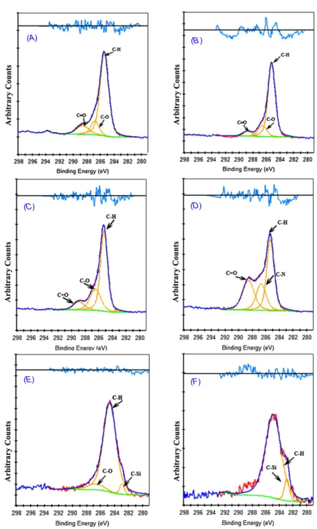
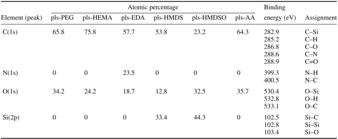
High-resolution scans of surfaces were taken at the C1s spectrum, to determine the types of carbon species of the surfaces. Figures 1(A)–(F) show the high-resolution C1s scans for the various modified stent surfaces. All species contain C–H aliphatic hydrocarbon bond at 285 eV due to carbon backbones. C1s scans of the HEMA-, PEG- and AA-modified surfaces show similar spectra and approximately the same amounts of C–H (285.2 eV), C–O (286.8 eV) and C=O (288.9 eV) [31]. The characteristic C–Si bond appeared after low-energy argon ion sputtering for the HMDS and HMDSO samples was carried out for 30 s at 282.9 eV [32] for each spectrum with a small C–O impurity in the HMDS coating at 286.8 eV. The EDA-modified surfaces have both a C–N peak at 288.6 eV in C1s scans and N–H bonds at 399.3 eV. Due to oxidation of the surfaces, oxygen was found in O1s scans in all species as O–Si, O–H and O–C at 530.4 eV, 532.8 eV and 533.1 eV, respectively. The atomic percentages of oxygen were found to be relatively larger when the polymerization precursor contained oxygen itself (PEG, HEMA, AA and HMDSO). The results are tabulated in table1.
Biomed. Mater. 5 (2010) 055007 C Bayram et al
(A) (B )
(C) (D)
(E) (F)
Figure 1.High resolution C1s spectra of the modified samples.
Contact angle measurements of SS 316L stents
Figure 2 shows the contact angles of the surfaces of SS 316L stents, which were determined by the sessile drop technique. The spreading drop shapes of water indicate the hydrophilicity of the surface. The individual measurements for each modification had been compared to the measurements of the bare SS 316L. Ten independent measurements had been utilized for the assessment of significance. The p cut-off value has been selected as 0.01 for significance (figure2).
The bare SS 316L plaque has a 91.6◦ water contact angle and has poor wettability properties. Surface wettability
was found to be changed by the used monomers in both an increasing and decreasing manner with respect to the different functional groups. The results show that the monomers which have polar and hydrophyl groups such as –OH, –NH2 and
C=O caused a decrease in the contact angle. –CH3, functional
group and Si-based structure containing plasma modifications generated more hydrophobic surfaces. The surface wettability increased with the decrease in contact angle [33].
The most wettable surface was found to be pls-PEG SS 316L with a discharge power of 50 W. The contact angle is 44.5◦ for the 50 W pls-PEG SS 316L, which means that it has a hydrophilic character. The functional group –OH is the most hydrophilic group among the used polymers. Other
Figure 2.Contact angle values of the samples.
Table 1.Elemental analysis of the samples.
Atomic percentage Binding
Element (peak) pls-PEG pls-HEMA pls-EDA pls-HMDS pls-HMDSO pls-AA energy (eV) Assignment
C(1s) 65.8 75.8 57.7 53.8 23.2 64.3 282.9 C–Si 285.2 C–H 286.8 C–O 288.6 C–N 288.9 C=O N(1s) 0 0 23.5 0 0 0 399.3 N–H 400.5 N–C O(1s) 34.2 24.2 18.7 12.8 32.5 35.7 530.4 O–Si 532.8 O–H 533.1 O–C Si(2p) 0 0 0 33.4 44.3 0 102.5 Si–C 102.8 Si–Si 103.4 Si–O
coatings which decreased the water contact angle of SS 316L are EDA and HEMA as 58.2◦ and 65.6◦, respectively, at 50 W discharge power. –NH2 in EDA and –OH and C=O in
HEMA play a role in increasing surface wettability. Surface wettability was decreased with the increase in discharge power in the EDA modification as can be seen in figure2.
The lowest wettable surface modification was obtained by the HMDSO plasma at 50 W discharge power. The contact angle of this modified surface is 103.7◦and it is more hydrophobic than the bare SS 316L itself. Also the plasma-assisted HMDS surface modifications generated hydrophobic surfaces with contact angles of 88.5◦ and 90.5◦ at 25 W and 50 W discharge powers, respectively. The results show that the silicone-based plasma-assisted modifications generate hydrophobic surfaces. The surface wettability order occurred
as
Pls− PEG SS 316L > Pls − EDA SS 316L
> Pls− AA SS 316L > Pls − HEMA SS 316L > Pls− HMDS SS316L > unmodified bare SS 316L > Pls− HMDSO SS 316L.
In vitro cytotoxicity assay
In vitro cell proliferation test was applied to the
plasma-modified SS 316L stents. The results are shown in figure3. There were no serious cytotoxic effects on the cell proliferation of the species that interacted with the surface-modified stents. Except for the drastic decrease in relative cell proliferation value of the AA–MMC stent, we can say that the antimitogenic
Biomed. Mater. 5 (2010) 055007 C Bayram et al
Figure 3.Cytotoxicity results of the samples.
Figure 4.Prothrombin time results of the samples.
effect of mitomycin-C drug had inhibited the mitogenic procedure as a potent DNA crosslinker (figure3).
Blood coagulation tests of SS 316L stents
PT, APTT and fibrinogen time measurements are essential haemocompatibility tests for the behaviour of a biomaterial that is in contact with blood [18,34].
These tests were applied to the materials to evaluate the effect of modifications in the coagulation cascade.
Figures4–6show the values of PT, APTT and fibrinogen time of the plasma in contact with the samples.
Prothrombin, which is a precursor to thrombin, is a coagulation (clotting) factor needed for the normal clotting of blood. The prothrombin time is commonly used as a method of monitoring the accuracy of the blood-thinning treatment with anticoagulants. The reference range for this test time is generally around 12–15 s. The values measured for the materials were close to the control shown in figure4, but pls-HEMA SS 316L have an increasing effect with respect to bare SS 316L on PT, which belongs to the extrinsic coagulation pathway about 0.5 s (p < 0.05). Pls–EDA SS 316L have a decreasing effect on PT about 0.4 s with respect to SS 316L, which means the surface has some activating properties on the extrinsic coagulation pathway (p < 0.05).
APTT is a blood test that measures how long it takes for blood to clot. It is the main test that reveals the clotting time problems. The test is essential for the biocompatibility of material. The biomaterials that trigger host response shorten the test time. In the values of the APTT pls-HMDS SS 316L,
Figure 5.Activated partial thromboplastin time results of the samples.
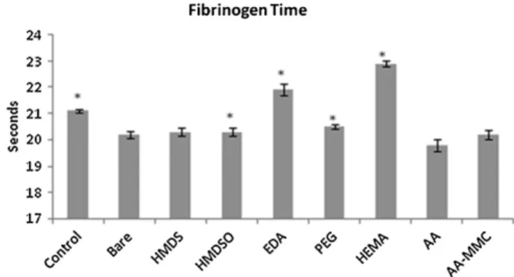
Figure 6.Fibrinogen time results of the samples.
pls-HMDSO SS 316L and pls-HEMA SS 316L, there are elevations in clotting time with respect to the values of the unmodified SS 316L stents as 1.3, 1.4 and 2.6 s, respectively (figure5) (p < 0.05). The results indicate an adsorption of proteins related to the intrinsic coagulation pathway [18].
In figure 6, there are some differences in clotting time in the measurements of the fibrinogen test. Fibrinogen is usually carried out with other blood clotting tests. Fibrinogen levels are a reflection of the clotting ability and activity in the body. For pls-HEMA SS 316L with pls-EDA SS 316L, which again refer to an adsorption of plasma proteins that are involved with the intrinsic coagulation pathway [18], the clotting time increases by 2.1 and 1.1 s, respectively, with respect to unmodified bare SS 316L stents (p < 0.05). In contrast, the clotting time shortened in pls-HMDSO and pls-AA modifications by 0.5 and 0.8 s, respectively, which indicates that the surface characteristics of these materials have inducing properties of fibrin formation (p < 0.05).
Conclusion
In this work, we reported that bare stents would be more effective and biocompatible with the surface treatment technique and further biomodifications. For altering the host response, mitomycin-C was selected as the model drug. Mitomycin-C is a well-known anticancer drug and also has some potent inhibitor effects against vascular smooth muscle
cell proliferation and neointima formation after arterial injuries [35,36].
The coating of the SS 316L stents with polymeric materials was carried out in the study. The new surface properties showed good compatibility with an increase in wettability of some modifications with no cytotoxic effect on cell proliferation. Although different gas plasma treatments were applied to the material surfaces to incorporate different functional groups, there is no clear correlation between them and the blood surface reactions. However, our results demonstrate that there are some relative chemical changes for reducing protein activation events that are involved with the coagulation cascade. The resulting coatings herein provide a form of materials that stands between the bare metal stents and drug-eluting stents. With the functional groups that exist on the surface of the coating, we expect that it will be possible to bind any anticoagulant or antimitogenic agents related with CAD and have an opportunity to develop intracoronary stents in an alternative way.
Acknowledgments
This work was funded by The Scientific and Technological Research Council of Turkey, Health Sciences Research Group (SBAG) project number 106S182. Additionally, the stents used in this study were kindly gifted by Alvimedica Medical Inc., Turkey.
References
[1] Okrainec K, Banerjee D K and Eisenberg M J 2004 Coronary artery disease in the developing world Am. Heart J.
1487–15
[2] Stary H C, Chandler A B, Dinsmore R E, Fuster V, Glagov S, Insull W, Rosenfeld M E, Schwartz C J, Wagner W D and Wissler R W 1995 A definition of advanced types of atherosclerotic lesions and a histological classification of atherosclerosis—a report from the committee-on-vascular-lesions of the council-on-arteriosclerosis
Circulation 92 1355–74
[3] Ross R 1993 The pathogenesis of atherosclerosis—a perspective for the 1990s Nature362801–9
[4] Joung Y K, Kim H I, Kim S S, Chung K H, Jang Y S and Park K D 2003 Estrogen release from metallic stent surface for the prevention of restenosis J. Controlled Release
9283–91
[5] Michaels A D and Chatterjee K 2002 Angioplasty versus bypass surgery for coronary artery disease Circulation
106E187–90
[6] Bennett M R and O’Sullivan M 2001 Mechanisms of angioplasty and stent restenosis: implications for design of rational therapy Pharmacol. Ther.91149–66
[7] Fattori R and Piva T 2003 Drug-eluting stents in vascular intervention Lancet361247–9
[8] Moreno R, Fernandez C, Hernandez R, Alfonso F, Angiolillo D J, Sabate M, Escaned J, Banuelos C, Fernandez-Ortiz A and Macaya C 2005 Drug-eluting stent thrombosis—results from a pooled analysis including 10 randomized studies J. Am. Coll. Cardiol.45954–9 [9] Steffel J and Tanner F C 2007 Biological effects of
drug-eluting stents in the coronary circulation Herz
32268–73
[10] Kirkpatrick C J, Wagner M, Kohler H, Bittinger F, Otto M and Klein C L 1997 The cell and molecular biological approach to biomaterial research: a perspective J. Mater.
Sci. Mater. Med.8131–41
[11] Wei J H, Igarashi T, Okumori N, Igarashi T, Maetani T, Liu B L and Yoshinari M 2009 Influence of surface wettability on competitive protein adsorption and initial attachment of osteoblasts Biomed. Mater.4045002 [12] Cokeliler D, Caner H, Zemek J, Choukourov A, Biederman H
and Mutlu M 2007 A plasma polymerization technique to overcome cerebrospinal fluid shunt infections Biomed.
Mater.239–47
[13] Cokeliler D, Erkut S, Shard A G, Akdogan E, Ozden N, Imirzalioglu P and Mutlu M 2008 A novel approach for improvement of the interfacial binding of ceramics for dental materials: chemical treatment and oxygen plasma etching J. Appl. Polym. Sci.1102656–64
[14] Haidopoulos M, Turgeon S, Laroche G and Mantovani D 2005 Surface modifications of 316 stainless steel for the
improvement of its interface properties with
RFGD-deposited fluorocarbon coating Surf. Coat. Technol.
197278–87
[15] Riepe G, Heintz C, Kaiser E, Chakfe N, Morlock M, Delling M and Imig H 2002 What can we learn from explanted endovascular devices? Eur. J. Vasc. Endovasc. Surg.
24117–22
[16] Bertrand O F, Sipehia R, Mongrain R, Rodes J R, Tardif J C, Bilodeau L, Cote G and Bourassa M G 1998
Biocompatibility aspects of new stent technology J. Am.
Coll. Cardiol.32562–71
[17] Indolfi C, Mongiardo A, Curcio A and Torella D 2003 Molecular mechanisms of in-stent restenosis and approach to therapy with eluting stents Trends Cardiovasc. Med.
13142–8
[18] Yoshioka T, Tsuru K, Hayakawa S and Osaka A 2003 Preparation of alginic acid layers on stainless-steel substrates for biomedical applications Biomaterials
242889–94
[19] Windecker S, Mayer I, De Pasquale G, Maier W, Dirsch O, De Groot P, Wu Y P, Noll G, Leskosek B and Meier B 2001 Stent coating with titanium-nitride-oxide for reduction of neointimal hyperplasia Circulation104928–33
[20] Okner R, Oron M, Tal N, Mandler D and Domb A J 2007 Electrocoating of stainless steel coronary stents for extended release of Paclitaxel Mater. Sci. Eng. C
27510–3
[21] Ratner B D 1992 Plasma deposition of biomedical
applications—a brief review J. Biomater. Sci.– Polym. Ed. 4 3–11
[22] Saito T, Hasebe T, Yohena S, Matsuoka Y, Kamijo A, Takahashi K and Suzuki T 2005 Antithrombogenicity of fluorinated diamond-like carbon films Diamond Relat.
Mater.141116–9
[23] Okamoto K, Nakatani T, Yamashita S, Takabayashi S and Takahagi T 2008 Development of
surface-functionalized drug-eluting stent with diamond-like carbon nanocoated by using PECVD method Surf. Coat.
Technol.2025750–2
[24] Tseng D Y and Edelman E R 1998 Effects of amide and amine plasma-treated ePTFE vascular grafts on endothelial cell lining in an artificial circulatory system J. Biomed. Mater.
Res.42188–98
[25] Park K, Shim H S, Dewanjee M K and Eigler N L 2000 In
vitro and in vivo studies of PEO-grafted blood-contacting
cardiovascular prostheses J. Biomater. Sci.– Polym. Ed.
111121–34
[26] Kiaei D, Hoffman A S and Hanson S R 1995 Ex vivo and in
vitro platelet adhesion on RFGD-deposited polymers J. Biomed. Mater. Res.26357–72
Biomed. Mater. 5 (2010) 055007 C Bayram et al [27] Nho Y C and Kwon O H 2003 Blood compatibility of AAc,
HEMA, and PEGMA-grafted cellulose film Radiat. Phys.
Chem.66299–307
[28] Chaudhuri V, Potts B R and Karasek M A 2006 Mechanisms of microvascular wound repair I, role of mitosis, oxygen tension, and I-kappa B In Vitro Cell. Dev. Biol.– Anim. 42 308–13
[29] Strauss B H, Wilson R A, van Houten R, van Suylen R J, Murphy E S, Escaned J, Verdouw P D, Serruys P W and van der Giessen W J 1994 Late effects of locally delivered mitomycin C on formation of neointima and on vasomotor response to acetylcholine Coron. Artery Dis. Ther. Prev.
5633–41
[30] Kim Y J, Kang I K, Huh M W and Yoon S C 2000 Surface characterization and in vitro blood compatibility of poly(ethylene terephthalate) immobilized with insulin and/or heparin using plasma glow discharge Biomaterials
21121–30
[31] Valdes T I, Ciridon W, Ratner B D and Bryers J D 2008 Surface modification of a perfluorinated ionomer using a glow discharge deposition method to control protein adsorption Biomaterials291356–66
[32] Shivaraman S, Chandrashekhar M V S, Boeckl J J and Spencer M C 2009 Thickness estimation of epitaxial graphene on SiC using attenuation of substrate Raman intensity
J. Electron. Mater.38725–30
[33] Mahapatro A, Johnson D M, Patel D N, Feldman M D, Ayon A A and Agrawal C M 2006 Surface modification of functional self-assembled monolayers on 316L stainless steel via lipase catalysis Langmuir22901–5
[34] Tan Q G, Ji J, Barbosa M A, Fonseca C and Shen J C 2003 Constructing thromboresistant surface on biomedical stainless steel via layer-by-layer deposition anticoagulant
Biomaterials244699–705
[35] Tang L L, Chen X C, Tang S B, LaLonde T and
Gardin J M 2007 Granulation encapsulated stent: a new therapeutic approach for vascular implantation Heart
93238–43
[36] Granada J F, Ensenat D, Keswani A N, Kaluza G L, Raizner A E, Liu X M, Peyton K J, Azam M A, Wang H and Durante W 2005 Single perivascular delivery of mitomycin C stimulates p21 expression and inhibits neointima formation in rat arteries Arterioscler. Thromb. Vasc. Biol.
252343–8