The Effect of a Special Amino Acid
Mixture on Healing of Left Colonic
Anastomosis: an Experimental Study
AABBSSTTRRAACCTT OObbjjeeccttiivvee:: Arginine, glutamine, and Beta-hydroxy-beta-methylbutyrate were com-bined in a dietary supplement. This specialized amino acid mixture enhances wound collagen ac-cumulation and increases wound healing. We aimed to investigate the effects of this special amino acid mixture on the healing process of experimental left colonic anastomosis. MMaatteerriiaall aanndd M Meetthh--ooddss:: The study included 20 adult male Wistar-Albino rats. The study group (n=10) received 685 mg/kg/day specialized amino acid mixture for 7 days until 12 h before surgery and was maintained throughout the study. Following midline laparatomy a 1-cm segment of the left colon was resected. Bowel continuity was restored with an end-to-end anastomosis. The animals were re-anesthetized on day 7 after the operation and anastomotic bursting pressure was measured by passing a catheter per anum up to the area of anastomosis. Anastomotic segments were removed en bloc and were vertically divided into two. One was used for hydroxyproline measurement and the other for histopathological examination. Abramov’s histologic scoring system was used in this study. RReessuullttss:: The hydroxyproline levels and bursting pressures in the study group were superior compared to the control group. Collagen deposition and reepithelization scores of the study group were higher than that of the control group. CCoonncclluussiioonn: Results of the present study indicates that significantly en-hancing the anastomotic bursting pressure, hydroxyproline level and collagen deposition may im-prove anastomosis healing.
KKeeyy WWoorrddss:: Colorectal surgery; arginine; glutamine; beta-hydroxyisovaleric acid
Ö
ÖZZEETT AAmmaaçç:: Arjinin, glutamin ve beta-hidroksi-beta-metilbütirat, diyet desteği olarak kombine edilmiştir. Bu özel amino asit karışımı, yara yerinde kollajen birikimini artırır ve yara iyileşmesini geliştirir. Bu çalışmanın amacı, deneysel sol kolon anastomozunun iyileşmesinde özel amino asit karışımının etkilerini araştırmaktır. GGeerreeçç vvee YYöönntteemmlleerr:: Çalışmada 20 adet yetişkin Wistar-Al-bino sıçan kullanıldı. Çalışma grubu (n=10) ameliyattan 7 gün öncesinden başlayarak tüm çalışma süresince 685 mg/kg/gün özel amino asit karışımı aldı. Orta hattan karın açıldı ve 1 cm’lik sol kolon segmenti rezeke edildi. Uç-uca anastomoz yapılarak barsak lümeninin devamlılığı sağlandı. Sıçanlara bu ameliyattan 7 gün sonra tekrar anestezi verildi. Anastomoz patlama basıncı, anüsten yerleştirilip anastomozun üzerine çıkan bir kateter ile ölçüldü. Anastomoz sahaları sağlam sınırlarla çıkarıldı ve dikey olarak iki parçaya bölündü. Bir parça hidroksiprolin ölçümünde, diğer parça his-tolojik değerlendirmede kullanıldı. Hishis-tolojik değerlendirme için Abramov’un skorlama sistemi kullanıldı. BBuullgguullaarr:: Hiroksiprolin düzeyleri ve patlama basınçları çalışma grubunda, kontrol gru-buna göre daha yüksekti. Kollajen birikimi ve reepitelizasyon skorları çalışma grubunda daha be-lirgindi. SSoonnuuçç:: Bu çalışmanın sonuçları, özel amino asit karışımının, anastomoz patlama basıncı, hidroksiprolin düzeyi ve kollajen birikimini arttırarak anastomoz iyileşmesinde yararlı olabile-ceğini göstermiştir.
AAnnaahhttaarr KKeelliimmeelleerr:: Kolorektal cerrahi; arjinin; glutamin; beta-hidroksivalerik asit
TTuurrkkiiyyee KKlliinniikklleerrii JJ MMeedd SSccii 22001133;;3333((33))::667788--8844
İsmail YAMAN,a
Cemal KARA,b
Hayrullah DERİCİ,a
Gülden DİNİZ,c
Ragıp ORTAÇ,c
Beyhan Cengiz ÖZYURTd
aDepartment of General Surgery,
Balıkesir University Faculty of Medicine, Balıkesir
bClinic of General Surgery,
İzmir Karşıyaka State Hospital,
cClinic of Pathology,
Dr. Behçet Uz Children's Hospital, İzmir
dDepartment of Public Health,
Celal Bayar University Faculty of Medicine, Manisa
Ge liş Ta ri hi/Re ce i ved: 11.05.2012 Ka bul Ta ri hi/Ac cep ted: 19.11.2012 Ya zış ma Ad re si/Cor res pon den ce: İsmail YAMAN
Balıkesir University Faculty of Medicine, Department of General Surgery, Balıkesir, TÜRKİYE/TURKEY
doi: 10.5336/medsci.2012-30385
nastomotic leakage (AL) is one of the most serious surgical complications after col-orectal surgery. Although many local and systemic factors affecting anastomotic healing have been determined, AL is still a serious problem that significantly increases morbidity and mortality rates.1The prevalence of AL varies in the literature
between 0.5% and 30%.2,3Therefore, research has
focused on various systemically or locally applied materials that can improve anastomotic healing.1-3
Arginine (ARG) is a dietary semi essential amino acid that becomes conditionally indispensa-ble in trauma and surgical patients.4,5Dietary ARG
supplementation induces positive nitrogen balance, increases hydroxyproline (HP) content, wound col-lagen accumulation, shows antioxidative effect and enhances wound healing.4-7Glutamine (GLU) is a
preferred energy source for cells of the intestinal mucosa and of the immune system; furthermore GLU and ARG have immune stimulatory effects.5,8
GLU is the most abundant amino acid, but its con-centration falls after injury, surgery, or infection.3In
various experimental and clinical studies, GLU had a positive effect on both wound and anastomotic heal-ing.9,10Beta-hydroxy-beta-methylbutyrate (HMB) is
a metabolite of the essential amino acid leucine.4It
reduces muscle proteolysis, improves nitrogen bal-ance and wound collagen deposition.4,7,11 ARG,
GLU and HMB have been combined in a dietary supplement specifically designed for patients with catabolic diseases.7Williams et al.4reported that
this special amino acid mixture enhanced wound collagen accumulation and increased wound heal-ing. With these findings in mind, we aimed to ex-amine the therapeutic efficacy of the special amino acid supplement on the healing process of experi-mental left colonic anastomosisby measuring HP level, bursting pressure (BP) and inflammatory changes in anastomosis.
MATERIAL AND METHODS
The study included 20 adult male 5-month-old Wistar-Albino rats weighing between 250 and 300 g. The procedures were run at the Ege University Faculty of Medicine animal research laboratory. All animals were housed in cages under standard
con-ditions (room temperature 22°-24°C, 12-hour light/dark cycle). All experimental manipulations and postoperative care were undertaken in accor-dance with the National Institutes of Health Guide for the Care and Use of Laboratory Animals. The study was also approved by the Animal Ethics Com-mittee of the Balıkesir University Medical School. The rats were fed on standard laboratory diet and water ad libitum and had free access to water and standard rat chow until 12 h before surgery. Twenty rats were randomized into two groups of 10 each. The study group (SG, n=10) received 685 mg/kg/day specialized amino acid mixture (Abound®Abbott,
İstanbul, Turkey) dissolved in 1ml water per day for 7 days until 12 h before the surgery and was main-tained throughout the study while the control group (CG, n=10) received the same amount of saline orally with orogastric tube.
OPERATION PROCEDURE
Each rat was anesthetized with intramuscular injec-tion of ketamin hydrochloride (Ketalar, Eczacıbası, Warner-Lambert Laboratories, Levent, İstanbul, Turkey) 60 mg/kg and xylazine hydrochloride (Rompun, Bayer Laboratories, Şişli, İstanbul, Turkey) 10 mg/kg. All procedures were performed under ster-ile conditions. All animals were allowed to breathe spontaneously during the experiments. After the ab-dominal wall was shaved and was disinfected with a 10% solution of povidone-iodine, a 3-cm midline la-paratomy was made. Body temperature was main-tained between 36° and 38°C using a heating lamp. The intestines were covered with sterile gauze pads soaked with saline at 37°C to minimize evaporation from the tissue. A 1-cm segment of the left colon was resected aproximately 3 cm proximal to the peri-toneal reflection. Bowel continuity was restored with an end-to-end anastomosis of nine or ten interrupted sutures (6/0 monofilament polypropylene, Ethicon, UK). The abdominal fascia and skin were closed in a continuous fashion with running 3/0 silk sutures. One operator performed all procedures. Five ml saline solution was given subcutaneously to prevent dehydration in the animals during the experimental period. Animals were fed with standard rat chow and water starting at six hours after the laparotomy.
The animals in each group were re-anes-thetized on day seven after the operation for in vivo analytic procedures. After relaparotomy, the abdomen and anastomoses were examined macro-scopically. Wound complications, intestinal ob-structions, anastomotic complications (macroscopic abscess, and dehiscence), and abscess formation were recorded. Intestinal obstruction was recorded as present when the diameter of the segment prox-imal to the anastomosis was twice the diameter of the segment distal to the anastomosis.
BURSTING PRESSURE MEASUREMENT
Seven days after the surgery, the animals under-went re-laparotomy to determine the in vivoBP prior to death (by cardiac puncture) without de-taching adhesions. Anastomotic BP was measured by passing a catheter per anum up to the area of anastomosis. Fecal content of the bowel was cleared by gentle washout with saline. Without disturbing the adhesions, the bowel (2 cm above and below the anastomosis) was tied with a 0 silk ligature. The distal catheter was connected via a pressure transducer to the recorder (Abbott, Mon-itoring Kit, Ireland). The bowel was infused with a continuous flow of physiological saline (5 ml/min). BP (mmHg) was recorded as the highest figure reached before evident saline leakage or sudden loss of pressure. It was measured by two surgeons blind to the group assignment.
After sacrification, anastomotic segments, ap-proximately 4 cm in length with the suture line in the middle, were carefully removed en bloc with adhered tissues and were vertically divided into two. One was used for HP measurement and the other was placed in 10% formaline for histopatho-logical examination. All representative anastomotic segment sections in each rat were examined histo-logically under a light microscope by two patholo-gists in blinded fashion.
MEASURING HYDROXYPROLINE
Another 2×1-cm portion of the sample, including the anastomotic segment in the middle, was frozen in liquid nitrogen and was stored at −80°C for fur-ther biochemical analysis. After the samples had been thawed, dried, weighed, and homogenized
separately, the HP levels were determined accord-ing to the method of Prochop and Kivirikko as mg/100 g of tissue.12
HISTOLOGIC GRADING
Biopsy specimens from each anastomotic segment wounds were obtained as described above. The samples were immediately fixed in formalin, em-bedded in paraffin, sectioned, and stained with hematoxylin & eosin and Gomori’s trichrome stains and were examined under ×100- ×400 magnifica-tion. The main histologic outcome measures in-cluded the amount of acute and chronic inflammatory infiltrates, the amount and matura-tion of granulamatura-tion tissue, collagen deposimatura-tion, reepithelialization, and neovascularization. Acute inflammation was defined as the presence of neu-trophils, while chronic inflammation was defined as the presence of plasma and monocytic cells. We used the Abramov’s histologic scoring system13for
this study. Abramov’s system assessed each param-eter independently and scored it from 0-3. Acute and chronic inflammatory infiltrates, the amount of granulation tissue and collagen deposition were graded as: 0 (none), 1 (scant), 2 (moderate), 3 (abundant). The maturation of granulation tissue was graded as: 0 (immature), 1 (mild maturation), 2 (moderate maturation), 3 (fully matured). Reep-ithelialization was graded as: 0 (none), 1 (partial), 2 (complete but immature or thin), 3 (complete and mature). Neovascularization was graded as: 0 (none), 1 [up to five vessels per high-power field (HPF)], 2 (6-10 vessels per HPF), 3 (more than 10 vessels per HPF).12
STATISTICAL ANALYSIS
The results were expressed as median (min-max). Comparisons between two groups were performed using the Mann–Whitney U and Fisher exact test. Differences were considered statistically significant when p<0.05.
RESULTS
A rat in SG died one hour after the surgery due to anesthesia complication. Another rat in CG died on day three after the surgery due to peritoneal sepsis. These two rats were excluded from any further
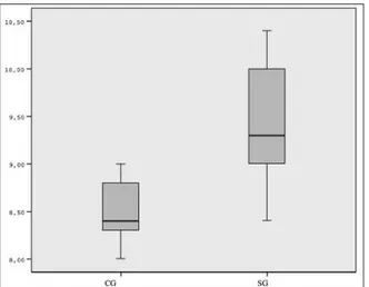
analysis. The remaining 18 rats (SG=9, CG=9) sur-vived the surgical procedures with no complica-tions (wound infection, anastomotic dehiscence, intraabdominal abscesses, and intestinal obstruc-tion) during the study. The mean values of HP lev-els and BPs of the anastomotic segments for both groups and the statistical comparisons of the groups were shown in Table 1. The HP levels, and BPs in SG were superior compared to the the CG, and the difference was statistically significant (p<0.001, Figure 1, 2 respectively).
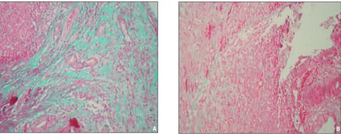
Collagen deposition and reepithelization scores of the SG were higher than that of the CG (p=0.029, collagen deposition: Figure 3). There was no significant difference between the two groups in terms of acute inflammation, chronic inflamma-tion, the amount of granulation tissue, fibroblast mat-uration and neovascularization (p=0.637, p=1.000, p=1.000, p=1.000 and p=1.000 respectively). Histo-logical comparisons of SG and CG for anastomotic segments were shown in Table 2.
DISCUSSION
Although preoperative preparation and surgical techniques have improved significantly, leakage of colonic anastomoses is still a serious problem in surgery and causes increased mortality and mor-bidity.1,2Anastomotic colon healing is affected by
many local and systemic factors. Materials such as GLU, short-chain fatty acids, erythropoietin, fibrin glue and peritoneal graft have been experimentally reported to be beneficial in the healing of colonic anastomoses.1-3,10,14,15
ARG is a dietary semi-essential amino acid and it is metabolized to ornithine and subsequently to proline and polyamines both known to interact in collagen synthesis.4,16Dietary ARG
supplementa-tion increases wound collagen accumulasupplementa-tion.4,17
GLU is a preferred energy source for cells of the in-testinal mucosa.8It has the potential to stimulate
wound healing, because one of the products of GLU metabolism is proline.3GLU enhances gut mucosal
Scores CG Median (min-max) SG Median (min-max) p
Hydroxyproline levels 8.4 (8.0-9.0) 9.3 (8.4-10.4) <0.001
Bursting pressures 140.0 (105.0-170.0) 185.0 (110.0-270.0) <0.001
TABLE 1: Comparison of tissue hydroxyproline levels (mg/100 gr tissue) and
bursting pressures (mmHg) of the colon anastomosis between two groups.
CG: Control groups, SG: Study groups
FIGURE 1: Comparison of tissue hydroxyproline levels (mg/100 gr tissue) of
the colon anastomosis between two groups.
CG: Control groups; SG: Study group; HP: Hydroxyproline.
FIGURE 2: Comparison of bursting pressures (mmHg) of the colon
anasto-mosis between two groups.
growth, repair, and improves intestinal injuries of animals and humans.3Postoperative GLU-enriched
diet was shown to improve wound healing in rats.18
HMB is the bioactive metabolite of the essential amino acid leucine.4HMB inhibits muscle
proteol-ysis, and enhances wound collagen deposition.4
Specialized amino acid mixture was reported to en-hance wound collagen accumulation and increase wound repair.4To our knowledge, there is no study
that has investigated the effect of special amino acid mixture supplementation on the healing of
colonic anastomosis. We hypothesized that special amino acid mixture might improve the healing of colonic anastomosis.
Anastomotic BP and HP levels have been widely used to assess the intrinsic resistance of anastomoses to rupture.19Collagen is important in
all phases of wound healing and critical for the re-turn of tissue integrity and strength.20-22Between
the fifth and seventh days after surgery, collagen synthesis peaks and the wound strength depends mainly on these newly formed, organized collagen
Scores CG (n%) SG (n%) p
Acute inflammation Scant 3 (33.3%) 5 (55.6%) 0.637
Moderate-abundant 6 (66.7%) 4 (44.4%)
Chronic inflammation None-Scant 8 (88.9%) 8 (88.9%) 1.000
Moderate 1 (11.1%) 1 (11.1%)
The amounts of granulation tissue Scant 4 (44.4%) 3 (33.3%) 1.000
Moderate 5 (55.6%) 6 (66.7%)
Fibroblast maturation Immature-Mild 8 (88.9%) 7 (77.8%) 1.000
Moderate 1 (11.1%) 2 (22.2%)
Collagen deposition Scant 5 (55.6%) 0 (0%) 0.029
Moderate-abundant 4 (44.4%) 9 (100.0%)
Reepithelization None-Partial 4 (44.4%) 9 (100.0%) 0.029
Complete immature-mature 5 (55.6%) 0 (0.0%)
Neovascularization < 5/HPF 8 (88.9%) 9 (100%) 1.000
6-10/HPF 1 (11.1%) 0 (0%)
TABLE 2: Comparison of acute inflammation, chronic inflammation, the amounts of granulation tissue,
fibroblast maturation, collagen deposition, reepithelization, neovascularization scores between two groups.
CG: Control groups; HPF: High-power field; SG: Study groups.
FIGURE 3: Histologic picture of SG (A) and CG (B): prominent collagen deposition (A: score 3 out of 3; B: score 2 out of 3, Gomori's trichrom X100).
(See color figure at http://tipbilimleri.turkiyeklinikleri.com/)
fibers.3Thus, postoperative day seen was chosen to
evaluate the anastomotic wound healing in our study. There is no study that has investigated the dose of special amino acid mixture supplementa-tion in rats. Williams et al.4reported the use of 14
g ARG, 3 g HMB, and 14 g GLU in humans. This dose equals to 685 mg/kg/day of special amino acid mixture. Therefore, we based the doses 685 mg/kg/day in our study on this trial in humans. We used the Abramov’s histologic scoring system13for
this study. Correlating this scoring system with ob-jective measures, such as HP level for collagen dep-osition, polarized light microscopy for assessment of granulation tissue maturation, and immunohis-tochemistry for characterization of inflammatory cells were all beyond the scope of this study. In our study, the investigated outcome measures were gross anastomotic healing, BP, HP level, and pa-rameters of histopathological healing. HP levels were significantly higher in SG. Barbul et al.17also
reported that injured rodents given a perioperative dietary supplementation of ARG had significantly improved wound healing, as assessed by wound breaking strength and the HP content. BPs, fi-broblast maturation and collagen deposition scores were significantly higher in SG. Da Costa et al.23
studied the effects of oral GLU supplementation on the healing of colonic anastomosis in rats and they also reported improvement in BP and increased mature collagen in the GLU group. Rolandelli et al.24investigated the effects of butyrate infusion on
the healing of colonic anastomosis in rats and the authors observed improvement of BP in the bu-tyrate group. Williams et al.4 also reported that
healthy elderly volunteers given a dietary supple-mentation of specialized amino acid mixture accu-mulated 67% more collagen, as assessed by the HP content and had significantly improved wound healing. It seems that high HP levels, and collagen deposition strengthens the anastomosis mechani-cally, resulting in increased BP of the anastomosis.
CONCLUSION
In conclusion, our findings show that special amino acid mixture can be used as a supporting factor for the healing of colonic anastomosis. Results of the present study indicates that it could improve anas-tomosis healing by significantly enhancing the anastomotic BP, HP level and collagen deposition. Further clinical studies are needed to clarify the usefulness of special amino acid mixture for healing of colonic anastomosis.
REFERENCES 1. Uludag M, Citgez B, Ozkaya O, Yetkin G,
Ozcan O, Polat N, et al. Effects of amniotic membrane on the healing of normal and high-risk colonic anastomoses in rats. Int J Col-orectal Dis 2009;24(7):809-17.
2. Irkorucu O, Comert M. Effects of intraperi-toneal sildenafil administration on healing of left colonic anastomoses and intra-abdominal adhesion formation in the presence of intra-abdominal infection. Dis Colon Rectum 2009; 52(5):1026-7.
3. Girgin S, Gedik E, Ozturk H, Akpolat V, Akbu-lut V, Kale E, et al. Effects of combined pulse electromagnetic field stimulation plus gluta-mine on the healing of colonic anastomosis in rats. Dig Dis Sci 2009;54(4):745-50. 4. Williams JZ, Abumrad N, Barbul A. Effect of a
specialized amino acid mixture on human col-lagen deposition. Ann Surg 2002;236(3):369-75.
5. May PE, Barber A, D’Olimpio JT, Hourihane A, Abumrad NN. Reversal of cancer-related
wasting using oral supplementation with a combination of β-hydroxy-β-methylbutyrate, arginine, and glutamine. Am J Surg 2002; 183(4):471-9.
6. Ozdemir G, Inanç Tolun F, Imrek S. The ef-fect of L-NAME treatment on leptin associated retinal nitrosylation in hypercarbic oxygen ın-duced retinopathy in newborn rats. Turkiye Klinikleri J Med Sci 2011;31(4):780-4. 7. Marcora S, Lemmey A, Maddison P. Dietary
treatment of rheumatoid cachexia with β-hy-droxy-β-methylbutyrate, glutamine and argi-nine: a randomised controlled trial. Clinical Nutrition 2005;24(3):442-54.
8. De-Souza DA, Greene LJ. Pharmacological nutrition after burn ınjury. J Nutr 1998;128(5): 797-803.
9. Jian ZM, Cao JD, Zhu XG, Zhao WX, Yu JC, Ma EL, et al. The impact of alanyl-glutamine on clinical safety, nitrogen balance, intestinal permeability, and clinical outcome in postop-erative patients: a randomized, doubleblind,
controlled study of 120 patients. JPEN J Par-enter Enteral Nutr 1999;23(5 Suppl):62-6. 10. Guven A, Pehlivan M, Gokpinar I, Gurleyik E,
Cam M. Early glutamine-enriched enteral feed-ing facilitates colonic anastomosis healfeed-ing: light microscopic and immunohistochemical evalua-tion. Acta Histochem 2007;109(2):122-9. 11. Clements RH, Saraf N, Kakade M,
Yelluma-hanthi K, White M, Hackett JA. Nutritional ef-fect of oral supplement enriched in beta-hydroxybeta-methylbutyrate, glutamine and arginine on resting metabolic rate after la-paroscopic gastric bypass. Surg Endosc 2011; 25(5):1376-82.
12. Prockop DJ, Kivirikko KI. Relationship of hy-droxyproline excretion in urine to collagen me-tabolism. Ann Intern Med 1967;66(6):1243-66. 13. Abramov Y, Golden B, Sullivan M, Botros SM, Miller JJ, Alshahrour A, et al. Histologic char-acterization of vaginal vs. abdominal surgical wound healing in a rabbit model. Wound Re-pair Regen 2007;15(1):80-6.
14. Topçu O, Karadayı K, Kuzu MA, Ulukent S, Erkek B, Alacayır I. Enteral and intraluminal short-chain fatty acids improves ischemic left colonic anastomotic healing in the rat. Int J Colorectal Dis 2002;17(3):171-6.
15. Giuratrabocchetta S, Rinaldi M, Cuccia F, Lemma M, Piscitelli D, Polidoro P, et al. Pro-tection of intestinal anastomosis with biological glues: an experimental randomized controlled trial. Tech Coloproctol. 2011;15(2):153-8. 16. Witte MB, Vogt N, Stuelten C, Gotoh T, Mori
M, Becker HD. Arginase acts as an alternative pathway of L-arginine metabolism in experi-mental colon anastomosis. J Gastrointest Surg 2003;7(3):378-85.
17. Barbul A, Rettura G, Levenson SM, Seifter E. Arginine: a thymotropic and wound-healing promoting agent. Surg Forum 1977;28:101-3.
18. Tekin E, Taneri F, Ersoy E, Oguz M, Eser E, Tekin I, et al. The effects of glutamine-en-riched feeding on incisional healing in rats. Eur J Plast Surg 2000;23(2):78-81.
19. Dinc S, Ozbirecikli B, Gulcelik MA, Erdem E, Korukoglu B, Alagol H. [The effects of intra-operative radiation on the bowel anastomosis in rats]. Turkiye Klinikleri J Med Sci 2002; 22(2):148-51.
20. Aytekin FO, Teke Z, Aydin C, Kabay B, Yenisey C, Sacar S, et al. Effects of a mem-branepermeable radical scavenger, tempol, on healing of colonic anastomoses in the cecal ligation and pucture model of polymicrobial sepsis in rats. Am Surg 2007;193(6): 723-9.
21. Colak T, Nayci A, Polat G, Polat A, Comelekoglu U, Kanik A, et al. Effects of
tra-pidil on the healing of colonic anastomoses in an experimental rat model. ANZ J Surg 2003; 73(11):916-21.
22. Gunal O, Ghandouri S, Aslaner A. The effect of intrarectal paclitaxel lavage on the healing of rectal anastomosis: a comparison with povi-done iodine. Turkiye Klinikleri J Med Sci 2005;25(5):653-7.
23. da Costa MA, Campos AC, Coelho JC, de Barros AM, Matsumoto HM. Oral glutamine and the healing of colonic anastomoses in rats. JPEN J Parenter Enteral Nutr 2003;27(3): 182-5.
24. Rolandelli RH, Buckmire MA, Bernstein KA. Intravenous butyrate and healing of colonic anastomoses in the rat. Dis Colon Rectum 1997;40(1):67-70.