Turk J Gastroenterol 2016; 27: 477-9
Giant but silent lesion of the stomach in a geriatric patient
Question:
A 65-year-old female patient with a complaint of dyspep-sia aggravating after meals approximately for 1 month was seen in the gastroenterology clinic. At presenta-tion, her physical examination was normal without con-sidering her obesity. Her medical background revealed nothing remarkable. A mild normochromic, normocytic anemia (Heamoglobin: 11.5 g/dL, Mean corpuscular vol-ume: 82 fL, Mean corpuscular heamoglobin: 27.9 pg) was
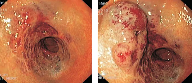
found on complete blood count. Other than her post-menopausal period, she was healthy and did not have any alarming symptoms. Upper gastrointestinal endos-copy showed a submucosally located nodular erythema-tous lesion starting from the antrum–corpus junction on the lesser gastric curvature and extending to the pylo-rus (Figure 1). The first appearance of this lesion almost resembled multiple vascular ectasia, but this lesion had mass-like appearance during exsufflation and deflated during air insufflation (Figure 2).
477
Imag
e of the Is
sue
What is the diagnosis? Answer: Gastric hemangioma
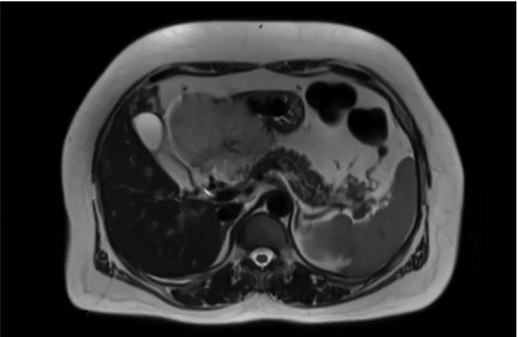
After upper gastrointestinal endoscopy, the patient underwent radiologic imaging for assessment of gastric mass. A lesion was observed as a well-circumscribed giant mass, 9×9×5.5 cm in diameter, with hypointensity on T1-weighted images and hy-perintensity on T2-weighted images, related to the antrum and lesser curvature of the stomach and extended to the liver on abdominal magnetic resonance imaging (Figure 3). During laparotomy, exophytic gastric mass resection was performed (Figure 4). When the surgical specimen was studied, it was seen to have explicit characteristics consistent with cavernous hem-angioma. During the 12-month follow-up period, the patient was asymptomatic and had no recurrence. A written informed consent from this patient was obtained before this report was prepared.
Gastric hemangioma, a rare hamartomatous tumor located mostly in the antrum, represents only 1.7% of all gastric benign tumors (1). Clinically, onset of the gastric hemangioma varies from bleeding abruptly to slowly growing over a period of
years (2). An increase in the size of the lesion depends on the anatomic location and relationship with neighboring tissues (2). In our patient, there was no clinical sign until this lesion came up to the giant size.
Diagnostic work-up for gastric hemangiomas frequently starts with an endoscopic assessment because the majority of the patients have upper gastrointestinal hemorrhage or anemia (2,3). A biopsy might be employed for both diagnosis and dif-ferentiation from other gastric lesions (3). The angiodysplasia, arteriovenous malformations, gastric antral vascular ectasia, or “watermelon stomach” should be kept in mind in order to dis-tinguish the vascular lesions of the stomach (3). In this case, instead of biopsy, because of the risk of serious bleeding of this compact vascular lesion and limited benefit owing to the sub-mucosal localisation, radiologic imaging was preferred to com-prehend the nature and extension of this mass. The endoscopy demonstrated its vascular texture and MRI helped describing its size, border and spread in abdominal cavity in our case. Due to the lack of endoscopic ultrasound in our hospital, this lesion could not be evaluated by this aspect.
Complete surgical excision is one of the reasonable treatment choices for gastrointestinal hemangioma (4). Some authors sug-gest the other options such as radiotherapy, cryotherapy, injec-tion of sclerotic agents, and endoscopic submucosal dissecinjec-tion (4). However, these procedures might be utilized for gastric lesions that can be excised adequately by endoscopy and smaller ones. In our patient, this hemangioma was very large in size and almost overflowed from the stomach and extended to the liver; and that is why, the surgical treatment strategy was more suitable. This case has two important points: First, a gastric hemangioma might be asymptomatic until the age of 65, even if it reaches a giant size. Second, the endoscopist must always be alert to recog-nize gastric vascular malformations and prefer firstly non-invasive methods for diagnosis of unknown lesions of stomach in order to avoid some complications especially like hemorrhage by due to endoscopic interventions.
Ethics Committee Approval: Ethics committee approval was received
for this study from the ethics committee of Acıbadem Hospital.
Informed Consent: Written informed consent was obtained from
pa-tients/patients’ parents/ the parents of the patients/patient who partici-pated in this study.
Peer-review: Externally peer-reviewed.
Author Contributions: Concept - D.Ö.E.; Design - D.Ö.E.; Supervision -
M.M., A.Z.; Funding - D.Ö.E.; Materials - D.Ö.E., V.T., H.A.; Data Collection and/or Processing - D.Ö.E., V.T., H.A.; Analysis and/or Interpretation - D.Ö.E., M.M., A.Z.; Literature Review - D.Ö.E., V.T., H.A.; Writer - D.Ö.E.; Critical Review - D.Ö.E., M.M.
Conflict of Interest: No conflict of interest was declared by the authors. Financial Disclosure: The authors declared that this study has received
no financial support.
478
Etik et al. Gastric hemangioma Turk J Gastroenterol 2016; 27: 477-9
Imag
e of the Is
sue
Figure 3. Radiologic assessment of gastric mass at axial T1-weighted MR image
Diğdem Özer Etik1, Volkan Tümay2, Hikmet Aktaş2, Mehmet Meriç3,
Abdullah Zorluoğlu2
1Department of Gastroenterology, Başkent University School of Medicine,
Ankara Hospital, Ankara, Turkey
2Department of General Surgery, Acıbadem Hospital, Bursa, Turkey 3Department of Gastroenterology, Acıbadem Hospital, Bursa, Turkey
REFERENCES
1. Zong L, Chen P, Shi GH, Wang L. Gastric cavernous hemangioma: A rare case with upper gastrointestinal bleeding. Oncology Let-ters 2011, 2: 1073-5.
2. Attash SM, Ali MS, Al-Nuaimy HA. Isolated cavernous hemangi-oma of the sthemangi-omach in a 3-year-old child: an unusual cause of upper GI bleeding. BMJ Case Reports 2012; 10: 1136.
3. Handra-Luca A, Montgomery E. Vascular malformations and hemangiolymphangiomas of the gastrointestinal tract: morpho-logical features and clinical impact. Int J Clin Exp Pathol. 2011; 4: 430-43.
4. Kye BH, Kim SH, Lee JI, et al. Hemorrhage from a jejunal polypoid hemangioma: single incisional laparoscopic approach. J Korean Surg Soc 2011; 80: 362-6. [CrossRef]
Address for Correspondence: Diğdem Özer Etik E-mail: [email protected]
Received: June 1, 2016 Accepted: July 13, 2016
© Copyright 2016 by The Turkish Society of Gastroenterology • Available online at www.turkjgastroenterol.org • DOI: 10.5152/tjg.2016.16329
479
Etik et al. Gastric hemangioma
Turk J Gastroenterol 2016; 27: 477-9
Imag
e of the Is