R E S E A R C H
Open Access
Epicardial adipose tissue thickness and plasma
homocysteine in patients with metabolic
syndrome and normal coronary arteries
Akif Serhat Balcio
ğlu
1*, Murtaza Emre Durako
ğlugil
2, Davran Çiçek
1, U
ğur Abbas Bal
3, Bülent Boyaci
4and Haldun Müderriso
ğlu
3Abstract
Background: Increased epicardial adipose tissue thickness and plasma homocysteine levels are associated with Metabolic Syndrome (MS) and coronary artery disease. The majority of patients with MS have subclinical or manifest coronary artery disease. The aim of this study was to evaluate the relationship between MS and plasma
homocysteine levels and epicardial adipose tissue thickness in subjects without epicardial coronary artery disease. Methods: Patients who underwent coronary angiography due to angina or equivocal symptoms and/or abnormal stress test results and were found to have normal coronary arteries were evaluated for the presence of MS. The study group comprised 75 patients with normal coronary arteries and MS, and the control group included 75 age-gender matched subjects without coronary artery disease or MS.
Results: Epicardial adipose tissue thickness (5.8 ± 1.9 mm vs. 4.3 ± 1.6 mm, p <0.001) and plasma homocysteine levels (21.6 ± 6.1μmol/L vs. 15.1 ± 5.8 μmol/L, p <0.001) were significantly higher in the MS group. Body mass index, triglyceride level, weight, age and waist circumference were positively and HDL cholesterol level were negatively correlated with both epicardial adipose tissue thickness and plasma homocysteine level. Epicardial adipose tissue thickness had the strongest correlation with plasma homocysteine level (r = 0.584, p < 0.001). For each 1 mm increase in epicardial adipose tissue thickness, an increase of 3.51μmol/L (95% CI: 2.24-4.79) in plasma homocysteine level was expected.
Conclusions: We observed a close relationship between MS and epicardial adipose tissue thickness and plasma homocysteine levels, even in the absence of overt coronary artery disease.
Keywords: Angina pectoris, Coronary angiography, Epicardial fat, Homocysteine Background
Metabolic syndrome (MS) is a combination of disorders that increase the risk of cardiovascular disease and dia-betes, including abdominal obesity, raised fasting plasma glucose, atherogenic dyslipidemia and high blood pressure [1]. Approximately 20 to 25 percent of the global popula-tion has MS, which is associated with a twofold increase in cardiovascular mortality and a threefold increase in the risk of myocardial infarction or stroke. In addition, the risk of developing type 2 diabetes is five times higher in people
with MS [2]. Abdominal obesity and insulin resistance play a key role in the development of MS and data sug-gests that prothrombotic and proinflammatory states are also involved [3].
Homocysteine, a sulfur-containing amino acid, is pro-duced through the metabolism of methionine. Elevated plasma homocysteine is associated with cardiovascular disease and stroke [4]. Studies have demonstrated that homocysteine induces endothelial cell injury and impairs endothelium-dependent vasodilation by decreasing the generation of nitric oxide and increasing the inactivation of nitric oxide [5-7]. The relationship between increased plasma homocysteine level and insulin resistance/hyper-insulinemia has been previously demonstrated [8,9]. In
* Correspondence:[email protected]
1
Department of Cardiology, Medical and Research Center of Alanya, Başkent University, Saray Mh. Yunus Emre Cad. No:1, 07400, Alanya, Antalya, Turkey Full list of author information is available at the end of the article
© 2014 Balcioğlu et al.; licensee BioMed Central Ltd. This is an Open Access article distributed under the terms of the Creative Commons Attribution License (http://creativecommons.org/licenses/by/2.0), which permits unrestricted use, distribution, and reproduction in any medium, provided the original work is properly credited. The Creative Commons Public Domain Dedication waiver (http://creativecommons.org/publicdomain/zero/1.0/) applies to the data made available in this article, unless otherwise stated.
addition, higher plasma homocysteine concentrations have been detected in patients with MS and clinically manifest atherosclerotic vascular disease [10].
Epicardial adipose tissue (EAT), deriving from the same embryological origin as mesenteric and omental fat, is de-posited around the heart, particularly around the coronary vessels [11]. EAT may influence coronary atherosclerosis and myocardial function due to the lack of a fibrous barrier blocking diffusion of free fatty acids and adipokines from the EAT to the underlying vessels and myocardium [11]. A re-cent meta-analysis demonstrated significantly higher echo-cardiographic EAT thickness in patients with MS [12]. EAT thickness is also related to epicardial obstructive coronary ar-tery disease, myocardial ischemia, major adverse cardiac events and subclinical coronary artery disease [13-16].
It is known that the majority of patients with MS have subclinical or manifest coronary artery disease. As both plasma homocysteine and EAT are associated with coron-ary artery disease, the presence of concomitant coroncoron-ary artery disease may influence the level of plasma homocyst-eine and EAT thickness in patients with MS. Therefore, the aim of this study was to evaluate the relation between MS and plasma homocysteine level and EAT thickness in subjects without epicardial coronary artery disease. Methods
Patients who underwent coronary angiography due to an-gina or equivocal symptoms and/or abnormal stress tests and who were found to have normal coronary arteries were evaluated for the presence of MS according to Inter-national Diabetes Federation criteria [17].
The study group comprised of 75 patients without cor-onary artery disease diagnosed with MS and the control group included 75 age-gender matched subjects without coronary artery disease or MS. The anthropometric char-acteristics of the control group were similar to those of the study group. All subjects in the control group had a risk factor of 0 or 1 for MS, mostly enlarged waist circum-ference (Table 1). The International Diabetes Federation defines MS as central obesity plus any two of the following four factors: raised fasting plasma glucose (≥100 mg/dL) or treatment of previously diagnosed type 2 diabetes, raised blood pressure (systolic blood pressure≥130 or dia-stolic blood pressure≥85 mmHg) or treatment of previously diagnosed hypertension, raised triglycerides (≥150 mg/dl), and reduced HDL cholesterol (<40 mg/dL in males, <50 mg/ dL in females). Central obesity was accepted as a waist cir-cumference of≥94 cm in males and ≥80 cm in females in our study group [17]. Subjects with coronary artery disease, diabetes mellitus, folic acid deficiency, malabsorption syn-dromes, hypo-hyperthyroidism or chronic systemic illness were excluded. Patients using drugs that can affect the level of plasma homocysteine and those with a poor acoustic win-dow were also excluded. The study was performed in
accordance with the principles stated in the Declaration of Helsinki and was approved by the Medical Ethics Committee of Başkent University. All subjects gave informed consent prior to enrollment.
Venous blood samples were collected after fasting for a minimum of twelve hours. Thyroid function, plasma glucose, total cholesterol, HDL cholesterol, LDL cholesterol, triglycer-ide, folic acid and homocysteine were measured. Plasma homocysteine assay was performed using the AxSYM im-munoassay according to the manufacturer’s instructions. Plasma homocysteine levels between 3.36 and 20.44μmol/L in men and 5.9 and 16μmol/L in women were considered normal according to the kit used in the study.
Anthropometric measurements were obtained by well-trained medical staff using the same methods and instruments for all participant. Waist circumference was measured at the midpoint between the lower rib margin and the iliac crest.
Blood pressure values were obtained in the echocardiog-raphy room using the traditional auscultatory method with a sphygmomanometer. Patients were advised to refrain from smoking, consuming coffee or tea and physical exer-cise for 30 minutes and were seated for rest for five minutes prior to the measurement. Three separate measurements were averaged to determine office blood pressure.
Subjects underwent transthoracic echocardiography in the left lateral decubitus position using a General Electric Vivid e (General Electric HealthCare Ultrasound Cardi-ology, Horton, Norway) and a 3.5 MHz transducer ac-cording to the guidelines of the American Society of Echocardiography by the same cardiologist specialized in echocardiography [18]. EAT thickness was measured on the free wall of the right ventricle at the end-diastole using both parasternal long and short axis views on M-mode echocardiography in three cardiac cycles, as de-scribed by Iacobellis [19]. Maximum values at any site were measured and the average value was used for stat-istical analyses. Intraobserver agreement was tested using repeated measures and no significant differences were seen for randomly selected subjects (intraclass cor-relation analysis was used and the strength of the con-cordance was 0.94).
Statistical methods
The Statistical Program for Social Sciences (SPSS for win-dows 15, Inc., Chicago, IL, USA) was used for all statistical calculations. Continuous variables are given as mean ± standard deviation; categorical variables are defined as numbers and percentages. Data were tested for normal distribution using the Kolmogorov-Smirnov test. Continu-ous variables were compared using the Student t test and Mann Whitney U test where appropriate. The chi-square test was used for comparison of the categorical variables between the two groups. Univariate correlation analysis
was performed using the Pearson’s test to identify variables that potentially affected EAT thickness and plasma homo-cysteine level. Variables with a p value of <0.05 in univariate correlation analysis were included in a multivariate stepwise linear regression analysis model in order to assess the inde-pendent determinants of EAT thickness and plasma homo-cysteine level. All tests of significance were two-tailed. Statistical significance was defined as p <0.05.
Results
Study population
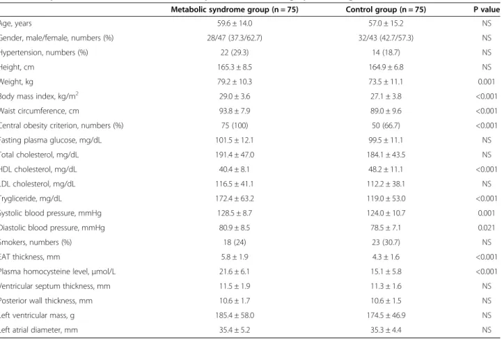
General characteristics of the study population are pre-sented in Table 1. As expected, weight, waist circumfer-ence, body mass index and systolic and diastolic blood pressure were higher in the MS group. Height was not dif-ferent between groups. The study group had higher plasma triglyceride levels and lower HDL levels. Plasma fasting glucose, total cholesterol and LDL cholesterol were similar in both groups.
EAT thickness
EAT thickness was significantly higher in patients with MS (Table 1) and was similar between gender groups (5.3 ± 1.9 mm in men, 4.9 ± 1.9 mm in women, p = 0.357). In the
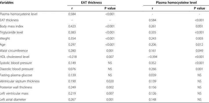
univariate correlation analysis, body mass index, triglyceride level, weight, age and waist circumference were correlated positively whereas HDL cholesterol level was correlated negatively with EAT thickness. However, plasma homocyst-eine levels had the strongest correlation with EAT thickness (Table 2). Moreover, EAT thickness was found to be related to some echocardiographic measures, including ventricular septum thickness, posterior wall thickness, left ventricular mass and left atrial diameter (Table 2).
Homocysteine concentrations
Plasma homocysteine levels were higher in patients with MS (21.6 ± 6.1 μmol/L vs. 15.1 ± 5.8 μmol/L, p < 0.001) and hypertension (21.7 ± 7.3μmol/L vs. 17.3 ± 6.3 μmol/L, p = 0.002). There was no significant difference between groups according to gender (19.2 ± 6.9 μmol/L in men, 17.8 ± 6.7 μmol/L in women, p = 0.195). EAT thickness, HDL cholesterol (reversely), systolic blood pressure, trigly-ceride level, diastolic blood pressure, body mass index, weight, age and weight circumference were significantly correlated with plasma homocysteine levels (Table 2). No association was found between homocysteine levels and echocardiographic measurements.
Table 1 Comparison of characteristics, blood samples and echocardiographic findings
Metabolic syndrome group (n = 75) Control group (n = 75) P value
Age, years 59.6 ± 14.0 57.0 ± 15.2 NS
Gender, male/female, numbers (%) 28/47 (37.3/62.7) 32/43 (42.7/57.3) NS
Hypertension, numbers (%) 22 (29.3) 14 (18.7) NS
Height, cm 165.3 ± 8.5 164.9 ± 6.8 NS
Weight, kg 79.2 ± 10.3 73.5 ± 11.1 0.001
Body mass index, kg/m2 29.0 ± 3.6 27.1 ± 3.8 <0.001
Waist circumference, cm 93.8 ± 7.9 89.0 ± 9.6 <0.001
Central obesity criterion, numbers (%) 75 (100) 50 (66.7) <0.001
Fasting plasma glucose, mg/dL 101.5 ± 12.1 99.5 ± 11.1 NS
Total cholesterol, mg/dL 191.4 ± 47.0 184.1 ± 43.5 NS
HDL cholesterol, mg/dL 40.4 ± 8.1 48.2 ± 11.1 <0.001
LDL cholesterol, mg/dL 116.5 ± 41.1 112.2 ± 38.1 NS
Trygliceride, mg/dL 172.4 ± 63.2 119.0 ± 53.0 <0.001
Systolic blood pressure, mmHg 128.5 ± 8.7 124.0 ± 10.7 0.001
Diastolic blood pressure, mmHg 80.9 ± 8.5 78.5 ± 7.1 0.021
Smokers, numbers (%) 18 (24) 23 (30.7) NS
EAT thickness, mm 5.8 ± 1.9 4.3 ± 1.6 <0.001
Plasma homocysteine level,μmol/L 21.6 ± 6.1 15.1 ± 5.8 <0.001
Ventricular septum thickness, mm 11.5 ± 1.9 11.3 ± 1.6 NS
Posterior wall thickness, mm 10.6 ± 1.7 10.6 ± 1.5 NS
Left ventricular mass, g 185.4 ± 58.0 174.5 ± 46.9 NS
Left atrial diameter, mm 35.4 ± 5.2 35.3 ± 4.4 NS
Multivariate analyses
The multivariate stepwise linear regression analysis revealed that plasma triglyceride level, age, body mass index, left atrial diameter and diagnosis of MS were the independent determinants of EAT thickness (Table 3). The independent determinants of plasma homocysteine level were systolic blood pressure, plasma triglyceride and HDL cholesterol levels and the diagnosis of MS (Table 4). For each 1 mm in-crease in thickness of EAT, a 3.51 μmol/L (95% CI: 2.24-4.79) increase in plasma homocysteine level was expected.
Discussion
In this study, we observed that plasma homocysteine levels and EAT thickness were increased in patients with MS. MS is a combination of several disorders, in which insulin
resistance and prothrombotic and proinflammatory states are common pathophysiological mechanisms [1].
Many previous studies and meta-analyses have demon-strated that EAT thickness and plasma homocysteine were increased in patients with MS and/or coronary artery dis-ease [4,12,20,21]. Ding et al. showed that an incrdis-eased quantity of EAT is associated with incident coronary artery disease and major adverse cardiac events independent of conventional risk factors [22]. There have been several meta-analyses investigating the association between homo-cysteine and coronary artery disease. One meta-analysis, including 72 case–control studies investigating the effect of methylentetrahydrofolate reductase gene mutation and 20 prospective studies evaluating the relation of serum homo-cysteine with coronary artery disease, demonstrated an odds ratio of 1.42 (95% CI 1.11-1.84) in case-controls and Table 2 Results of univariate correlation analysis for epicardial adipose tissue thickness and homocysteine levels
Variables EAT thickness Plasma homocysteine level
r P value r P value
Plasma homocysteine level 0.584 <0.001 -
-EAT thickness - - 0.584 <0.001
Body mass index 0.423 <0.001 0.261 0.001
Triglyceride level 0.383 <0.001 0.335 <0.001
Weight 0.354 <0.001 0.243 0.003
Age 0.297 <0.001 0.206 0.012
Waist circumference 0.280 0.001 0.161 0.049
HDL cholesterol level −0.218 0.007 −0.394 <0.001
Systolic blood pressure 0.149 NS 0.352 <0.001
Diastolic blood pressure 0.076 NS 0.266 0.001
Fasting plasma glucose 0.139 NS 0.039 NS
Ventricular septum thickness 0.190 0.020 0.139 NS
Posterior wall thickness 0.249 0.002 0.156 NS
Left ventricular mass 0.219 0.007 0.126 NS
Left atrial diameter 0.267 0.001 0.148 NS
EAT = epicardial adipose tissue, NS = not significant.
Table 3 Multivariate stepwise linear regression analysis for epicardial adipose tissue
B SRC t P value 95% CI for B
Lower bound Upper bound
Age 0.029 0.229 3.578 <0.001 0.013 0.046
Triglyceride level 0.008 0.262 3.737 <0.001 0.004 0.012
Body mass index 0.156 0.315 4.774 <0.001 0.092 0.221
Diagnosis of MS 0.741 0.197 2.734 0.007 0.205 1.278
IVS thickness −0.028 −0.026 −0.186 0.853 −0.324 0.269
PW thickness 0.226 0.193 1.313 0.191 −0.114 0.566
LVM −0.006 −0.165 −1.181 0.240 −0.016 0.004
Left atrial diameter 0.088 0.222 2.724 0.007 0.024 0.151
Dependent variable: Epicardial adipose tissue thickness, B = coefficient of regression, CI = confidence interval, IVS = interventricular septum, LVM = left ventricular mass, MS = Metabolic Syndrome, PW = posterior wall, SRC = standardized regression coefficient (r2= 0.405).
1.32 (95% CI 1.19-1.45) in the prospective studies for an in-crease of 5μmol/L in plasma homocysteine [23]. Similarly, a homocysteine level 25% higher than usual had an odds ratio of 1.12 (95% CI 1.04-1.20) in a meta-analysis involv-ing 5073 cardiac events [4].
The relationship between EAT and MS is also well known. In a recent meta-analysis consisting of 2027 subjects of whom 1030 had MS, EAT thickness was sig-nificantly higher in patients with MS (standardized differ-ence in means 1.15 mm, 95% CI 0.78 to 1.53, p =0.0001) [12]. Cut-off points for EAT thickness have been proposed to diagnose MS. Okyay et al. reported that EAT thickness of >4.35 mm indicated MS according to International Dia-betes Federation criteria [21]. Although there is conflicting data regarding the association between plasma homocys-tene level and MS, several studies support this relationship [8-10,24,25]. In addition, in patients with coronary artery disease, plasma homocysteine levels were found to be higher in subjects with MS than in those without [10]. This association between homocysteine level and MS was also observed in patients with prior myocardial infarction [26]. However, Hajer et al. reported that while not related to increased cardiovascular risk, plasma homocystein levels were high in patients with atherosclerotic vascular disease and MS [10].
When all of these complex and entwined relations are considered, the absence of epicardial coronary artery disease in our study population distinguishes as the presence of concomitant coronary artery disease may influence the amount of plasma homocysteine level and EAT thickness. Even so, we detected significantly higher plasma homocysteine and EAT thickness in subjects with MS compared with control subjects.
EAT has various endocrine and inflammatory func-tions and produce mediators such as interleukin-6, interleukin-1b, tumor necrosis factor-α and monocyte chemotactic protein-1, which can cause endothelial dys-function [27]. Unlike white adipose tissue, brown fat ex-presses adiponectin, which has a protective effect on endothelial function [28,29]. Several studies have shown an inverse association between brown adipose tissue and obesity, impaired glucose tolerance, and type 2 diabetes mellitus [29]. Accordingly, the dysregulated production of
adiponectin may contribute to endothelial dysfunction in subjects with MS [30].
Consistent with our results, a previous study on 68 women with chest pain and angiographically normal cor-onary arteries revealed that EAT thickness related to microvascular dysfunction, an indicator of endothelial dysfunction [31]. Even though the authors did not meas-ure plasma homocysteine, we may speculate that this ef-fect is in part due to homocysteine mediated endothelial dysfunction by both inducing endothelial cell injury and impairing nitric oxide levels [5-7].
Results of our study may be interpreted as showing that plasma homocysteine and EAT thickness begin to increase before angiographically detectable coronary artery disease in patients with MS, which represent a population at high risk for coronary artery disease. Moreover, as both EAT and homocysteine lead to endothelial dysfunction, a key event in the development of atherosclerosis predating clin-ically obvious vascular disease [32], these two factors may contribute to the initiation, progression and acceleration of coronary artery disease in patients with MS. For this rea-son, plasma homocysteine level and EAT thickness may be used as additional and easy diagnostic tools for the pres-ence of endothelial dysfunction and the necessity for follow-up for possible future overt coronary artery disease. In a recent study, Iacobellis et al. demonstrated that EAT thickness was found to be a good predictor of steatosis of non-cardiac organs such as the liver [33]. We previously showed that a relationship exists between nonalcoholic fatty liver disease and the presence of coronary artery disease [34]. These findings may also support the role of EAT on the process of coronary artery disease.
EAT volume was associated with MS and cardiometabolic risk factors, including plasma homocysteine, in patients with rheumatoid arthritis [35]. Our study also confirms the association between plasma homocysteine and EAT thick-ness in patients with MS. Moreover, the correlation be-tween EAT thickness and left ventricular mass in our study is consistent with previous studies [36,37].
Study limitations
The use of conventional coronary angiography for diag-nosis of normal coronary arteries may be considered a Table 4 Multivariate stepwise linear regression analysis for plasma homocysteine level
Variable B SRC t P value 95% CI for B
Lower bound Upper bound
HDL cholesterol level −0.151 −0.232 −3.299 0.001 −0.241 −0.060
Diagnosis of MS 3.229 0.239 3.028 0.003 1.121 5.337
Systolic blood pressure 0.130 0.191 2.651 0.009 0.033 0.227
Triglyceride level 0.015 0.144 2.009 0.046 0.000 0.030
Dependent variable: Plasma homocysteine level, B = coefficient of regression, CI = confidence interval, EAT = epicardial adipose tissue, MS = Metabolic Syndrome, SRC = standardized regression coefficient (r2
limitation of this study. Detection of early forms of cor-onary artery disease (plaque formation) and better deter-mination of the atherosclerotic process may be possible if coronary angiography had been supported with intra-vascular ultrasound. In addition, we did not measure additional indicators of atherosclerosis, such as carotid intima-media thickness or ankle brachial indices. Lastly, we do not have long-term follow-up data on cardiovas-cular events.
Conclusions
Our study demonstrated the close relationships between MS and EAT thickness and plasma homocysteine level in the absence of overt coronary artery disease. Although we cannot reveal the underlying pathological process of these findings, we think that increased EAT and homocysteine may constitute a higher cardiovascular risk in patients with MS and can be used to help diagnosis of endothelial dysfunction in patients complaining of chest pain with normal coronary arteries.
Abbreviations
EAT:Epicardial adipose tissue; MS: Metabolic syndrome. Competing interests
The authors declare that they have no competing interests. Authors’ contributions
Dr. ASB gathered data, performed the echocardiography and drafted the manuscript. Dr. MED helped with the drafting and interpretation of data. Dr.’s DÇ and UAB analyzed data and assisted in drafting the manuscript. Dr.’s BB and HM designed the study and helped in data interpretation. All authors contributed to the preparation and critical revision of the final manuscript. All authors read and approved the final manuscript.
Author details
1Department of Cardiology, Medical and Research Center of Alanya, Başkent
University, Saray Mh. Yunus Emre Cad. No:1, 07400, Alanya, Antalya, Turkey.
2Department of Cardiology, Faculty of Medicine, Recep Tayyip Erdoğan
University, Rize, Turkey.3Department of Cardiology, Faculty of Medicine, Başkent University, Ankara, Turkey.4Department of Cardiology, Faculty of
Medicine, Gazi University, Ankara, Turkey.
Received: 30 January 2014 Accepted: 20 May 2014 Published: 26 May 2014
References
1. Grundy SM, Becker D, Clark LT, Cooper RS, Denke MA, Howard WJ, Hunninghake DB, Illingworth DR, Luepker RV, McBride P, McKenney JM, Pasternak RC, Stone NJ, Van Horn L: Third report of the National Cholesterol Education Program (NCEP) expert panel on detection, evaluation, and treatment of high blood cholesterol in adults (Adult Treatment Panel III) final report. Circulation 2002, 106:3143–3421. 2. Stern MP, Williams K, Gonzalez-Villalpando C, Hunt KJ, Haffner SM: Does the
metabolic syndrome improve identification of individuals at risk of type 2 diabetes and/or cardiovascular disease? Diabetes Care 2004, 27:2676–2681. 3. Grundy SM, Brewer HB Jr, Cleeman JI, Smith SC Jr, Lenfant C, American
Heart A, National Heart L, Blood I: Definition of metabolic syndrome: report of the National Heart, Lung, and Blood Institute/American Heart Association conference on scientific issues related to definition. Circulation 2004, 109:433–438.
4. Clarke R, Collins R, Lewington S, Donald A, Alfthan G, Tuomilehto J, Arnesen E, Bonaa K, Blacher J, Boers GHJ, Bostom A, Bots ML, Grobbee DE, Brattström L, Breteler MMB, Hofman A, Chambers JC, Kooner JS, Coull BM, Evans RW, Kuller LH, Evers S, Folsom AR, Freyburger G, Parrot F, Genest J, Dalery K, Graham IM, Daly L,
Hoogeveen EK, et al: Homocysteine and risk of ischemic heart disease and stroke: a meta-analysis. JAMA 2002, 288:2015–2022.
5. Stamler JS, Osborne JA, Jaraki O, Rabbani LE, Mullins M, Singel D, Loscalzo J: Adverse vascular effects of homocysteine are modulated by
endothelium-derived relaxing factor and related oxides of nitrogen. J Clin Investig 1993, 91:308–318.
6. Tawakol A, Omland T, Gerhard M, Wu JT, Creager MA: Hyperhomocyst(e) inemia is associated with impaired endothelium-dependent vasodilation in humans. Circulation 1997, 95:1119–1121.
7. Wall RT, Harlan JM, Harker LA, Striker GE: Homocysteine-induced endothelial cell injury in vitro: a model for the study of vascular injury. Thromb Res 1980, 18:113–121.
8. Meigs JB, Jacques PF, Selhub J, Singer DE, Nathan DM, Rifai N, D’Agostino RB Sr, Wilson PW, Framingham Offspring S: Fasting plasma homocysteine levels in the insulin resistance syndrome: the Framingham offspring study. Diabetes Care 2001, 24:1403–1410.
9. Oron-Herman M, Rosenthal T, Sela BA: Hyperhomocysteinemia as a component of syndrome X. Metabolism 2003, 52:1491–1495. 10. Hajer GR, van der Graaf Y, Olijhoek JK, Verhaar MC, Visseren FL, Group SS:
Levels of homocysteine are increased in metabolic syndrome patients but are not associated with an increased cardiovascular risk, in contrast to patients without the metabolic syndrome. Heart 2007,
93:216–220.
11. Sacks HS, Fain JN: Human epicardial adipose tissue: a review. Am Heart J 2007, 153:907–917.
12. Pierdomenico SD, Pierdomenico AM, Cuccurullo F, Iacobellis G: Meta-analysis of the relation of echocardiographic epicardial adipose tissue thickness and the metabolic syndrome. Am J Cardiol 2013, 111:73–78.
13. Alam MS, Green R, de Kemp R, Beanlands RS, Chow BJ: Epicardial adipose tissue thickness as a predictor of impaired microvascular function in patients with non-obstructive coronary artery disease. J Nucl Cardiol 2013, 20:804–812. 14. Bachar GN, Dicker D, Kornowski R, Atar E: Epicardial adipose tissue as a
predictor of coronary artery disease in asymptomatic subjects. Am J Cardiol 2012, 110:534–538.
15. Cheng VY, Dey D, Tamarappoo B, Nakazato R, Gransar H, Miranda-Peats R, Ramesh A, Wong ND, Shaw LJ, Slomka PJ, Berman DS: Pericardial fat burden on ECG-gated noncontrast CT in asymptomatic patients who subsequently experience adverse cardiovascular events. J Am Coll Cardiol Img 2010, 3:352–360.
16. Tamarappoo B, Dey D, Shmilovich H, Nakazato R, Gransar H, Cheng VY, Friedman JD, Hayes SW, Thomson LE, Slomka PJ, Rozanski A, Berman DS: Increased pericardial fat volume measured from noncontrast CT predicts myocardial ischemia by SPECT. J Am Coll Cardiol Img 2010, 3:1104–1112. 17. Alberti KG, Zimmet P, Shaw J: Group IDFETFC: the metabolic syndrome–a
new worldwide definition. Lancet 2005, 366:1059–1062.
18. Lang RM, Bierig M, Devereux RB, Flachskampf FA, Foster E, Pellikka PA, Picard MH, Roman MJ, Seward J, Shanewise JS, Solomon SD, Spencer KT, Sutton MS, Stewart WJ: Recommendations for chamber quantification: a report from the American Society of Echocardiography’s Guidelines and Standards Committee and the Chamber Quantification Writing Group, developed in conjunction with the European Association of
Echocardiography, a branch of the European Society of Cardiology. J Am Soc Echocardiogr 2005, 18:1440–1463.
19. Iacobellis G, Ribaudo MC, Assael F, Vecci E, Tiberti C, Zappaterreno A, Di Mario U, Leonetti F: Echocardiographic epicardial adipose tissue is related to anthropometric and clinical parameters of metabolic syndrome: a new indicator of cardiovascular risk. J Clin Endocrinol Metab 2003,
88:5163–5168.
20. Clarke R, Daly L, Robinson K, Naughten E, Cahalane S, Fowler B, Graham I: Hyperhomocysteinemia: an independent risk factor for vascular disease. N Engl J Med 1991, 324:1149–1155.
21. Okyay K, Balcioglu AS, Tavil Y, Tacoy G, Turkoglu S, Abaci A: A relationship between echocardiographic subepicardial adipose tissue and metabolic syndrome. Int J Cardiovasc Imaging 2008, 24:577–583.
22. Ding J, Hsu FC, Harris TB, Liu Y, Kritchevsky SB, Szklo M, Ouyang P, Espeland MA, Lohman KK, Criqui MH, Allison M, Bluemke DA, Carr JJ: The association of pericardial fat with incident coronary heart disease: the Multi-Ethnic Study of Atherosclerosis (MESA). Am J Clin Nutr 2009, 90:499–504.
23. Wald DS, Law M, Morris JK: Homocysteine and cardiovascular disease: evidence on causality from a meta-analysis. BMJ 2002, 325:1202.
24. Feng SQ, Ye P, Luo LM, Xiao WK, Xu RY, Wu HM: Relationship between serum homocysteine and metabolic syndrome: a cross-sectional study. Zhonghua Liu Xing Bing Xue Za Zhi 2012, 33:256–259.
25. Vaya A, Carmona P, Badia N, Perez R, Hernandez Mijares A, Corella D: Homocysteine levels and the metabolic syndrome in a Mediterranean population: a case–control study. Clin Hemorheol Microcirc 2011, 47:59–66. 26. Agoston-Coldea L, Mocan T, Dobie L, Marginean A, Lupu S: The association
between homocysteine level and metabolic syndrome in patients of prior myocardial infarction. Rom J Intern Med 2010, 48:151–158.
27. Mazurek T, Zhang L, Zalewski A, Mannion JD, Diehl JT, Arafat H, Sarov-Blat L, O'Brien S, Keiper EA, Johnson AG, Martin J, Goldstein BJ, Shi Y: Human epicardial adipose tissue is a source of inflammatory mediators. Circulation 2003, 108:2460–2466.
28. Iacobellis G, Di Gioia C, Petramala L, Chiappetta C, Serra V, Zinnamosca L, Marinelli C, Ciardi A, De Toma G, Letizia C: Brown fat expresses adiponectin in humans. Int J Endocrinol 2013, 2013:126751.
29. Lidell ME, Betz MJ, Enerback S: Brown adipose tissue and its therapeutic potential. J Intern Med 2014, in press. doi:10.1111/joim.12255.
30. Wang ZV, Scherer PE: Adiponectin, cardiovascular function, and hypertension. Hypertension 2008, 51:8–14.
31. Sade LE, Eroglu S, Bozbas H, Ozbicer S, Hayran M, Haberal A, Muderrisoglu H: Relation between epicardial fat thickness and coronary flow reserve in women with chest pain and angiographically normal coronary arteries. Atherosclerosis 2009, 204:580–585.
32. Munzel T, Sinning C, Post F, Warnholtz A, Schulz E: Pathophysiology, diagnosis and prognostic implications of endothelial dysfunction. Ann Med 2008, 40:180–196.
33. Iacobellis G, Barbarini G, Letizia C, Barbaro G: Epicardial fat thickness and nonalcoholic fatty liver disease in obese subjects. Obesity 2014, 22:332–336. 34. Arslan U, Turkoglu S, Balcioglu S, Tavil Y, Karakan T, Cengel A: Association
between nonalcoholic fatty liver disease and coronary artery disease. Coron Artery Dis 2007, 18:433–436.
35. Ormseth MJ, Lipson A, Alexopoulos N, Hartlage GR, Oeser AM, Bian A, Gebretsadik T, Shintani A, Raggi P, Stein CM: Association of epicardial adipose tissue with cardiometabolic risk and metabolic syndrome in patients with rheumatoid arthritis. Arthritis Care Res (Hoboken) 2013, 65:1410–1415.
36. Erdogan T, Cetin M, Kocaman SA, Durakoglugil ME, Ergul E, Ugurlu Y, Canga A: Epicardial adipose tissue is independently associated with increased left ventricular mass in untreated hypertensive patients: an observational study. Anadolu Kardiyol Derg 2013, 13:320–327.
37. Iacobellis G, Petramala L, Barbaro G, Kargi AY, Serra V, Zinnamosca L, Colangelo L, Marinelli C, Ciardi A, De Toma G, Letizia C: Epicardial fat thickness and left ventricular mass in subjects with adrenal incidentaloma. Endocrine 2013, 44:532–536.
doi:10.1186/1758-5996-6-62
Cite this article as: Balcioğlu et al.: Epicardial adipose tissue thickness and plasma homocysteine in patients with metabolic syndrome and normal coronary arteries. Diabetology & Metabolic Syndrome 2014 6:62.
Submit your next manuscript to BioMed Central and take full advantage of:
• Convenient online submission
• Thorough peer review
• No space constraints or color figure charges
• Immediate publication on acceptance
• Inclusion in PubMed, CAS, Scopus and Google Scholar
• Research which is freely available for redistribution
Submit your manuscript at www.biomedcentral.com/submit