Characteristics of the spike bursts of the duodenal minute rhythm in
fasted and non-fasted sheep
Krzysztof Waldemar ROMAŃSKI
Wroclaw University of Environmental and Life Sciences, Faculty of Veterinary Medicine, Department of Biostructure and Animal Physiology, Wrocław, Poland.
Summary: The ‘minute rhythm’ (MR), the common motility pattern, occurs in the small bowel, but its character has not been fully recognized. For this purpose, seven adult rams were supplied with seven bipolar electrodes attached to the pyloric (abomasal) antrum and the entire small bowel for the electromyographic recordings. The number of the spike bursts in one MR episode and the number of spikes in the given MR-spike-burst were counted. Furthermore, the duration of the overall periods of ten consecutive MR episodes, illustrating MR incidence, was calculated. In fasted and non-fasted animals, feeding shortened the MR period duration either in the duodenal bulb during phase 2a and 2b of the myoelectric complex (MMC) or in the duodenum, especially during phase 2b of the MMC. Feeding also caused the increased number of spikes in the MR-spike bursts in fasted animals. These changes were mostly denoted during phase 2b of the MMC. In non-fasted animals, the effects of feeding upon the number of the MR-related spike bursts were less pronounced. The number of the spikes in one spike burst was often greater in the duodenal bulb than in the duodenum. The results confirm the high variability of the MR and suggest that this motility pattern can finely be controlled to be well adapted to the current situation in the gut.
Keywords: Electromyography, feeding, motility patterns, sheep, small bowel.
Introduction
The various biological rhythms omnipresent in humans and animals also contribute to the gastrointestinal motility (9, 10). The motility patterns usually exhibit the specific rhythmicity. The minute rhythm (MR) and the migrating motor complex (MMC), visible mostly in the gut, belong to these events.
The MR represents the common motility pattern observed in man and animals. It can be defined as the single propagated spike burst recurring every about one minute (7). Its physiological role and character are still unclear (19). It appears, however, that the MR variability is greater than described before (7, 18). It can occur regularly in the digestive system, although it may not occur exclusively under physiological conditions (13, 14, 26). The pattern is present either during the phase 2 of the MMC or during the fed state. Therefore, careful exploration of this pattern may help the recognition of these two motility patterns.
The MR can arrive in various regions of the gastrointestinal tract, although the characteristics of the pattern have never been presented in detail (11, 14, 20). Sheep is one of the animal species in which occurrence of the MR is the most evident (2, 7). These and other in vivo studies showed that in sheep MR arrives most regularly in the upper small intestine in relation to other motility
pattern, i.e. the MMC (7, 15). However, the MR has also been characterized so far only in the relative part in this species and sometimes its identification remains questionable. The pattern seems to exhibit the broad variability. Therefore, more detailed information is desired to define it more precisely, which can help in better understanding of its physiological role and initiation mechanism in the gut. Careful characterization of the MR may facilitate the differentiation of the normal and abnormal upper gastrointestinal motility, not only in the ruminants, since the pattern does not seem to vary markedly between the species.
Thus, the aim of this study was to describe further some aspects of MR characteristics in order to show the high variability of the spike bursts creating the pattern and to demonstrate the modifying role of various feeding conditions more precisely.
Materials and Methods
Seven Polish Merino adult rams each weighing 38-44 kg were used. Before the experiments, the animals were kept in normal conditions including normal light cycle and regular feeding. Drinking water was given ad libitum. Just before the experiments, all rams were clinically examined and found healthy.
Animal preparation: Animals were fasted 24 h
before the surgery. Under general and local anesthesia and after preoperative shaving, the laparotomy (incision 10-12 cm) was performed with the electric knife. The sterile surgical technique was applied. No reflexes triggered from the operation site were observed. The electrocautery was applied for hemostasis. Seven bipolar platinum serosal electrodes, mounted on the Teflon plates were fixed (1) to the pyloric (abomasal) antrum (4 cm proximally from the pyloric ring), (2) the duodenal bulb (6 cm distally from the pyloric ring), (3) the duodenum (50 cm distally to the bulbar electrode) and to (4) the proximal jejunum (200 cm distally from the duodenal electrode). Additionally, the other serosal electrodes were attached: (5) to the ‘distal’ jejunum, 300 cm distally from the duodenal electrode, (6) to the ‘proximal’ ileum, 110 cm before the ileocecal sphincter and (7) to the distal ileum, 10 cm before the ileocecal sphincter. These electrodes were attached for more complete identification of the myoelectric activity patterns. Food restriction lasted up to two days. Other details of this experimental model were described earlier (16, 17).
Experimental design: After the surgery the rams
were allowed for 2-weeks recovery. The total of 42 randomized experiments, each lasting 2.5-4 h were conducted. Three groups of the randomized experiments were performed either in 48-h fasted or non-fasted animals. In the first group, after the initial recording period, one full MMC cycle was recorded until the arrival of phase 3 of the next MMC cycle. In the second group, the experiments were similar and feeding (200 g of the grain mixture, consumed during 2-3 min) was started during phase 2b of the MMC. The electromyographical recordings were continued until the appearance of the phase 3 of the next MMC cycle. Drinking water was continuously available except during the experimental periods. Following the experiments, the rams were sacrificed and during autopsy the electrode localizations were further controlled.
Electromyographical recordings: The electromyographical recordings were performed throughout all the experiments using the multichannel electroencephalograph Reega Duplex TR XVI, Alvar Electronic, Montreuil, France. Upon the tracings, the MMC phases were identified with visual inspection along with further division of the longest phase 2 of the MMC into phase 2a and 2b (5, 6, 17). In the course of phase 2 of the MMC, the MR episodes were identified as well (7, 8). They were defined as the single or series of the spike bursts of various durations, recurring at around one-minute interval.
Elaboration of data: The data were derived either
from the duodenal bulb or duodenal recordings of all the experiments performed. Following MMC and MR
identification, ten last MR cycles observed during phase 2a, as well as ten first MR cycles during phase 2b MMC and also after feeding were selected and numbered consecutively. The duration of the period comprising these cycles was measured starting from the onset of the first spike burst of the initial MR episode No. 1 till the onset of the first spike burst of MR episode No. 10. Then, the number of spike bursts in one MR episode was counted. The number of spikes in the most prominent MR-spike burst was counted as well. Both figures show the representative examples.
Statistical analysis: All the numerical data were
grouped and the mean values with standard deviations were calculated. These results were compared to assess the statistical significances. Only the data obtained from the same MMC phase (2a or 2b) were compared. For period duration, the results obtained from fasted rams without feeding were compared with the data derived from fasted rams after feeding. The data obtained from non-fasted rams without feeding were compared with those also obtained from non-fasted rams after feeding. For the numbers of the spike bursts in one MR episode and for the numbers of the spikes in one MR-spike burst, two ways of comparisons were conducted. In the first one, the values obtained in two groups of fed animals were compared with the relevant values obtained from the experiments performed on fasted or not fasted rams without feeding during these experiments. In the second way of comparisons, the data obtained from the MR episodes No. 2-10 were compared with the relevant data from the MR episode No. 1 or with the relevant MR-spike burst. Statistical significances were calculated using the Student t-test for paired values preceded by analysis of variance (24).
Ethical aspects: The experimental model, applied in
this paper, was approved by the II Local Ethical Committee for Experimental Animals in Wroclaw, Poland.
Results
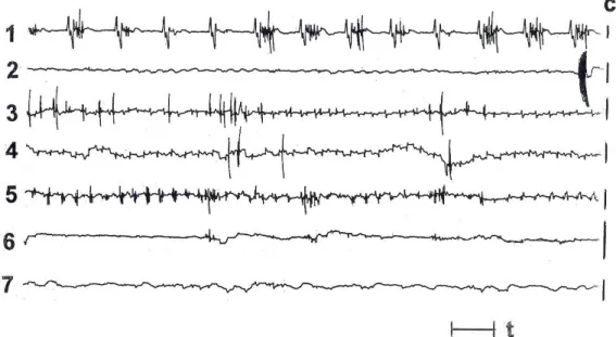
There were no meaningful differences in the MMC phases and in the MR pattern identifications although occasionally the border between phase 2a and phase 2b of the MMC was less clear. In the pyloric antrum, the MR pattern was observed infrequently. Sometimes the given MR episode was not evident in this region, especially when spiking activity was less intense (Figure 1). At the beginning of the phase 2a MMC, the pattern was often observed exclusively in the duodenal bulb during short period. Occasionally, the MR was absent in the duodenal bulb. In the distal jejunum and in the ileum, the identification of MR was often uncertain or virtually not possible.
Figure 1. The ‘minute rhythm’ (MR) on the electromyographical recordings from the small bowel of fasted sheep. Explanations. The figure shows the representative example. At least two propagated MR episodes in the duodenum and jejunum might be observed. Identification of MR episodes in the ileum is uncertain. No MR is present in duodenal bulb and in lower jejunum. 1 – pyloric antrum, 2 – duodenal bulb, 3 – duodenum, 4 – proximal jejunum, 5 – distal jejunum, 6 – proximal ileum, 7 – distal ileum; c – electrode calibration, each bar means 100 μV; t – time: 10s.
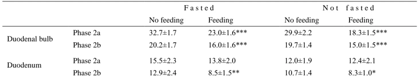
Table 1. Duration of periods containing ten selected MR episodes in duodenal bulb and duodenum of fasted and non-fasted rams. F a s t e d N o t f a s t e d
No feeding Feeding No feeding Feeding
Duodenal bulb Phase 2a 32.7±1.7 23.0±1.6*** 29.9±2.2 18.3±1.5***
Phase 2b 20.2±1.7 16.0±1.6*** 19.7±1.4 15.0±1.5***
Duodenum Phase 2a 15.5±2.3 13.8±2.0 12.0±1.9 12.4±2.1
Phase 2b 12.9±2.4 8.5±1.5** 10.7±1.4 8.3±1.0*
Explanations: Phase 2a, Phase 2b – phases of the migrating myoelectric complex. Values expressed in minutes, mean±SD, n=7. Statistical significances: *P<0.05, **P<0.01, ***P<0.001
In the duodenal bulb, the duration of the periods comprising ten consecutive MR episodes in fasted-not fed vs. fasted-fed rams and in fasted-not fed vs. non-fasted-fed rams was statistically significant either during phase 2a or 2b of the MMC. In the duodenal period duration however, the statistical significance was found only during phase 2b of the MMC (Table 1).
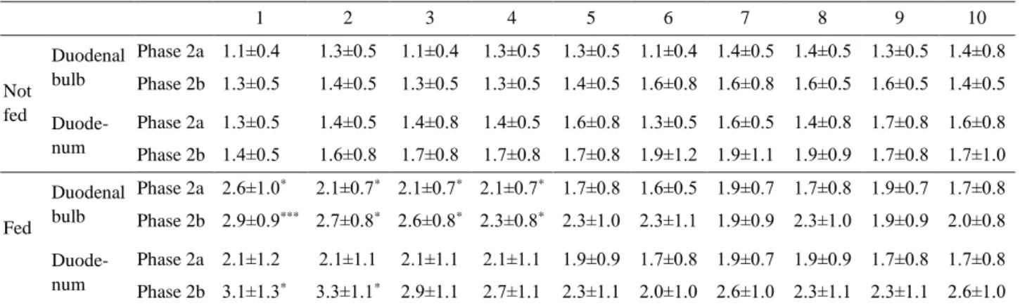
In the duodenal bulb, the number of the spike bursts observed in one MR episode during phase 2a and 2b of the MMC was significantly greater in the initial MR episodes observed after feeding in fasted animals. In the duodenum, these values were significantly greater only in the first two MR episodes identified during phase 2b of the MMC (Table 2). In non-fasted rams the increasing tendency was denoted during phase 2b of the MMC of fed and not fed animals in the duodenal bulb and duodenum. In fed rams, the number of the spike bursts in the first two MR episodes in the duodenum was significantly greater than in not fed animals (Table 3). In the course of the same MMC phase, the numbers of the spike bursts during last two MR episodes were significantly lower than during the first episode in fed animals.
Considering the number of spikes in one MR-spike burst in the duodenal bulb recorded during phase 2a of the MMC, the relevant values characterizing last three spike bursts were significantly lower after feeding than those in fasted-not fed animals (Table 4).
When the subsequent spike bursts were compared with the first one, several significant differences comprising the number of the spikes in one spike burst were noticed. In the duodenal bulb of fasted not fed animals, the last three mean values obtained during phase 2a and the last value obtained during phase 2b of the MMC were significantly higher or lower than the relevant first value, respectively. Especially, the clear tendency to change (increase) was observed during phase 2a of the MMC. In the duodenal bulb of fed animals, the values comprising last four spike bursts and obtained during phase 2b MMC, were significantly lower from the initial value. In the duodenum of the fed rams, more evident tendency (decrease) was observed thus the last five values were lower than the first one. In non-fasted animals, significant differences between not fed and fed rams were noticed in the duodenal bulb, where – during phase 2b of
Table 2. The number of the spike bursts in each of ten consecutive ‘minute rhythm’ episodes in the duodenal bulb and duodenum of fasted rams, with and without feeding.
1 2 3 4 5 6 7 8 9 10 Not fed Duodenal bulb Phase 2a 1.1±0.4 1.3±0.5 1.1±0.4 1.3±0.5 1.3±0.5 1.1±0.4 1.4±0.5 1.4±0.5 1.3±0.5 1.4±0.8 Phase 2b 1.3±0.5 1.4±0.5 1.3±0.5 1.3±0.5 1.4±0.5 1.6±0.8 1.6±0.8 1.6±0.5 1.6±0.5 1.4±0.5 Duode- num Phase 2a 1.3±0.5 1.4±0.5 1.4±0.8 1.4±0.5 1.6±0.8 1.3±0.5 1.6±0.5 1.4±0.8 1.7±0.8 1.6±0.8 Phase 2b 1.4±0.5 1.6±0.8 1.7±0.8 1.7±0.8 1.7±0.8 1.9±1.2 1.9±1.1 1.9±0.9 1.7±0.8 1.7±1.0 Fed Duodenal bulb Phase 2a 2.6±1.0* 2.1±0.7* 2.1±0.7* 2.1±0.7* 1.7±0.8 1.6±0.5 1.9±0.7 1.7±0.8 1.9±0.7 1.7±0.8 Phase 2b 2.9±0.9*** 2.7±0.8* 2.6±0.8* 2.3±0.8* 2.3±1.0 2.3±1.1 1.9±0.9 2.3±1.0 1.9±0.9 2.0±0.8 Duode- num Phase 2a 2.1±1.2 2.1±1.1 2.1±1.1 2.1±1.1 1.9±0.9 1.7±0.8 1.9±0.7 1.9±0.9 1.7±0.8 1.7±0.8 Phase 2b 3.1±1.3* 3.3±1.1* 2.9±1.1 2.7±1.1 2.3±1.1 2.0±1.0 2.6±1.0 2.3±1.1 2.3±1.1 2.6±1.0 Explanations: 1 – 10 – consecutive ‘minute rhythm’ episodes; Phase 2a, Phase 2b – phase 2a and 2b of the migrating myoelectric complex; mean±SD, n=7. Statistical significances: *P<0.05; ***P<0.001 vs. relevant value in not fed rams.
Table 3. The number of the spike bursts in each of ten consecutive ‘minute rhythm’ episodes in the duodenal bulb and duodenum of non-fasted rams, with and without feeding
1 2 3 4 5 6 7 8 9 10 Not fed Duodenal bulb Phase 2a 1.3±0.5 1.4±0.5 1.3±0.5 1.4±0.5 1.4±0.5 1.6±0.5 1.4±0.5 1.6±0.8 1.7±0.8 1.9±0.7 Phase 2b 1.4±0.8 1.6±0.5 1.6±0.8 1.7±0.8 1.6±1.0 1.6±0.8 1.7±1.0 1.4±0.5 2.1±0.9 1.6±0.8 Duode- num Phase 2a 1.6±0.5 1.6±0.5 1.6±0.5 1.7±0.8 1.9±0.7 1.6±0.5 1.7±1.0 1.7±0.8 1.4±0.5 1.6±0.8 Phase 2b 1.7±0.8 1.9±0.7 1.7±0.8 2.0±1.2 1.7±0.8 1.7±0.8 2.3±1.1 1.7±0.8 2.3±1.3 2.4±1.3 Fed Duodenal bulb Phase 2a 1.3±0.5 1.4±0.5 1.6±0.8 1.6±0.5 1.4±0.8 1.4±0.8 1.9±0.7 1.7±0.8 1.7±0.8 1.7±0.8 Phase 2b 1.7±0.5 1.9±0.7 1.7±0.8 1.9±0.9 1.7±1.1 1.7±0.8 2.0±1.2 2.6±1.3 2.4±1.3 2.1±0.9 Duode- num Phase 2a 2.4±1.0 2.3±1.0 2.3±1.1 2.3±1.1 2.1±1.1 2.0±1.0 1.9±0.9 1.6±0.8 1.9±0.7 1.9±0.9 Phase 2b 3.9*±1.3 3.7*±1.0 2.9±0.9 2.7±1.1 2.3±1.1 2.3±1.1 2.3±1.1 2.0±0.8 1.7±0.8a 1.7±0.8 Explanations: 1 – 10 – consecutive ‘minute rhythm’ episodes; Phase 2a, Phase 2b – phase 2a and 2b of the migrating myoelectric complex; mean±S.D., n=7. Statistical significances:
Table 4. The number of spikes in the spike bursts of ten consecutive ‘minute rhythm’ events in the duodenal bulb and duodenum of fasted rams, with and without feeding.
1 2 3 4 5 6 7 8 9 10 Not fed Duodenal bulb Phase 2a 4.3±1.0 4.6±0.5 4.6±0.8 4.7±0.8 4.9±0.9 5.3±0.8 5.4±1.3 6.3±1.5 6.4±1.0 7.0±1.2 Phase 2b 5.3±1.0 4.9±0.9 4.6±1.0 4.1±1.1 4.3±1.1 4.0±0.8 4.4±0.8 4.0±0.8 4.6±0.5 3.9±0.7 Duode- num Phase 2a 4.9±1.3 4.9±1.1 4.4±1.0 4.6±1.1 4.3±1.1 3.9±0.7 3.7±0.8 3.9±0.9 3.7±0.8 3.4±1.0 Phase 2b 3.7±0.8 3.7±0.8 4.0±0.8 3.6±0.8 3.7±1.1 4.0±0.8 3.4±0.8 3.9±0.7 4.0±1.2 4.0±1.2 Fed Duodenal bulb Phase 2a 5.4±1.1 5.7±1.4 5.3±1.0 4.4±1.0 4.3±1.1 4.1±0.7 4.0±1.2 4.0±1.2* 4.0±0.8** 3.9±0.9*** Phase 2b 6.4±1.0 6.0±1.4 5.4±1.0 5.7±1.4 5.6±1.0 5.9±1.3 4.9±0.7a 4.4±1.0a 4.4±1.1a 4.4±1.0 Duode- num Phase 2a 4.1±0.7 4.6±0.8 3.7±0.5 3.4±0.5 3.6±0.8 3.6±0.5 3.7±0.8 3.9±0.7 3.6±0.8 3.6±1.1 Phase 2b 5.7±1.3* 5.3±1.1 4.1±1.1 3.7±0.8 3.9±0.7 3.4±0.8a 3.3±0.5c 3.6±0.8a 3.1±0.4c 3.3±0.5 Explanations: 1 – 10 – consecutive ‘minute rhythm’ spike bursts; Phase 2a, Phase 2b – phase 2a and 2b of the migrating myoelectric complex, mean±SD, n=7.
Table 5. The number of spikes in the spike bursts of ten consecutive ‘minute rhythm’ events in the duodenal bulb and duodenum of non-fasted rams, with and without feeding.
1 2 3 4 5 6 7 8 9 10 Not fed Duodenal bulb Phase 2a 6.6±1.0 6.0±0.8 4.9±1.2 4.9±1.1 5.0±0.8 4.6±0.8 4.1±0.7 4.1±1.1 4.4±0.5 4.1±0.7 Phase 2b 4.3±0.8 4.3±1.0 4.6±1.0 4.6±1.3 3.9±0.9 4.1±0.7 4.0±0.8 4.4±0.5 4.0±0.8 4.0±0.6 Duode- num Phase 2a 4.6±0.8 3.9±0.7 4.4±1.0 3.7±0.8 3.9±0.7 3.4±0.5 3.4±0.5 3.4±0.8 3.1±0.7 3.4±0.5 Phase 2b 3.7±0.8 4.0±0.8 3.6±1.0 3.4±0.5 3.4±0.8 3.1±0.7 3.4±1.0 3.6±1.1 4.0±1.2 3.4±0.5 Fed Duodenal bulb Phase 2a 5.1±0.9 5.7±1.7 4.3±1.0 4.3±1.1 4.3±1.1 4.1±1.0 4.0±0.8 4.6±0.8 4.3±0.8 4.7±1.4 Phase 2b 7.6±1.1*** 6.3±1.1* 5.9±1.6 5.4±1.3 5.1±0.9 4.6±1.0 4.4±1.0 4.3±0.5 4.1±1.3 4.1±1.1 Duode- num Phase 2a 3.7±0.8 3.4±1.0 4.3±1.0 3.7±0.8 3.6±0.8 3.4±1.4 3.7±1.1 3.4±0.5 3.6±0.8 3.6±0.8 Phase 2b 4.7±1.1 4.4±0.5 3.9±0.9 3.4±1.0 3.7±1.0 3.4±0.5 3.3±1.1 3.1±0.7 3.1±0.4 3.3±0.5 Explanations: 1 - 10 – consecutive ‘minute rhythm’ spike bursts; Phase 2a, Phase 2b - phase 2a and 2b of the migrating myoelectric complex, mean±S.D., n=7. Statistical significances:
Figure 2. The ‘minute rhythm’ (MR)-spike bursts in duodenum and jejunum of non-fasted sheep. Explanations. The figure shows the example from the recording obtained from different sheep than Figure 1. Two MR episodes could be seen. Their spike bursts contain only 1-3 spikes each. c – calibration 50 μV; t – time in seconds. Other explanations as in Figure 1.
the MMC – the number of spikes in the first and second spike burst increased after feeding (Table 5). When the spike bursts No. 2-10 were compared to the first relevant spike burst, the statistical significances were depicted in not fed animals in the duodenal bulb, during phase 2a of the MMC where the last five values were lowered (Table 5). In fed animals (duodenal bulb, phase 2b of the MMC), the mean spike numbers in the spike bursts No. 5-10 were significantly lower from the spike burst No. 1, exhibiting decreasing tendency in the whole spike burst series examined under these conditions.
Generally, the number of spikes in one MR-spike burst was greater in the duodenal bulb than in the duodenum. In a few cases, however the very short MR-spike bursts, containing 1-3 MR-spikes each, were found in the
duodenum and jejunum, even in non-fasted rams (Figure 2).
Discussion and Conclusion
The MR is one of the well-recognized motility patterns in sheep while its detailed character is still under investigation. Its variability seems greater than it could be expected. The MR has not been characterized precisely also in monogastrics. However, it seems likely that their small-intestinal motility, including the MR, does not differ much from this in sheep. Fine characteristics of the MR, described by numerical data should allow determine better whether or not the gastrointestinal motility in various situations is normal. The well-characterized MR can thus serve as a reference point for investigators to evaluate the effect of drugs, feeding procedures or the surgical-type
experiments upon the gastrointestinal motility more precisely. When the gastrointestinal motility is disturbed, the evaluation of the MR normality may help to achieve more precise diagnosis. The detailed characteristics of this rhythmic pattern can further allow compare the gastrointestinal motility in various animal species in order to recognize the animal species differences, the usefulness of the animal models for human studies and to recognize better the detailed motility controlling mechanisms. Furthermore, the recognition of the incidence of propagating MR episodes may facilitate to predict the role of the MR on the flow of digesta in the small bowel, especially in the duodenum, which may be important for evaluation of the digestive process, but also in part, of the absorptive processes. These purposes can prompt the further appropriate investigations.
The numerous, more or less regular and pronounced alterations in the MR incidence and the number of the spike bursts forming the pattern indicate that its arrival is not incidental and suggest its meaningful physiological role. The character of the MR may depend upon the current situation in the intestinal lumen and, therefore, the pattern can be adapted to it well. Still little is known about its physiological role so far. Some authors clearly stated that the role is unknown (7, 8, 28). The mechanism of spontaneous initiation and migration of the MR is also unknown (20), though it appears that the MR is initiated in response to the gut stimuli. There were meaningful differences, regarding the MR, especially between the duodenal bulb and the duodenum. At the beginning of phase 2a of the MMC, the MR was often present exclusively in the duodenal bulb, and then it arrived in the duodenum where the pattern arrived most frequently. Thus it is possible that the pattern could be initiated not in the pyloric region but in this segment. However, in few cases, the MR was absent in the duodenal bulb. In spite of that, the MR occurred most clearly in the duodenal bulb as well as in the duodenum and the easiest identification of it could be done there. As it can be derived from the previous reports, the MR is often absent either in the pyloric antrum or in the jejunum (2, 7, 8). Usually it occupies the relatively short intestinal segment including the duodenum (22). That is why the current investigations concerned mostly the duodenal region.
Obtained results indicate that the number of the spike bursts in one MR episode observed in the duodenal bulb increased after feeding, mostly in fasted rams. When ten consecutive MR episodes were analyzed in non-fasted rams, slight increasing tendency was observed in the duodenal bulb, mostly during phase 2a of the MMC. After feeding, following the immediate increase, the decreasing tendency was denoted. All these findings suggest that in the duodenal bulb the number of the spike bursts in one MR cycle was related to the intensity of spiking activity in
this region. In the duodenum, such a tendencies were less clear although feeding increased the number of the spike bursts, at least in the first MR episodes during phase 2b of the MMC in fasted rams. Therefore, in the duodenum the number of the spike bursts in one MR episode was also somehow related to the intensity of spiking activity, mostly in dependence of feeding conditions. It is possible that these changes were also related to the character of neural stimuli evoking in the stomach and affecting the gut. It is well known that feeding itself is the strong natural stimulus for the gastrointestinal motility, even in sheep (1, 3, 4, 12, 23, 25). It can stimulate motility in both the neural and endocrine pathway. The greater number of the spike bursts in the MR patterns may increase the efficiency of digesta mixed by the MR in the intestinal lumen.
The number of spikes in one spike burst forming the MR pattern reflects the duration of this spike burst. It was often longer in the duodenal bulb than in the duodenum. Occasionally in the duodenal bulb, the number of spikes in one MR-spike burst exceeded six which can imply that the duration of the spike burst in this segment examined could be longer than 3 s (i.e. the average duration of the plateau phase of the slow wave in the stomach, see also 27). In the duodenum, duration of the spike bursts was shorter since they were superimposed upon the slow waves in which the duration of the plateau phase was even shorter than 3 s. However, in the first 8 cm of the ovine duodenal bulb, no slow waves were observed (21), thus the initiation of the longer spike bursts appears to require stronger stimuli. In spite of it, in a few cases very short spike bursts were observed in the duodenojejunum, even in non-fasted rams. The mixing and propulsive role of such a spike bursts is minimal.
The results provided the new data which confirm the high variability of the MR in the ovine duodenum and suggest that the given form of this motility pattern was precisely controlled in order to be adapted adequately to the current situation in the gut.
References
1. Ahluwalia NK, Thompson DG, Barlow J, et al. (1994):
Human small intestinal contractions and aboral traction forces during fasting and after feeding. Gut, 35, 625-630.
2. Bueno L, Fioramonti J (1975): Rhythms of
Abomaso-Intestinal Motility. 69-85. In: Y Ruckebusch, P Thivend
(Eds), Digestive Physiology and Metabolism in Ruminants. MTP Press Limited, Lancaster.
3. Bueno L, Fioramonti J, Ruckebusch Y (1975): Rate of
flow of digesta and electrical activity of the small intestine in dogs and sheep. J Physiol (Lond), 249, 69-85.
4. Bühner S, Ehrlein HJ (1989): Characteristics of
postprandial duodenal motor patterns in dogs. Dig Dis Sci,
34, 1873-81.
5. Code CF, Marlett A (1975): The interdigestive myoelectric
complex of the stomach and small bowel of dogs. J Physiol
6. Dent J, Dodds WJ, Sekiguchi T, et al. (1983):
Interdigestive phasic contractions of the human lower esophageal sphincter. Gastroenterology, 84, 453-460.
7. Fleckenstein P, Bueno L, Fioramonti J, et al. (1982):
Minute rhythm of electrical spike bursts of the small intestine in different species. Am J Physiol, 242,
G654-G659.
8. Fleckenstein P, Øigaard A (1978): Electrical spike activity
in the human small intestine. A multiple electrode study of fasting diurnal variations. Dig Dis, 23, 776-780.
9. Gobello C, Corrada YA, Castex GL, et al. (2002):
Secretory patterns of growth hormone in dogs: circannual, circadian, and ultradian rhythms. Can J Vet Res, 66,
108-111.
10. Hoogerwerf WA (2010): Role of clock genes in
gastrointestinal motility. Am J Physiol, 299, G549-G555.
11. Kellow JE, Borody TJ, Phillips SF, et al. (1986): Human
interdigestive motility. Variations in patterns from esophagus to colon. Gastroenterology, 91, 386-395.
12. McCoy EJ, Baker RD (1968): Effect of feeding on
electrical activity of dog’s small intestine. Am J Physiol,
214, 1291-1295.
13. Primi MP, Bueno L (1987): Effects of centrally
administered naloxone on gastrointestinal myoelectric activity in morphine-dependent rats. J Pharmacol Exp Ther,
240, 320-326.
14. Quigley EMM, Phillips SF, Dent J (1984): Distinctive
patterns of interdigestive motility at the canine ileocolonic junction. Gastroenterology, 87, 836-844.
15. Romański KW (2002): Characteristics and cholinergic
control of the ‘minute rhythm’ in ovine antrum, small bowel and gallbladder. J Vet Med A, 49, 313-320.
16. Romański KW (2003): Character and cholinergic control
of myoelectric activity in ovine duodenal bulb: relationships to adjacent regions. Vet Arhiv, 73, 1-16.
17. Romański KW (2007): Regional differences in the effects
of various doses of cerulein upon the small-intestinal migrating motor complex in fasted and non-fasted sheep. J
Anim Physiol Anim Nutr, 91, 29-39.
18. Romański KW (2009). Cholecystokinin-dependent selective inhibitory effect on ‘minute rhythm’ in the ovine small intestine. Animal, 3, 275-286.
19. Romański KW (2016) The diversity of ‘minute rhythm’
forms in the ovine small bowel: relationship to feeding and to the phase of the migrating myoelectric complex. Vet
Arhiv, 86, 351-362.
20. Romański KW, Rudnicki J, Sławuta P (2001): The
myoelectric activity of ileum in fasted and fed young pigs. J
Physiol Pharmacol, 52, 851-862.
21. Ruckebusch Y, Pairet M (1984): Duodenal bulb motor
activity in sheep. J Vet Med A, 31, 401-413.
22. Sarna SK, Otterson MF (1989): Small intestinal
physiology and pathophysiology. Gastroenterol Clin North
Am, 18, 375-405.
23. Schwartz MP, Samsom M, Smout AJPM (2001): Human
duodenal motor activity in response to acid and different nutrients. Dig Dis Sci, 46, 1472-1481.
24. Snedecor GW, Cochran WG (1971): Statistical Methods. The Iowa State University Press, Ames.
25. Soffer EE, Adrian TE (1992): Effect of meal composition
and sham feeding on duodenojejunal motility in humans.
Dig Dis Sci, 37, 1009-1014.
26. Summers RW, Anuras S, Green J (1983): Jejunal
manometry in health, partial intestinal obstruction, and pseudoobstruction. Gastroenterology, 85, 1290-1300.
27. Szurszewski JH (1987): Electrical Basis for Gastrointestinal Motility. 383-422. In: LR Johnson (Ed),
Physiology of the Gastrointestinal Tract. Raven Press, New York.
28. Weisbrodt NW (1981): Patterns of intestinal motility. Ann Rev Physiol, 43, 21-31.
Geliş tarihi: 05.12.2016 / Kabul tarihi: 06.04.2017
Address for correspondence:
Prof. Dr. Hab. Krzysztof Waldemar ROMAŃSKI, ul. Kordiana 26/2, 51-348 Wrocław, Poland, tel. +48 71 3305656