Ankara Üniv Vet Fak Derg, 58, 209-212, 2011 DOI: 10.1501/Vetfak_0000002476
Short Communication / Kısa Bilimsel Çalışma
Systemic toxoplasmosis in a kangaroo (Macropus sp.)
Yonca Betil KABAK1, Tolga GÜVENÇ1, Oğuz KUL2, Ahmet DENİZ3, M.Yavuz GÜLBAHAR1 1Ondokuz Mayıs Üniversitesi, Veteriner Fakültesi, Patoloji Anabilim Dalı, Samsun; 2Kırıkkale Üniversitesi, Veteriner Fakültesi,
Patoloji Anabilim Dalı, Kırıkkale; 3Etlik Veteriner Kontrol ve Araştırma Enstitüsü, Ankara.
Summary: This paper describes systemic toxoplasmosis in a brush kangaroo died after diagnosed with chronic diarrhoea in a
local zoo. Macroscopically, widespread subcutaneous petechial haemorrhagies, 100 ml sero-sanguineous fluid in the abdominal cavity, many nodular structures on omentum and fat tissues with dilatation in subserosal vessels of gastric and small intestines were observed. Microscopically, necrotic bronchointerstitial pneumonia in the lungs and multiple areas of necroses with pyogranulomatous inflammation in the myocardium, adrenal glands and tunica muscularis of stomach and small intestines were detected. The pancreas and surrounding abdominal adipose tissues had multifocal coalescing acellular necroses conjoined with pyogranulomatous inflammation. Free and/or intracellularly located Toxoplasma gondii-like granular structures showed co-association with necrotic lesions. These granular structures and necrotic areas exhibited strong immunoreactivity to polyclonal
anti-T.gondii antibodies whereas reaction was negative for Neospora caninum and Leishmania sp.. Nested-PCR designed to amplify a 97
bp long specific part in B1 gene of T. gondii gave positive results. In conclusion, the first case of systemic toxoplasmosis in a kangaroo from a local zoo in Turkey was diagnosed by detailed histochemical, immunoperoxidase technique and PCR.
Key words: Immunoperoxidase, kangaroo, PCR, Toxoplasma gondii, toxoplasmosis.
Bir kanguruda (Macropus sp.) sistemik toksoplasmozis
Özet: Bu çalışma, yerel hayvanat bahçesinde kronik diyare teşhisinden sonra ölen bir çalı kangurusunda (Macropus sp.)
sistemik toxoplazmozisi tanımlanmaktadır. Makroskopik olarak, deri altında yaygın peteşiyal kanamalar, karın boşluğunda 100 ml kanlı sıvı, omentum ve yağ dokuda pek çok nodüler yapılar ile mide ve ince bağırsakların subserozal damarlarında dilatasyon gözlendi. Mikroskopik olarak, akciğerde nekrotik bronkointerstisyel pnömoni ve miyokardium, adrenal bezler ile mide ve ince bağırsakların tunika muskularisinde pyogranülomatöz yangı ve çok sayıda nekrotik alan belirlendi. Pankreas ve çevresindeki abdominal yağ dokuda pyogranülomatöz yangı ile birleşmiş multifokal asellüler nekroz vardı. Serbest ve /veya intrasellüler yerleşimli Toxoplasma gondii benzeri granüler yapılar nekrotik alanlar ile birlikte görüldü. Bu granüler yapılar ve nekrotik alanlar poliklonal anti-Toxoplasma gondii antikoru ile şiddetli immunoreaksiyon gösterirken, Neospora caninum ve Leishmania sp. için reaksiyon negatifti. Toxoplasma gondii B1 geninin 97 bp uzunluğunda spesifik bir bölgesine yapılan nested-PCR ile toksoplazmozisin teşhisi doğrulandı. Sonuç olarak, Türkiye’de local bir hayvanat bahçesindeki kanguruda sistemik toksoplazmozis vakası histopatolojik, immunoperoksidaz teknik ve PCR kullanılarak ilk kez teşhis edildi.
Anahtar sözcükler: İmmunoperoksidaz, kanguru, PCR, Toxoplasma gondii toksoplazmozis.
Toxoplasma gondii (T gondii) is a protozoan
parasite of wild and domestic felids with an unusually wide range of intermediate host. Toxoplasmosis is common in many warm-blooded animals and human (4,6,13,15,17). It is not only a major cause of congenital disease and abortion in human and domestic animals, but also a life-threatening opportunistic infection for immunologically vulnerable hosts (2,3). This is a widespread protozoon in Turkey, therefore, several experimental studies nearby studies representing pathologic and epidemiologic findings of naturally infected animals are present (10,11,12,14). Wallabies and Australian marsupials are considered among the most susceptible species to
toxoplasmosis (2,5,7,17,18). Their vulnerability to toxoplasmosis is explaining with increased stress factors and immunsupression due to zoo conditions and/or their possibly infrequent contact with feline species during evolution processes (5,6,18). Toxoplasmosis can cause sudden deaths with the absence of prominent clinical findings in captive and wild Australian marsupials, is not always fatal (1,3,4,7,9,18). Clinical findings can be associated with respiratoric, neurologic and gastrointestinal disorders (9,17,18). In the differential and definitive diagnosis of T. gondii infections, serologic (e.g. IFA, Sabin Feldman Dye test) (3,4,7,9), ultrastructural (2,8), immunohistochemical (7,8,9,11,12), molecular examinations
Yonca Betil Kabak - Tolga Güvenç - Oğuz Kul - Ahmet Deniz -M. Yavuz Gülbahar 210
(1) and bioassay studies in experimental animals (14,18) generally take place among the main diagnostic tools.
In the present report, fatal toxoplasmosis in a zoo kangaroo was investigated by detailed macroscopic, histopathologic, immunohistochemical and PCR examinations. According to literature, this study is suggested to be the first example of toxoplasmosis in wallabies in Turkey.
A dead adult female brush kangaroo (Macropus irma/ Western brush wallaby) was introduced to Department of Pathology, Faculty of Veterinary Medicine, Ondokuz Mayis University, Turkey, from a local zoo. In necropsy, samples were taken from brain, heart, lung, liver, spleen, kidney, pancreas, adrenal glands, stomach, small and large intestines, regional lymph nodes, omentum, trachea, oesophagus, tongue and skeletal muscles. All tissue samples were fixed in 10% neutral buffered formalin for 24 hours, embedded in paraffin wax, and sectioned at 5 µm thickness. Prepared sections were stained with hematoxylin&eosin (HE), periodic acid-Schiff (PAS), Ziehl- Neelsen (ZN) and Grocott’s methenamine silver (GMS) methods (16). Additional sections were immunostained for T. gondii, Neospora caninum and
Leishmania sp. using polyclonal antibodies and standard
streptavidin-biotin peroxidase complex method (ABC-P) with a commercial kit (Zymed, USA). The reaction product was visualized by aminoethylcarbazole (AEC) chromogen (Zymed, USA) and counterstained with Gill's haematoxylin. Nested-PCR was performed to confirm T.
gondii infection using 2 primer sets. In the first step; 194
bp part of B1 gene of T.gondii (Accession number; AF179871) was amplified using Toxo1 for: 5’- GGAACTGCATCCGTTCATGAG-3’ and Toxo2 rev: 5’- TCTTTAAAGCGTTCGTGGTC-3’ and in the second step a specific 97 bp part of the T. gondii B1 gene was targeted in previously amplified template DNA using Toxo3 for: 5’- TGCATAGGTTGCCAGTCACTG-3’ and Toxo4 rev 5’-GGCGACCAATCTGCGAATACACC-3’ primers. Parasitic DNA was extracted from formalin fixed paraffin embedded tissues as follows; serial 10 µm paraffin tissue sections were cut and 25 µm tissue was collected in a microsentrifuge tube for each paraffin block. Microtome blade was changed in each new sample to prevent cross contamination. After 5 minutes deparaffinization in xylene, tissues were allocated in alcohol and tissue digestion was performed by incubation in 400 µg proteinase K in 500 µl of 10 mM Tris HCI (pH 7.5). PBS was used as negative control, T. gondii RH strain tachyziotes obtained from effluent periton of mouse was used as positive control. Consequently, all usual steps of spin colon-centrifuge DNA isolation method were followed according to commercial DNA isolation kit protocol (Qiagen, FFPE DNA extraction kit, Cat. # 51304).
In macroscopic examination, subcutaneous petechial haemorrhagies at the lateral of abdominal and thoracal regions and approximately 100 ml sero-sanguineous fluid were observed in abdominal cavity. There were mesenterial and pancreatic lymph node edema, nodular structures scattered throughout the omentum and petechial haemorrhagies on the gastric and intestinal serosa. Cranial lobes of the lungs were consolidated.
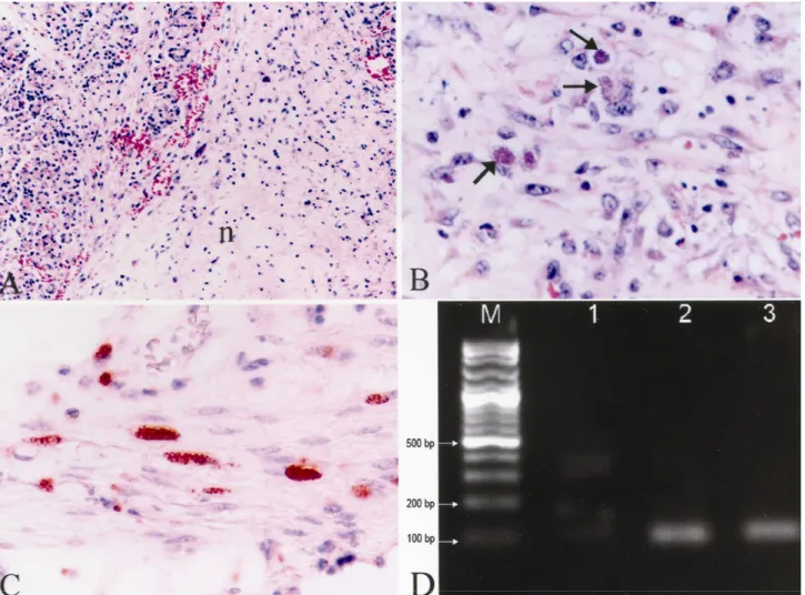
Microscopically, severe macrophage and lymphocyte infiltrations were present in the lamina propria and submucosa of the stomach and the small intestines in addition to mononuclear cell infiltrations in the areas adjacent to foci of necroses in some muscle fibers. Inflammation and focal necrotic areas were determined in the adrenal medulla, pancreatic parenchyma, peripancreatic (Fig. 1A) and abdominal fat tissues. Steatitis in fat tissues was characterized by multifocal to coalescing acellular necrosis, pyogranulomatous inflammation and peripherally active chronic fibrosis. Inflammatory cells consisted mostly of macrophages, lymphocytes with a few neutrophil leucocytes. Coagulation necrosis and lymphocyte infiltration in portal triad were also observed. There were necrosis and lymphoid depletion in the germinal centers of lymphoid follicules. Additionally, sinusoidal macrophage infiltrations in mesenterial lymph nodes and spleen were noticed. Severe hyperemia in the lungs, oedema, lymphocyte and macrophage infiltrations in interalveolar septal tissue and necrotic brochiolitis were observed. A focal calcification area confined by a delicate fibrous connective tissue containing lymphocytes and extra and/ or intracellular T.
gondii tachyziotes were detected. There were multifocal
necroses, focal gliosis and meningitis in the brain. Focal necrosis, interstitial macrophages and lymphocytes infiltration were present in the heart and skeletal muscles. Some necrotic areas were calcified in the heart. Free and/or intracellular tachyzoites of the parasite were readily detected in sections of the small intestine, gut, heart, skeletal muscle, pancreatic lymph node and omentum. Tachyzoites were also detected in the granulomatous inflammation occurred in pancreas and adipose tissue around the mesenterium (Fig. 1B), pancreatic lymph nodes and kidneys. PAS and GMS stains were applied to the tissue sections containing various forms of the parasite and results appeared negative. Acid fast bacteria were not observed at the Ziehl Neelsen staining of the lung section showing necroses and calcifications. Immunoperoxidase tests were positive for T. gondii (Fig. 1C) but negative for N.
caninum and for Leishmania sp.. T. gondii tissue
cyst-like structures, free or intracellular tachyzoites showed red in colour granular and cluster-like T.gondii specific immunoreactivities with a varying degree depending on the antigenic density. Molecular confirmation was also established by the observation of T. gondii specific DNA
Ankara Üniv Vet Fak Derg, 58, 2011 211
bands (97 bp) in agarose gel after two steps nested-PCR procedure (Fig. 1D).
Toxoplasmosis has been reported in captive wallabies and kangaroos from zoos of various parts of the world (1,2,3,6). Disease generally progresses subclinically in mankind and animals (6,10,13,14,15), while it causes sudden deaths in kangaroos (3,4,9,18). Oocyte shedding subclinically infected stray cats are suspected to be the source of toxoplasmosis in kangaroos (1,4,6).
Toxoplasmosis results with sudden deaths in kangaroos; nevertheless, toxoplasmosis can also show clinical symptoms of diarrhoea, weight gain, tiredness, respiratory and neurologic disorders (2,4,5,6,17). In this case, the zoo veterinarian reported that the kangaroo exhibited only diarrhea symptom prior to its death.
Lesions are determined in many tissues as a result of the systemic infections occurred by toxoplasmosis in
kangaroo. Concordant with literature, performed necropsy revealed congestion, edema and consolidation in lungs (1,2,3,7,9), lymph node enlargement (3,5,18) and sero-sanguineous fluid accumulation in the abdominal cavity (1,3,17,18). However, it was not noticed, many gross findings related with toxoplasmosis reported in literature such as, splenomegaly (3,5,7,18), sero-sanguinous fluid accumulation in the thoracic cavity and in pericardium (1,2,7,17), focal ulcers and haemorrhagia at the small intestines (1,2,5,17,18), multiple pale foci at myocardium (1, 9, 17,18), enlargement and nutmeg scene at liver (1, 17), haemorrhage at heart, kidneys and lymph nodules (1), malasia at brain (5).
Moreover, the present case showed steatitis in abdominal fat tissues. In kangaroo, steatitis associated with protozoan parasites has been rarely encountered in veterinary literature. Dubey and Hartley (8) reported
Figure 1. A) Peripancreatitis, (n= necrotic area), Hematoksilen&Eozin x80. B) The phagocyted T. gondii tachyzoites in the necrotic adipose tissue (arrows), Hematoksilen&Eozin x400. C) Immunopositive T. gondii tachyzoites in the Tunica muscularis of the stomach, ABC-P x400. D) Agarose gel electrophoresis image of oxidized PCR products, M: DNA marker 100 bp DNA ladder, 1; Negative control, 2; Positive control, 3; Kangaroo pancreas tissue.
Şekil 1. A) Peripankreatitis, (n= nekrotik alan), HE x80. B) Nekrotik yağ dokuda fagosite edilmiş T. gondii takizoitleri (oklar), HE x400, C) Midenin Tunika muskularis katında immunopozitif T. gondii takizoitleri, ABC-P x400, D) Yükseltgenen PCR ürünlerinin agaroz jel elektroforez görüntüsü. M; DNA marker 100 bp DNA ladder, 1; Negatif kontrol, 2; Pozitif kontrol, 3; Kanguru, pankreas dokusu.
Yonca Betil Kabak - Tolga Güvenç - Oğuz Kul - Ahmet Deniz -M. Yavuz Gülbahar 212
steatitis in adipose tissue of a red kangaroo associated with a coccidian-like protozoon. However, they could not identify the parasite by immunohistochemical and ultrasutructural examinations. The present case was positive for T. gondii antiserum. Moreover, grossly nodular structures were observed in the omentum. In addition to these findings, microscopic detection of multifocal granulomatous inflammation in the lung was firstly diagnosed in this study.
In conclusion, toxoplasmosis in a zoo kangaroo in Turkey is firstly reported by detailed description of its histopathologic, immunohistochemical, and PCR findings.
Acknowledgment
This paper was presented as a poster in IV. National Congress of Veterinary Pathology, 29 October- 02 November 2008, Antalya, Turkey.
References
1. Basso W, Venturini MC, More G, Quiroga A, Bacigalupe D, Unzaga JM, Larsen A, Laplace R, Vebturini L (2007): Toxoplasmosis in captive Bennett’s
wallabies (Macropus rufogriseus) in Argentina. Vet
Parasitol, 144, 157-161.
2. Bermudez R, Failde LD, Losada AP, Nieto JM, Quiroga MI (2009): Toxoplasmosis in Bennett’s wallabies
(Macropus rufogriseus) in Spain. Vet Parasitol, 160,
155-158.
3. Bettiol SS, Obendorf DL, Nowarkowski M, Goldsmid JM (2000): Pathology of experimental toxoplasmosis in
eastern barred bandicoots in Tasmania. J Wildlife Dis, 36,
141-144.
4. Boorman GA, Kollias GV, Taylor RF (1977): An
outbreak of toxoplasmosis in wallaroos (Macropus robustus) in a California Zoo. J Wildlife Dis, 13, 64-68.
5. Canfield PJ, Hartley WJ, Dubey JP (1990): Lesions of
Toxoplasmosis in Australian Marsupials. J. Comp. Path,
103, 159-167.
6. Dubey JP, Beattie CP (1988): Toxoplasmosis of Animals
and Man. CRC Press, Florida.
7. Dubey JP, Ott-Joslın J, Torgerson RW, Topper MJ, Sundberg JP (1988): Toxoplasmosis in Black-faced
Kangaroos (Macropus fuliginosus melanops). Vet
Parasitol, 30, 97-105.
8. Dubey JP, Hartley WJ (1992): Steatitis in a red kangaroo
(Macropus rufus) associated with a coccidia-like protozoon. J Vet Diagn Invest, 4, 93-96.
9. Hartley MP (2006): Toxoplasma gondii infection in two
common wombats (Vombatus ursinus). Aust Vet J, 84,
107-109.
10. Hazıroğlu R, Altınsaat MS, Atasever A, Akın G (1988):
Kedilerde fatal toksoplazmozis. AÜ Vet Fak Derg, 35(2-3),
330-340.
11. Hazıroğlu R, Gülbahar MY, Altıntaş K (1994):
Demonstration of toxoplasma gondii antigen in naturally-infected cats using immunoperxidase technique. Isr J Vet
Med, 49(1), 28-30.
12. Hazıroğlu R, Altıntaş K, Atasever A, Gülbahar MY, Kul O, Tunca R (2003): Pathological and
immunohistochemical studies in rabbits experimentally infected with Toxoplasma gondii. Turk J Vet Anim Sci, 27,
285-293.
13. Hazıroğlu R, Milli Ü (2000): Veteriner Patoloji, I Cilt, 1. Baskı, Medipres Yayıncılık, Ankara.
14. Hökelek M, Kul O, Altıntaş K, Hazıroğlu R (2002):
Deneysel toxoplasmosiste patolojik bulgular. T Parazitol
Derg, 26(1), 17-19.
15. Jubb KVF, Kennedy PC, Palmer N (2007): Pathology of
domestic animals. Fifth edition, Vol 2, Elsevier Saunders,
China.
16. Luna LG (1968): Manual of Histologic Metods of the Armed Forcs Institute of Pathology. Third ed., McGraw-Hill, NewYork.
17. Miller MA, Ehlers K, Dubey JP, Van Steenbergh K (1992): Outbreak of toxoplasmosis in wallabies on exotic
animal farm. J Vet Diagn Invest, 4, 480-483.
18. Reddaclıff GL, Hartley WJ, Dubey JP, Cooper DW (1993): Pathology of experimentally-induced, acute
toxoplasmosis in macropods. Aust Vet J, 70, 4-6. Geliş tarihi: 30.06.2009 / Kabul tarihi: 29.09.2010
Address for correspondence:
Yonca Betil Kabak
Ondokuz Mayis University, Faculty of Veterinary Medicine Department of Pathology
55139 Kurupelit- SAMSUN, TURKEY. e-mail: [email protected]