Dear Editor,
We have read the article numbered doi: 10.5152/jarem.2012.09 with the title “A rare case: gluteal muscular metastasis of blad-der cancer” presented unblad-der Case Presentation by Akın et al. in the April 2012 issue of your journal. We would like to mention a case of multiple organ metastasis of bladder cancer, which we consider would contribute to this valuable article. Bladder cancer is most commonly metastatic to the lymph nodes, lung, bone, liver, and adrenal glands (1, 2). Orbita and pituitary gland metastases are very rare. There are cases in the literature that presented with orbital metastasis findings before bladder can-cer diagnosis. Orbital metastases are considered to be indica-tors of poor prognosis. The mean survival time is reported to be 3 months (3). Pituitary metastases are generally rare clinically. They have been detected as occurring between 1% and 25% in the autopsies of those who had had malignancy at the terminal stage. Pituitary gland metastases generally originate from the breast and lung. Pituitary gland metastasis from the bladder is rare. The cases are generally asymptomatic, and diabetes
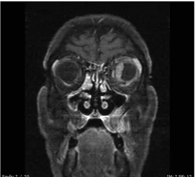
insipidus or oculomotor nerve palsies can be seen (4). In our case, a 69-year-old male patient presented with intermittent gross hematuria and irritative micturation. There was no medical feature in his medical history. Urothelial carcinoma was detected in the bladder. Mass lesion was observed in the right lung in chest radiography. Orbital magnetic resonance (MR) imaging was performed because of redness in the left eye in fundus examination. In orbital MR, the mass lesion was observed to not extend out of the globus at the medial in the left globus, which had a hypointense feature in T2-weighted series and a mild hy-perintense feature in T1-weighted series, with contrast involve-ment restricted in diffusion in diffusion-weighted examination and with a diameter of approximately 12×8×17 mm at its widest (Figure 1). Pituitary and brain MR were conducted on observing the mass in cella in the sections examined. In the pituitary MR, a mass lesion with an approximate diameter of 27×33 mm at its widest was observed, which had sellar–suprasellar localization, generated pressure on the optic chiasm, had thickened infun-dibulum, and had extension into the sphenoid sinus. The mass leaned to the cavernous portion of the bilateral internal carotid
Multiple Systems Metastasis Bladder Cancer with Imaging
Findings
Sermin Tok
1, Naime Altınkaya
1, Gül Nihal Nursal
21Department of Radiology, Başkent University, Adana Practice and Research Hospital, Adana, Turkey 2Department of Nuclear Medicine, Başkent University, Adana Practice and Research Hospital, Adana, Turkey
65
Letter to the Editor
Received Date: 10.08.2015 Accepted Date: 24.08.2015
© Copyright 2016 by Gaziosmanpaşa Taksim Training and Research Hospital. Available on-line at www.jarem.org DOI: 10.5152/jarem.2015.893
Address for Correspondence: Dr. Sermin Tok, E-mail: [email protected]
Figure 1. Mass lesion having contrast involvement not extending
out of the globus in the medial and left globus in orbital MR. MR: Magnetic resonance
Figure 2. Mass lesion having sellar–suprasellar localization, generating
pressure on the optic chiasm, a thickening infundibulum, and having extension into the sphenoid sinus in pituitary MR. MR: Magnetic resonance
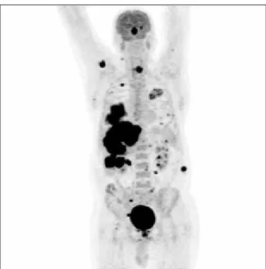
artery and extended to the cavernous sinus (Figure 2). No other mass lesion was detected in brain MR. Bladder Ca metastasis was verified in the biopsy. In the positron emission tomog-raphy/computerized tomography examinations, widespread hypermetabolic metastatic lesions were detected in the right lung lower lobe superior segment and right lung middle and lower lobe bronchus neighborhood; in both the lungs and the subpleural region; in the left orbita, hypophysis, and in a large number in the liver; in the left adrenal gland and bilateral renal neighborhood and fat tissue; in the bilateral anterior peridia-phragmatic region and neighborhood of the lower end anterior esophagus; in the neighborhood of the pancreatic tail and the right external iliac region; and in the right scapula posterior and the right abdominal wall between the muscular structures and musculoskeletal system (Figure 3). The case died a month later. Consequently, striated muscular structures, pituitary gland, and orbita metastasis are rare, and pituitary gland metastases may not have clinical findings. These metastases are indicators of poor prognosis.
REFERENCES
1. Akın Y, Birkan Z, Başara I, Bozkurt A, Nuhoğlu B. Nadir bir olgu: mesane kanserinin gluteal kas metastazı. JAREM 2012; 2: 27-8.
(CrossRef)
2. Wettach GR, Steele EA. Urothelial Cell Carcinoma of the bladder presenting as orbital metastasis. Arch Pathol Lab Med 2008; 132: 1224.
3. SooHoo JR, Gonzalez OM, Slomos VJ, Durairaj VD. Urothelial carci-noma with orbital metastasis. Urology 2012; 80: 4. (CrossRef)
4. Furuta S, Hatakeyama T, Zenke T, Fukumoto S. Pituitary metastasis from carcinoma of the urinary bladder mimicking pituitary apoplexy. Neurol Med Chir 1999; 39: 165-8. (CrossRef)
66
Bladder Cancer Metastazis. JAREM 2016; 6: 65-6Tok et al.Figure 3. Widespread hypermetabolic metastatic lesions in the right
lung lower lobe superior segment and right lung middle and lower lobe bronchi neighborhood; in both of the lungs and the subpleural region; in the left orbita, hypophysis and a large number in the liver; in the left adrenal gland and bilateral renal neighborhood and fat tissue; in the bilateral anterior peridiaphragmatic region and neighborhood of the lower end anterior esophagus; in the neighborhood of the pancreatic tail and the right external iliac region; and in the right scapula posterior and the right abdominal wall between the muscular structures and musculoskeletal system in PET/BT examination.