Case report
Pseudoduplication of the optic disc in moderate myopia
Zeynep Eylül Ercana,⇑; Aylin Karalezlib; Gokcen CobancAbstract
We report a case of optic disc pseudoduplication without pathological myopia. A 79-year-old woman presented with gradually decreased vision. Fundus examination showed macular drusen in both eyes and subretinal haemorrhage in the left eye. Both optic discs appeared to be tilted and the left optic disc was seen to be inferiorly duplicated.
Pseudodoubling of the optic discs can be caused by optic disc and peripapillary chorioretinal colobomas and pathological myopia. Considering that our patient did not have enough axial length to merit a diagnosis of pathological myopia and no pathological fundus lesions typical of elongated sclera, we suggest that acquired pseudoduplication is not a condition unique to pathological myopia, but can occur in all myopic patients. The clinical materials, including ophthalmological examination, fundus fluorescent angiography and MRI scan of the orbit, were reported, and its pertinent literatures were reviewed.
Keywords: Pseudoduplication, Optic disc, Coloboma, Myopia
Ó 2016 The Authors. Production and hosting by Elsevier B.V. on behalf of Saudi Ophthalmological Society, King Saud University. This is an open access article under the CC BY-NC-ND license (http://creativecommons.org/licenses/by-nc-nd/4.0/).
http://dx.doi.org/10.1016/j.sjopt.2016.11.003
Introduction
Doubling of the optic disc is rare and is seen as true dou-bling or pseudodoudou-bling. True duplication of the optic disc occurs with separation of the optic nerve into two or more strands and the presence of a single optic nerve is the key feature distinguishing pseudoduplication from true duplica-tion of the optic disc.1Pseudoduplication of the optic disc shows a well-circumscribed disc-like coloboma with vascula-ture and surrounding chorioretinal atrophy, which is usually inferior to the normal optic disc. Typical colobomas are located inferiorly and slightly nasally, resulting from failure of closure of the foetal fissure. It is also shown that lesions due to pathological myopia can cause acquired pseudodupli-cation.2We present a case with pseudodoubling of the optic disc of a patient with age related macular degeneration without pathological myopia.
Case report
A 79-year-old woman presented with gradually decreased vision. Her best corrected vision acuity was 30/100 and counting fingers at 1 m right and left eye respectively. Axial length of the right eye was 24.66 mm with the spherical
equivalent of 2.50D. Axial length of the left eye was
24.71 mm and its spherical equivalent to 3.00D. Bilateral grade two nuclear cataracts were also present. Fundus exam-ination showed macular drusen in both eyes and subretinal haemorrhage in the left eye. Both optic discs appeared to be tilted and the left optic disc was seen to be inferiorly duplicated (Fig. 1). Optical coherence tomography and
fluorescent fundus angiography showed left choroid
neovascularization, intraretinal and subretinal oedema. Intravitreal Anti-VEGF treatment was started for age related macular degeneration. Fundus fluorescent angiography
Peer review under responsibility of Saudi Ophthalmological Society,
King Saud University Production and hosting by Elsevier
Access this article online: www.saudiophthaljournal.com www.sciencedirect.com Received 2 August 2015; accepted 2 November 2016; available online 10 November 2016.
a
Department of Ophthalmology, Baskent University School of Medicine, Ankara, Turkey
bDepartment of Ophthalmology, Mug˘la Sıtkı Koçman University School of Medicine, Mug˘la, Turkey cDepartment of Radiology, Baskent University School of Medicine, Konya, Turkey
⇑ Corresponding author at: Baskent Universitesi Hastanesi, Fevzi Cakmak Cd. 10. Sk. No: 45, Bahçelievler/Ankara, Turkey. e-mail address:[email protected](Z.E. Ercan).
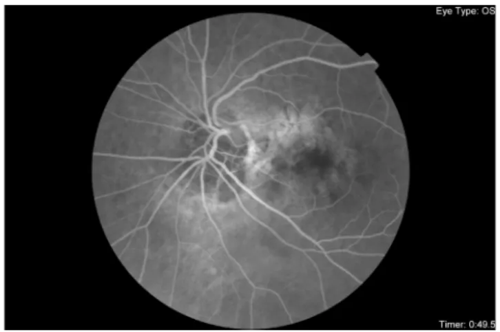
showed inferior retinal artery and vein making a kink on pseudodoublication (Fig. 2). Visual field test was not per-formed due to the poor sight of the left eye. Although the lesion showed bridging of retinal vessels, no doubling of reti-nal artery and no late hyperfluorescence expected from a
true optic disc, orbital MRI was performed to definitely exclude a secondary optic nerve. Orbital MRI (Fig. 3) was reported as left glob lateral radius asymmetric lengthening with no optic nerve doubling or pathological optic nerve lesions, hence establishing the diagnosis of pseudodoubling of the left optic disc.
Discussion
True duplication of the optic disc is a rare anomaly where there are two discs with separate retinal vessels. It can be diagnosed by imaging techniques revealing two optic nerves leaving the eye or fundus fluorescent angiography showing separate retinal vasculature.3 Pseudodoubling of the optic discs, on the other hand, is more common and can be caused by optic disc and peripapillary chorioretinal colobomas.4The coloboma involves an injury to the proximal embryonic fis-sure and typically occurs in the inferonasal quadrant. They are asymptomatic lesions and require no treatment. How-ever, they may extend to include the optic nerve and give rise to subretinal neovascularisation, especially if involving the optic nerve head.5,6It can be differentiated from true dou-bling with imaging techniques. Pseudoduplications also have bridging retinal vessels, originating from the true optic disc to the second pseudo disc, which can be seen in fundus fluo-rescent angiography.5
However, all pseudoduplications are not results of colobo-mas. In a study by Ahn et al, it was shown that 2.3% of patients with pathological myopia had acquired pseudodu-plication of the optic disc. They concluded acquired pseu-doduplication of the optic disc may be one of the presentations of pathological myopia due to extensive eye elongation, myopic chorioretinal atrophy and scleral excava-tion.2Supporting this, Ohno-Matsui et al. suggest that peri-papillary lesions develop in eyes with pathological myopia from a stretch-associated scleral schisis in areas of openings of posterior emissary for the short posterior ciliary arteries.7 Ahn. et al. also concluded, unlike pseudodublications due to colobomas that have abnormal vascular anastomosis with the optic disc, acquired pseudoduplication has no vascular relation with the true disc. Their cases had ciliary artery in the pseudo disc, so they suggested that pathogenesis of a Figure 2. Fundus fluorescent angiography: vascular kink on
pseudoduplication.
Figure 3. (A–B): On axial T1 weighted (A) image, bilateral optic nerves are symmetric in terms of shape, thickness and intensity. Lateral length of the left glob was slightly extended in comparison with right side. Bilateral periorbital fat tissue, lacrimal glands and glob muscles are normal. On thin sectional fat suppressed sagittal T2 weighted (B) image, one optic nerve is seen on left side in normal appearance.
Figure 1. Fundus colour photo of the left eye: Tilted optic disc with the inferionasal pseudodublicated disc.
myopic pseudo disc may result from myopic chorioretinal atrophy and scleral excavation or defects caused by the pen-etration of a ciliary artery through the staphyloma.2
In our case, pseudodoubling location was inferionasal to the optic disc, typical of chorioretinal colobomas. However since both eyes had tilted discs and left glob length was found extended in magnetic resonance images, we con-cluded that this case was an example of acquired pseudodu-plication due to myopia. Considering our patient did not have enough axial length to merit a diagnosis of pathological myopia, a refractive error of 3.0D and no pathological fun-dus lesions typical to elongated sclera, we suggest that acquired pseudoduplication is not a condition unique to pathological myopia, but can occur in all myopic patients. Conflict of interest
The authors declared that there is no conflict of interest.
References
1. McLoone EM, Buchanan TA. Duplication of the optic disc: true
or pseudo? A coloboma or not a coloboma? Eur J Ophthalmol 2004;14(2):163–5.
2. Ahn SJ, Woo SJ, Hwang JM. Acquired pseudoduplication of the optic
disc in pathologic myopia. Optom Vis Sci 2014;91(7):e177–84.
3. Brink JKLFE. Pseudodoubling of the optic disc: a fluorescein
angiographic study of a case with coloboma. Acta Ophthalmol 1977;55(862):70.
4. Kamath G, Prasad S, Patwala Y, Watts M. Peripapillary coloboma
simulating double optic disc. J Ophthalmol 1999;83(10):1207.
5. Islam N et al. Optic disc duplication or coloboma? Br J Ophthalmol
2005;89(1):26–9.
6. Gregory-Evans CY, Williams MJ, Halford S, Gregory-Evans K. Ocular
coloboma: a reassessment in the age of molecular neuroscience. J Med Genet 2004;41:881–91.
7. Ohno-Matsui K et al. Acquired optic nerve and peripapillary pits in
pathologic myopia. Ophthalmology 2012;119(8):1685–92.