C L I N I C A L R E P O R T
A Rare Case: Cartilaginous Choristoma of the Soft Palate
S. Karatayli Ozgursoy1• H. Umudum2•G. K. Beriat1•S. Kaya1Received: 22 April 2015 / Accepted: 8 September 2015 / Published online: 11 September 2015 Ó Association of Otolaryngologists of India 2015
Abstract Cartilaginous choristomas occur in abnormal sites that usually do not contain chondrocytes. These lesions are very rarely seen in the soft palate. We present a 38 year-old patient with cartilaginous choristoma located in nasopharyngeal surface of soft palate. Histopathological examination demonstrated a well demarcated mature hya-line cartilage tissue which was surrounded with seromu-cous salivary glands under a benign respiratory epithelium.
Introduction
Choristomas are benign tissue islands of essentially normal tissue occurring in an abnormal location. They have been reported in the various parts of head and neck region including oropharynx, hypopharynx, oral cavity and middle ear [1,2]. These lesions can arise in cartilage, bone, sali-vary gland, glial tissue, and thyroid gland. We report a rare case of cartilaginous choristoma located on the upper (nasopharyngeal) surface of the soft palate.
Case Report
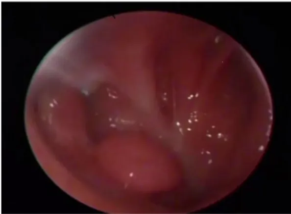
A 38 year-old male patient presented to our clinic with chronic post nasal discharge. During his ENT examination, nasal endoscopy revealed an approximately 1 9 0.5 cm incidental mass on the right side of upper surface of soft palate (Fig.1). No other pathological findings were noted in the oropharynx, larynx, ears and head and neck region. When questioned, he had neither swallowing difficulty, tickling sensation nor any other disturbance related to the lesion. He had no specific medical or past surgical history, other than adenoidectomy when he was 6 years old. The patient was informed about the lesion and after he gave informed consent, the lesion was excised under the guid-ance of an endoscope from the oral cavity while everting the soft palate under general anesthesia.
Macroscopic evaluation of excised specimen revealed a polypoid lesion in 1 9 0.4 9 0.4 cm dimensions. Histo-logical examination showed that excised tissue was cov-ered with benign respiratory epithelium (Fig.2). Beneath the epithelium there was a well demarcated mature hyaline cartilage tissue which was surrounded with sero-mucous salivary glands. A patchy chronic non-specific inflamma-tion was observed in both subepithelial areas and in sali-vary glands. Chondrocytes that form mature hyaline cartilage varied from small to large sizes however they were lack of significant nuclear atypia. Epithelial and cartilage tissue component of lesion did not contain sig-nificant nuclear pleomorphism, necrosis or mitotic activity that rendered malignant behavior. Regarding these find-ings, the lesion was diagnosed as ‘‘cartilaginous choris-toma’’ arising in minor salivary gland.
In 3 month follow up, no recurrence was seen. Postop-erative endoscopic examination revealed anatomically and functionally normal soft palate.
& S. Karatayli Ozgursoy [email protected]
1 Department of Otolaryngology, Ufuk University, Ankara,
Turkey
2 Department of Pathology, Ufuk University, Ankara, Turkey
123
Indian J Otolaryngol Head Neck Surg
Discussion
The nomenclature of the similar lesions with different levels of differentiation is sometimes confusing. These lesions were named variably, such as hairy polyp, epig-nathus, hamartoma, true teratoma, dermoid, teratoid and epignathus. Teratomas and teratoids contain parts from all three germ layers, teratoids are poorly differentiated [3]. Teratomas located in the oral cavity is called epignathus. Hamartomas are excessive and focal overgrowth of cells and tissues natural to the organ in which they occur. Der-moids are composed of epidermal and mesodermal germ layers. Choristoma is pointed out to be a more appropriate term for hairy polyp, in which mesoderm is surrounded by ectoderm. Recent reports also support that hairy polyp and choristoma are developmental malformations, more than teratoma [4,5].
Choristomas have been classified based on tissue type that they contain. According to this classification, we
classified current lesion as cartilaginous choristoma rather than mixed or salivary type choristoma, since minor sali-vary glands are expected to present on this anatomical location whereas a mature hyaline cartilage is not.
Several theories about the origin of cartilaginous cho-ristomas have been proposed. In embryonal theory, origin of heterotopic cartilage is fetal remnants [6]. Metaplastic theory postulates that intraoral trauma might lead to development of heterotopic cartilage from undifferentiated mesenchymal cells [7]. On the other hand, some authors claimed that these choristomas probably arise from the vestigial remnants [8]. The nasopharyngeal choristoma of presented patient was incidentally found in the adulthood, which might lead us think of metaplastic theory due to the trauma caused by the adenoidectomy operation in his childhood.
Zahn, in 1885 was first to describe the presence of cartilaginous tissue in the oral soft tissues [7]. Later, in 1892, Berry reported a case of fibrochondroma of the tongue. Since then, benign cartilaginous lesions of the oral soft tissues, which have been reported in the literature, have been named as cartilage, metaplastic cartilage, dys-trophic chondrometaplasia, osteocartilage, fibrochon-droma, chondroma, enchondroma and calcifying chondroma by various authors. It was in 1971 when Knoll and coworkers, in their review, favored the term ‘choris-toma’ for these lesions. Cartilaginous choristomas have been reported in 1-day old infants [6] to 90-year old female, with the mean age being 47 years [1]. The most common site is the tongue [9] followed by the gingiva, buccal mucosa and soft palate.
Congenital choristomas should be carefully distin-guished from cartilaginous metaplasia, which are charac-terized by scattered cartilaginous cells of different stage of maturation and diffuse deposits of calcium. Excision of the lesion including perichondrium and surrounding soft tissue is considered as curative. Removal of the perichondrium is essential in order to avoid recurrences, because perichon-drium may have the potential to develop new cartilage.
Congenital nasopharyngeal lesions mostly present as an emergency respiratory problem and require immediate surgery. In childhood emergency cases of the congenital nasopharyngeal lesions, immediate surgery should be carefully carried out by paying attention to spare velopharyngeal structure. If the lesion is large, velopha-ryngeal musculature might not always be spared. Local and free flaps have been described for reconstruction of palatal defects. In our case, the lesion was excised transorally and endoscopically by everting the soft palate, sparing all the neighbor musculature.
In conclusion, choristomas are benign, rare lesions that should be kept in mind among the oronasopharyngeal lesions. During the surgery, perichondrium should be Fig. 2 Histological examination of the lesion
Fig. 1 Preoperative endoscopic view of the lesion
Indian J Otolaryngol Head Neck Surg (Oct–Dec 2015) 67(4):444–446 445
excised, the neighbor anatomical structure should tried to be spared, and the lesion should be differentiated from true neoplasms.
References
1. Chou LS, Hansen LS, Daniel TE (1991) Choristomas of the oral cavity: a review. Oral Surg Oral Med Oral Pathol 72:584–593 2. Lee FP (2005) Cartilaginous choristoma of the bony external
auditory canal: a study of 36 cases. Otolaryngol Head Neck Surg 133:786–790
3. Calcaterra T (1969) Teratomas of the nasopharynx. Ann Otol Rhinol Laryngol 78(1):165–171
4. Burns BV, Axon PR, Pahade A (2001) ’Hairy polyp’ of the pharynx in association with an ipsilateral branchial sinus: evidence that the ‘hairy polyp’ is a second branchial arch malformation. J Laryngol Otol 115(2):145–148
5. Erdogan S, Tunali N, Canpolat T, Tuncer R (2004) Hairy polyp of the tongue: a case report. Pediatr Surg Int 20(11–12):881–882 6. Moore K, Worthington P, Campbell RL (1990) Firm mass of
tongue. J Oral Maxillofac Surg 48:1206–1210
7. Unal T, Ertu¨rk S (1994) Cartilaginous choristoma of the gingiva. Report of two cases; review of the literature of both gingival choristomas and intraoral chondromas. Ann Dent 53(1):19–27 (Review)
8. Samant HC, Gupta OP (1971) Chondroma of the tongue. Oral Surg Oral Med Oral Pathol 32(3):450–452
9. Desmedt M, Weynand B, Reychler H (2007) Cartilaginous choristoma of the oral cavity: a report of two cases. B-ENT 3(2):87–91
446 Indian J Otolaryngol Head Neck Surg (Oct–Dec 2015) 67(4):444–446