AGE AND GENDER ALTER SYNAPTIC PROTEINS IN
ZEBRAFISH (DANIO RERIO) MODELS OF NORMAL AND
DELAYED AGING
A THESIS SUBMITTED TO
THE GRADUATE SCHOOL OF ENGINEERING AND SCIENCE
OF BILKENT UNIVERSITY
IN PARTIAL FULFILLMENT OF THE REQUIREMENTS FOR THE DEGREE OF
MASTER OF SCIENCE IN
NEUROSCIENCE
By
Elif Tuğce Karoğlu July 2017
i
AGE AND GENDER ALTER SYNAPTIC PROTEINS IN ZEBRAFISH (DANIO
RERIO) MODELS OF NORMAL AND DELAYED AGING
By Elif Tuğce Karoğlu July 2017
We certify that we have read this thesis and that in our opinion it is fully adequate, in scope and in quality, as a thesis for the degree of Master of Science.
Approved for the Graduate School of Engineering and Science
Director of the Graduate School
Michelle Marie Adams (Advisor)
Özlen Konu Karakayalı
Çağdaş Devrim Son
ii
ABSTRACT
AGE AND GENDER ALTER SYNAPTIC PROTEINS IN ZEBRAFISH (DANIO RERIO) MODELS OF NORMAL AND DELAYED AGING
Elif Tuğce Karoğlu MSc in Neuroscience Advisor: Michelle Marie Adams
July 2017
Cognitive decline occurs during normal aging in some specific domains of cognitive abilities including but not limited to episodic memory, divided attention and executive functions, however, it is not a unitary decline since some cognitive domains, including vocabulary and implicit memory tend to be preserved and even improved at older ages. Normal aging is not associated with global and significant neuronal and synapse loss, yet subtle molecular alterations occurring in gene expression patterns, protein homeostasis, mitochondrial dynamics and hypofunction in the cholinergic system may account for the age related decline in some cognitive abilities. Additionally, males and females showed differential vulnerabilities against age-related alterations in the cognitive abilities, physiological integrity and subtle molecular dynamics. More direct relationships can be established between the age-related cognitive decline and subtle molecular changes by analyzing the elements of synaptic integrity, which could alter synaptic plasticity and result in the changes in learning and memory abilities. Post-synaptic 95 (PSD-95), gephyrin (GEP) and synaptophysin (SYP) are integral synaptic proteins and they could be attributed as
iii
indicators of excitatory post-synaptic, inhibitory post-synaptic and pre-synaptic integrities, respectively. The first aim of this study was to show effects of age and gender on the expression levels of PSD-95, GEP and SYP in young, middle-aged and old, female and male zebrafish cohorts. Significant age by gender interactions were revealed in the levels of PSD-95 and SYP. It was shown that PSD-95 and SYP levels tend to be preserved and increased in the female groups throughout the aging process, whereas, in male groups, expression levels of these proteins tend to be reduced at older ages. The second aim was to investigate whether ameliorating the cholinergic hypofunction might have beneficial effects on the aging-related protein expression alterations and check for sexually dimorphic patterns. For this aim old male and female zebrafish from a mutant line (ache), which has decreased levels of acetylcholinesterase and increased levels of acetylcholine, were compared with old male and female wildtype animals. In the ache old groups, significant increases in the expression levels of SYP and GEP were revealed compared to the wildtype, and also in the old ache females SYP expression was higher than the other groups. These studies emphasized the importance of gender and sexually dimorphic patterns in the context of aging andcholinergic manipulations could be a promising target of intervention to attenuate the effects of age-related synaptic alterations, which could have possible contributions to age-related cognitive decline.
.
Key words: zebrafish, aging, sexual dimorphism, synaptic proteins, acetylcholine, acetylcholinesterase
iv
ÖZET
YAŞ VE CİNSİYETİN NORMAL VE GECİKTİRİLMİŞ YAŞLANMA ZEBRABALIĞI (DANIO RERIO) MODELLERİNDE SİNAPTİK
PROTEİNLERİ DEĞİŞTİRMESİ
Elif Tuğce Karoğlu
Nörobilim Lisansüstü Programı, Yüksek Lisans Tez Danışmanı: Michelle Marie Adams
Temmuz 2017
Normal yaşlanmayla birlikte; epizodik hafıza, bölünmüş dikkat ve yürütme işlevleri de dahil olmak üzere bilişsel işlevlerin bazı belirli alanlarında bilişsel gerileme gözlenmektedir. Normal yaşlanma, büyük çaplı ve anlamlı nöronal kayıpla ve sinaps kaybıyla ilişkili değildir, ancak, gen ekspresyon motiflerinde, protein homeostazında, mitokondriyal dinamiklerde ortaya çıkan küçük moleküler değişiklikler ve kolinerjik sistem hipofonksiyonu yaşlanmaya bağlı bilişsel gerilemeyi fizyolojik düzlemde açıklayıcı role sahiptirler. Buna ek olarak, dişi ve erkeklerde yaşlanmanın fizyolojik ve bilişsel integriteye olan etkilerinin farklılık gösterdiği belirtilmiştir. Sinaptik integrite elementlerini analiz ederek, yaşlanmaya bağlı bilişsel gerileme ve küçük moleküler değişiklikler arasında daha doğrudan ilişkiler kurulabilir. Post-sinaptik 95 (PSD-95), gefirin (GEP) ve sinaptofizin (SYP) anahtar sinaptik proteinlerdir ve sırasıyla eksitatör sinaptik, inhibitör post-sinaptik ve pre-post-sinaptik integritelerin göstergeleri olarak atfedilebilirler. Bu proteinler nöral hücrelerin indüksiyon kapasitelerini değiştirerek öğrenme ve hafıza
v
gibi bilişsel işlevlere etki edebilirler. Bu çalışmanın ilk amacı yaş ve cinsiyetin PSD-95, GEP ve SYP'nin protein ekspresyonu düzeyleri üzerindeki etkilerini genç, orta-yaşlı ve orta-yaşlı, dişi ve erkek zebrabalığı kohortlarında göstermektir. Bulgularda, PSD-95 ve SYP düzeylerinde istatistiksel olarak anlamlı cinsiyet yaş etkileşimi gözlemlenmiştir. PSD-95 ve SYP düzeylerinin dişi gruplarda artan yaşla birlite seviyelerinin korunduğu ve arttığı gözlemlenirken, erkek gruplarda PSD-95 ve SYP ekspresyon seviyeleri yaşlanmayla birlikte azalma eğilimi göstermektedir. Çalışmanın ikinci amacı, kolinerjik hipofonksiyonun iyileştirilmesinin, yaşlanmaya bağlı protein ekspresyonu değişiklikleri üzerinde faydalı etkileri olup olmadığını araştırmak ve olası cinsel dimorfik farklılıkları kontrol etmektir. Bu amaçla, düşük asetilkolinesteraz seviyesine sahip olan erkek yaşlı mutant hattı (ache), dişi-erkek yaşlı yabanıl-tip zebrabalığı grupları ile karşılaştırıldı. Bulgularda, yaşlı ache mutantlarında yaşlı yabanıl–tip kontrol grubuna nazaran ,SYP ve GEP'in ekspresyon düzeylerinde istatistiksel olarak anlamlı bir artış gözlemlenmiştir. SYP ekspresyon seviyelerinin, dişi-yaşlı ache mutant grubunda diğer gruplara kıyasla daha yüksek olduğu gözlemlenmiştir. Bu çalışma yaşlanma bağlamında cinsiyetin etkilerin ve cinsel dimorfik farklılıkların önemini vurgulanmaktadır. Kolinerjik manipülasyonlar, yaşla ilişkili bilişsel gerilemeye olası katkılar sağlayabilecek yaşa bağlı sinaptik değişikliklerin etkilerini hafifletmek için umut verici bir hedef olabilir.
.Anahtar kelimeler: zebrabalığı, yaşlanma, seksüel dimorfizm, sinaptik proteinler,
vi
ACKNOWLEDGEMENTS
Foremost, I would like to thank my advisor Dr. Michelle Adams for accepting me into her laboratory, giving me a great opportunity to work in the field of the neuroscience, creating a really supportive and fruitful research environment, her academic guidance and giving me a great motivation and encouragement for all 5 years.
I would like to thank Dr. Özlen Konu, Dr. Hulusi Kafalıgönül and Dr. Çağdaş Devrim Son for taking part in my thesis committee and their help.
I would like to thank and express my sincere gratitude to Dr. Ayça Arslan Ergül, for supporting me scientifically and emotionally; and for teaching me the appreciation of effort and hard-working which was crucial for success and moving further. I would like to thank Dr. Füsun Doldur-Ballı for her patience and guidance during my internship periods.
I would like to thank all the former and current members of Adams Lab; who were far more than colleagues; I would like to thank Melek Umay Tüz-Şaşik, Narin Ilgım Ardıç, Göksemin Fatma Şengül, Özge Pelin Burhan, Naz Şerifoğlu, Meriç Kınalı and Begün Erbaba for their amazing support, friendship and compassion.
I would like to thank my dearest friends Ayşegül Düşündere, Fazilet Zeynep Yıldırım, Zeynep Çenesiz and Tuba Şahin for their endless support, compassion, understanding and love.
I would like to thank my dear mother Dr. Hülya Karoğlu, for her support, understanding and great love, without her I cannot achieve anything; and I would like to thank Karoğlu family, Eravşar family and my dear fiancée Ebubekir for their compassion, concerns, appreciation, patience and love.
vii
I would like to acknowledge Tülay Arayıcı for her help in the zebrafish facility; and Ayşe Gökçe Keşküş for her help during the genotyping experiments.
I would like to acknowledge The Scientific and Technological Research Council of Turkey (TUBITAK) with grant no 214S236 for financially supporting the preliminary experiments and establishing reliable experimental methodology mentioned in Chapter 2, with grant no 215S71 for financially supporting the experiments mentioned in Chapter 4; and European Molecular Biology Organization (EMBO) Installation Grant to Michelle M. Adams for financially supporting the experiments in Chapter 3.
viii
TABLE OF CONTENTS
ABSTRACT ... ii
ÖZET ... iv
ACKNOWLEDGEMENTS ... vi
TABLE OF CONTENTS ... viii
LIST OF FIGURES ... xi
LIST OF TABLES ... xiii
CHAPTER 1 ... 1
Introduction ... 1
1.1 Normal Aging vs. Pathological Aging ... 2
1.2 Cognitive and Neurocognitive Alterations in Normal Aging ... 4
1.3 Structural and Cellular-Molecular Alterations during Normal Aging ... 9
1.3.1 Structural Alterations ... 9
1.3.2 Cellular and Molecular Alterations... 9
1.4 Aging and Dysregulation of Synaptoproteome ... 11
1.4.1 Postsynaptic density-95 ... 11
1.4.2 Gephyrin ... 13
1.4.3 Synaptophysin ... 14
1.5 Sexual Dimorphism and Aging ... 15
1.6 Zebrafish as a Gerontological Model ... 17
1.7 Conclusion ... 19
CHAPTER 2 ... 21
Methods ... 21
2.1. Subjects ... 21
2.2 Dissections ... 22
2.3. Protein Isolation from Whole Brain of the Zebrafish ... 22
2.4. Bradford Assay ... 23
ix
2.6. Image Quantification ... 33
CHAPTER 3 ... 36
Expression of Postsynaptic Density-95, Gephyrin and Synaptophysin in Young, Middle-Aged and Old Male and Female Zebrafish ... 36
3.1. Introduction ... 36
3.2. Materials and Methods ... 37
3.2.1. Subjects ... 37 3.2.2. Dissections ... 38 3.2.3. Protein Isolation ... 40 3.2.4. Western Blot ... 40 3.2.5. Statistical Analysis ... 41 3.3. Results ... 42
3.3.1 Antibodies Yield Expected Bands in the Zebrafish Brain Protein Lysates 42 3.3.2. The Expression of the Neurotransmission Regulating Proteins were Affected Differentially by Age and Gender ... 44
3.3.2.1. Postsynaptic Density-95 (PSD-95) ... 44
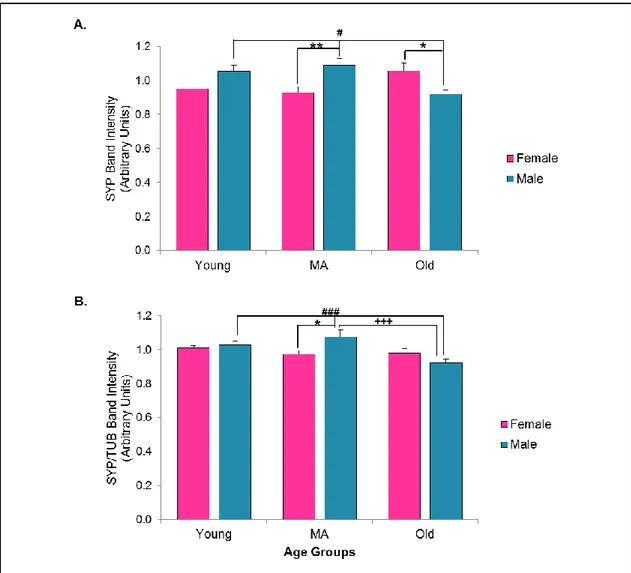
3.3.2.2. Synaptophysin ... 46
3.3.2.3. Gephyrin ... 48
3.3.3. Principle Component Analysis and Investigating Clustering Profiles ... 50
3.3.4. Expression Alterations at Very Old Ages in Female Group ... 57
3.3.5. Region-specific Alterations of the Key Synaptic Proteins According to Age and Gender ... 58
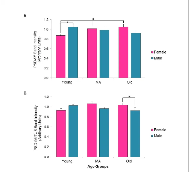
3.3.6. Alterations in the House-keeping Protein: β- Tubulin ... 62
3.4. Discussion ... 63
CHAPTER 4 ... 66
Expression of Postsynaptic Density-95, Gephyrin and Synaptophysin in Old Wild-type and Delayed Aging Mutant Zebrafish Models ... 66
4.1. Introduction ... 66
4.1.1 Cholinergic System ... 67
x
4.1.1.2 Cholinergic System and its Pathway ... 68
4.1.2 Cholinergic System and Normal Aging ... 68
4.1.3 Cholinergic System and Pathological Aging ... 70
4.1.4 Cholinergic System and Synaptic Alterations ... 71
4.1.5 Cholinergic System and Sexual Dimorphism ... 72
4.1.6. Zebrafish Mutant Line with Manipulated Cholinergic System Activity .. 73
4.2. Materials and Methods ... 74
4.2.1. Subjects ... 74
4.2.2. Dissections ... 76
4.2.3. Protein Isolation ... 77
4.2.4. Western Blot ... 77
4.2.5. DNA Extraction from Tail ... 78
4.2.6. Touch-down Polymerase Chain Reaction... 79
4.2.7. Statistical Analysis ... 83
4.3. Results ... 84
4.3.1. Postsynaptic Density-95 ... 84
4.3.3. Synaptophysin ... 85
4.3.3. Gephyrin ... 87
4.3.4. Analysis of the House Keeping Proteins ... 89
4.3.5. Principle Component Analysis and Investigating Clustering Profiles ... 91
4.3.6 Clustering Profiles of Combined Datasets from Chapter 3 and 4... 94
4.3.7 Screening the Mutation to Differentiate Wild-type and Heterozygous Mutants ... 97
4.4. Discussion ... 99
CHAPTER 5 ... 101
Conclusions and Future Prospects ... 101
xi
LIST OF FIGURES
Figure 2.1. Illustration of the standard curve ... 26
Figure 2.2. Illustration of the transfer assembly ... 31
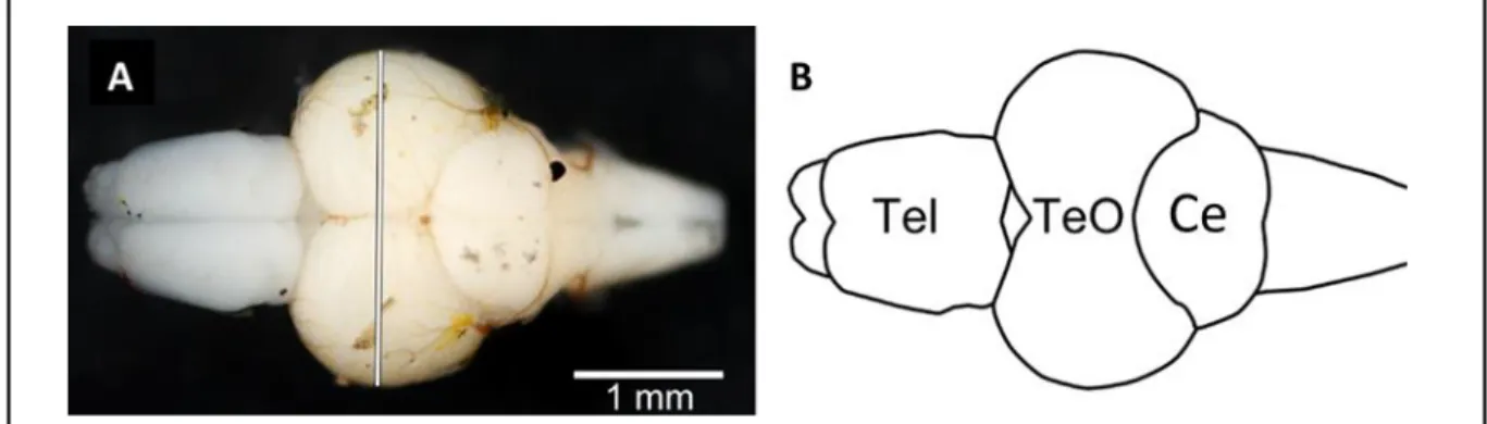
Figure 3.1. Illustration of the microdissection procedure on zebrafish brain, zebrafish brain ... 39
Figure 3.2. Observed bands for the protein samples extracted from zebrafish brain lysate and mouse cortical lysate as positive control ... 43
Figure 3.3. Illustration of the experimental cohorts in each western blot for PSD-95, SYP, GEP and TUB ... 44
Figure 3.4. PSD-95 expression across gender and age groups ... 46
Figure 3.5. SYP expression across gender and age groups ... 48
Figure 3.6. GEP expression across gender and age groups ... 50
Figure 3.7. Principal component analysis for overall data ... 51
Figure 3.8. Plotting each sample with respect to orthogonal components PC1 and PC2 ... 53
Figure 3.9. Principal component analysis for young group ... 54
Figure 3.10. Principal component analysis for middle aged group ... 55
Figure 3.11. Principal component analysis for old group ... 56
Figure 3.12. Expression of the synaptic proteins through aging in female group ... 58
Figure 3.13. Region-specific expression of the key synaptic proteins in gender and age groups ... 61
xii
Figure 4.2. Representation of the mutation and the primers on the partial
sequence ... 80
Figure 4.3. PSD-95 expression across gender and genotype groups ... 85
Figure 4.4. SYP expression across gender and genotype groups ... 87
Figure 4.5. GEP expression across gender and genotype groups ... 88
Figure 4.6. Analysis of the different housekeeping controls, on the one set of a blot ... 91
Figure 4.7. Principal component analysis for all groups together ... 93
Figure 4.8. Plotting each sample with respect to orthogonal components PC1 and PC2 ... 94
Figure 4.9. Principal component analysis for combined datasets from Chapter 3 and 4. ... 95
Figure 4.10. Plotting each sample with respect to orthogonal components PC1, PC2 and PC3 in 3D space ... 97
Figure 4.11. Gel image of the PCR products from wildtype and ache genomic DNA samples ... 98
xiii
LIST OF TABLES
Table 2.1. Preparation of 1 ml RIPA solution ... 23
Table 2.2. Preparations of the standards ... 25
Table 2.3. Preparation of the unknown protein samples ... 25
Table 2.4 Illustration of the template design for 96-well plate... 25
(loaded partially)... 25
Table 2.5. Illustration of the net absorbance values ... 26
for the standards and samples ... 26
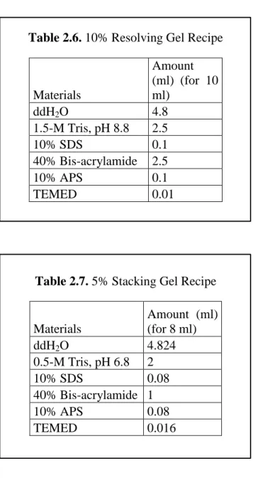
Table 2.6. 10% Resolving Gel Recipe ... 28
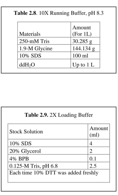
Table 2.7. 5% Stacking Gel Recipe ... 28
Table 2.8. 10X Running Buffer, pH 8.3 ... 29
Table 2.9. 2X Loading Buffer ... 29
Table 2.10. Transfer Buffer ... 30
Table 2.11. 10X TBS, pH 7.6 ... 32
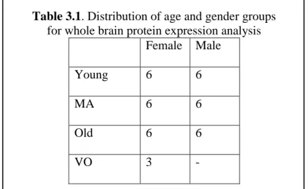
Table 3.1. Distribution of age and gender groups ... 38
for whole brain protein expression analysis ... 38
Table 3.2. Distribution of age and gender groups for the ... 38
protein expression analysis from micro-dissected regions ... 38
Table 3.3. Mean values of TUB for each group ... 62
Table 4.1. Distribution of the gender and the genotype groups ... 76
Table 4.2. Sequences of the primers ... 80
Table 4.3. Prepared PCR reactions for each sample... 80
Table 4.4. PCR mix for each sample ... 81
xiv
Table 4.6. Recipe of 50X TAE buffer ... 83 Table 4.7. Recipe of 1% Agarose gel ... 83 Table 4.8. Mean values of TUB and ACTIN for each group ... 90
1
CHAPTER 1
Introduction
Thanks to advances in technology, medicine and life styles, the older population, who are over 65 years old, is increasing. Reports indicated that, in 2010 the older population was estimated to be 40.2 million; and in 2050 a distinct increase was projected, with older individuals comprising 88.5 million in US [1]. Yet it is evident that increased lifespan does not always correspond to better health and mind-span, it is crucial to maintain them all to have a good quality of life and to achieve successful aging. Advancing age is associated with increased prevalence of cognitive impairments and approximately 50% of the older population suffers from cognitive decline associated with normal aging and age-related pathologies [2]. Therefore, studies to understand molecular mechanisms and high-level alterations in behavior and brain activity associated with aging; and interventions focusing on ameliorating those components become promising and required areas of research. To understand the mechanisms of normal aging, distinctions should be made between the normal aging-associated alterations and changes, which are the result of pathological age-related conditions including Alzheimer’s disease (AD), Parkinson’s disease (PD), frontotemporal dementia (FD) and so forth. After the distinction between normal and pathological brain aging; molecular and structural changes that support normal aging-related cognitive alterations that lead to modifications in the higher order brain systems will give a unified picture to understand the mechanisms of aging.
2
1.1 Normal Aging vs. Pathological Aging
Although pioneering studies indicated that there is significant neuronal loss in the brain with advancing age even in the absence of pathological conditions, subsequent studies reported that in normal aging there is no significant neuronal loss [3], [4]. Furthermore, it was indicated that there is no significant global synapse loss during normal aging [5], [6]. The most evident distinction between normal and pathological aging could be made based on selective neuronal and synaptic losses and vulnerabilities, specific for each pathological age-related condition neuropathological profile.
Alzheimer’s disease (AD) is one of the most common progressive neurodegenerative diseases affecting the older individuals. In AD impairments in memory and higher cognitive abilities are more prominent than the normal aging-related cognitive decline. Episodic memory impairments, delayed recall deficits, semantic coding and semantic memory deficits were evident in AD [7]. In terms of neurodegeneration, progressive neural loss occurs in the neocortex and hippocampus, which are crucial regions for memory encoding and higher level cognitive abilities like executive functioning. Also, global degeneration was observed in the interconnections and the projections between the association areas of the brain in AD patients [8], [9]. Additionally, cholinergic system has selective vulnerability in the context of AD, as degeneration was evident in the cholinergic projection from the nucleus basalis of Meynert [10]. Amyloid β (Aβ) protein accumulation in the extracellular space and the intracellular inclusions of the neurofibrillary tangles, which contain the abnormally hyper-phosphorylated tau proteins, are the neuropathological hallmarks of AD [8]. Although some non-demented older adults
3
also Aβ protein accumulation and the presence of neurofibrillary tangles, the distribution profile of those hallmarks differs between the AD patients and non-demented older adults. Those neuropathological hallmarks are present in restricted brain regions in the non-demented older adults, whereas they are wide-spread in the AD patients, which has synaptotoxic effects and eventually results in wide-spread synaptic loss and disturbances in axonal dynamics [11].
Parkinson’s disease (PD) is another age-related progressive neurodegenerative disease, which has devastating effects on the motor system. Clinical features of this disease include tremors, slowness of movement, abnormal posture like disturbed axial position of the body and rigidity of movement [12]. Besides the evident motor symptoms, cognitive impairments including dementia and affective disorders including anxiety, depression and anhedonia were observed in PD patients [12]. In PD dopaminergic neural loss occurs, especially in the substantia nigra of the basal ganglia, and this critical loss is associated with the motor symptoms during the onset of PD [13]. In terms of neuropathological profile, neural loss is accompanied with the aggregation and accumulations of Lewy bodies and neurites, which are the aggregated, as well as accumulated forms of alpha-synuclein [13].
Frontotemporal dementia (FD) is characterized by significant atrophy in the frontal and the temporal lobes of the brain. Compared to AD, in FD personality problems, which are due to the disturbed dynamics of the frontal lobe, are prominent. There are more impairments in executive functions, whereas less impairment in memory performance in FD [7]. In terms of the neuropathological profile, severe neural loss in the frontal and temporal lobes accompanied with proteinopathies are
4
present and not related with tau or alpha-synuclein proteins but rather ubiquitin. This distinguishes the pathology of the FD from the other age-related neuro-degenerative pathologies [14].
To summarize age-related pathological conditions like AD, PD and FD, have differential neurodegenerative profiles and specific brain regions are more vulnerable. This is not the case during normal aging, which is not accompanied by significant global neuronal loss. Furthermore, the pathological pattern and distribution of those pathologies across the brain regions differentiate neurodegenerative disorders among themselves and normal aging.
1.2 Cognitive and Neurocognitive Alterations in Normal Aging
Older adults experience cognitive decline without an existence of evident neuropathology or dementia. However, this decline is not a unitary process, rather multidimensional. While specific cognitive domains are declining with increased age; some cognitive domains are stable and even improved in older adults compared to the young group.
Cognitive abilities can be evaluated under the more general domains, which are crystallized and fluid intelligence. Crystallized intelligence is based on the well-learned and practiced information and access to that information; vocabulary and the general knowledge about the concepts could be examples in this category. On the contrary fluid intelligence requires more active monitoring, evaluation and the reasoning about the concepts which were novel and not based on the previous experiences; cognitive abilities, which require more flexibility, such as, working memory, processing speed and executive functions can be an examples for this category [15]. Pioneering studies indicated that fluid intelligence is more vulnerable
5
to normal aging, as normal functioning older adults tend to show lower performance with the tasks under this category compared to younger adults; whereas in the tasks that require crystallized intelligence, older adults showed similar and even improved performance compared to the young controls [16]. Therefore, cognitive abilities can be analyzed with respect to these two domains, to see the pattern of age-related cognitive alterations.
Processing speed refers to the speed of the cognitive skills in addition to the speed of the motor responses. This ability includes many components like speed of the decision making processes, perceptual processes, motor processes and reaction time. It was shown that processing speed is impaired in older adults compared to the younger subjects, and theories suggested that a slower speed of processing can be an underlying factor for decline in the varieties of the cognitive skills in older adults [17]. Processing speed requires evaluation and the reaction in unfamiliar tasks and the weight of the previous experiences is not that strong for this ability; so processing speed can be attributed to part of the fluid intelligence and it is plausible to expect age-related decline in this ability.
Attention basically refers to concentrating on a relevant stimulus while ignoring the irrelevant distractor stimuli in the context. Yet, this cognitive domain includes multiple components. Selective attention is the process in which a specific feature of a stimulus was attended to like the visual shape of the letter while ignoring the irrelevant sets of the other stimuli like the color of the letter. Older adults performed slower in the tasks requiring selective attention compared to the young groups, which can be attributed to impaired processing speed [18]. Another and the most affected component is divided attention; this ability requires processing two or
6
more information simultaneously. Divided attention is distinctly impaired with advancing age, which might be due to inappropriately and inefficiently allocating and dividing the attentional resources in the demanding tasks in older adults [19]. Sustained attention is another component and refers to focus and attention to certain stimuli over a period of time and studies indicated that the sustained attention performances of the older adults are similar as the young groups [20].
Memory is one of the most well-studied cognitive skills of the aging domain because of the distinguishing deteriorations that results from age-related pathological conditions or normal aging. Memory has many subcategories but firstly the processes of forming a memory can be analyzed with respect to aging. The first step is the acquisition, which requires encoding of the novel knowledge and information. Studies indicated that encoding declines with increasing age [21]. Retention, which is recalling the previously encoded information, is another process of memory, and in the older adults this process is stable with subtle problems in the source information [22]. Retrieval is recalling and accessing the newly encoded information and this process is also impaired in the older adults compared to young [21]. Memory can be subdivided into two categories, explicit and implicit memory. Explicit memory includes the collection of the information that is consciously acquired; and explicit memory is divided into two categories; episodic and semantic. Episodic memory is the collection of the memories of personal experience and this memory category declines with aging [23], whereas semantic memory refers to the information collection about facts, concepts, ideas and so forth, compared to episodic memory in which there is a life-long decline, in semantic memory there is a late-life decline in this ability [23]. Implicit memory refers to the unconscious collection of the
7
information; one of the examples could be priming; which occurs unconsciously and affects the successive encoding of the new information and manipulates the reaction, implicit memory skills are preserved at the older ages too compared to explicit memory [15].
Language abilities tend be stable and preserved in the older ages. Vocabulary knowledge is one of the constituents of the language and research indicates that it is stable and there are even improved patterns with an increasing age. Language processes, which depend on speed of processing, show subtle declines with increasing age in this category, verbal fluency can be an exemplar that requires active search for and relevant generation of a word [24], [15].
Additionally to the age-related cognitive alterations, thanks to the advancing technology in brain imaging and manipulation tools, neural correlates of cognitive decline have been investigated. It is evident that in the specific cognitive domains, aging is associated with deficits and impairments, yet it is crucial to understand what kind of activity alterations occurred in the brain in response to aging. Studies indicated that during the verbal tasks associated with long-term and working memory, younger adults have increased activity in the left prefrontal cortex. While older adults were engaging with the same task; different from the younger adults, bilateral activation in the prefrontal cortex was observed [25], [26]. The underlying reason for the recruitment of the additional regions and activity was speculated many times; the reason was attributed to the inefficient processing of the inhibitory mechanisms; while it was argued that this additional recruitment is supportive and functional (for review see [27]). It was revealed that older adults with better behavioral and cognitive performance showed significant bilateral activity, whereas
8
the neural activity in the low-performing older adults was unilateral and the activity pattern was similar as the young control groups [28]. Additionally repetitive transcranial magnetic stimulation (rTMS), which temporarily disrupts the neuronal function in the area of interests according to the applied signal frequency, showed that when the rTMS was applied to the left dorsolateral prefrontal cortex in the young adults, the performance and the accuracy in the memory tasks was significantly impaired compared to the condition when rTMS was applied to the right dorsolateral prefrontal cortex. However, in the older adults there was no difference in whether rTMS was applied to the right or left dorsolateral prefrontal cortex, behavioral performance is significantly impaired in both of the conditions [29]. Those studies indicated that increased bilateral activation and the recruitment of the additional brain regions during the task performance are functional and supportive against age-related cognitive decline.
To conclude, cognitive decline is experienced by older adults, yet there is no unitary decline. Cognitive domains based on the usage of well-learned knowledge and skills were preserved at even older ages, whereas decline is evident in the cognitive domains that are more flexible and require active reasoning and manipulation of the novel information. Additionally this decline is associated with the alterations in the brain activity, and the compensatory recruitment of the additional brain areas during the cognitive performance serve as scaffolding against aging.
9
1.3 Structural and Cellular-Molecular Alterations during Normal Aging 1.3.1 Structural Alterations
Age-related structural alterations should be investigated to have insights about underlying factors contributing to cognitive decline. Although, initial studies indicated that there is an age-related significant neural loss in the brain, later work demonstrated that there is no significant neural loss during the course of normal aging [3], [4]. Additionally, it was shown that there is no global synapse loss in the aging brain [5], [6], yet there are region specific synaptic vulnerabilities (for review see, [30]).
Although there is no neuronal and synaptic loss during normal aging, cortical volumetric measurements indicated that after age of 60, shrinkage was observed in the regions including frontal and parietal cortices, and white matter deficiencies were observed in the frontal regions [27]. A comprehensive study, revealed that volume loss without a neural loss can be explained by the reduction in the neuronal volume and disturbance in the dendritic architecture [31]. Therefore, it could be said that significant neuronal or synaptic loss are not underlying factors for cognitive decline, yet there is alterations in the cellular and synaptic dynamics which could give insights about it.
1.3.2 Cellular and Molecular Alterations
Due to the situation that there are no major structural changes or significant neuronal or synapse loss during aging, it is likely that subtle cellular and molecular alterations underlie the changes in cognitive abilities. So examining how physiological integrity is changed during the course of the normal aging would give
10
insight into possible contributors of cognitive alterations. Aging-related subtle cellular and molecular alterations may lead to impairments in the function of the cellular dynamics. Moreover, these targets could be used for future drug manipulations to improve the course of cognitive declines.
The first factor could be epigenetic alterations occurring throughout the aging process. Transcriptional changes are one of the contributors to epigenetic differences in aged brains and it was revealed that age-related alterations exist in the encoding of the genes that are the crucial elements of lysosomal clearance, inflammatory pathways and mitochondrial regulation. Moreover, overexpression in the genes, which are regulating the inflammatory and immune responses, was found, while there is a lower expression of the genes regulating energy metabolism and apoptosis in the older subjects including humans, rats and mice compared to the young groups [32]. Therefore, these kinds of common motifs in gene expression profile might give information about age-related alterations in the transcriptional integrity.
Another crucial component is protein homeostasis and aging can disturb the dynamics of it. Heat shock proteins, chaperones and ubiquitin proteasomes are the key elements of the control mechanism for protein folding and renewal processes; so disturbances in this control mechanism result in aggregated and abnormal protein accumulation. During aging there is a functional decline in the crucial components of this control machinery [33].
Mitochondrial function and reactive oxygen species (ROS) were broadly investigated in the aging domain. According to first observations, advancing age is associated with mitochondrial dysfunction and increased production of ROS, which eventually increases the mitochondrial damage and leads to disturbances in the
11
cellular dynamics [34]. However, studies indicated that increased production of ROS is for the compensation of the homeostatic disturbance, but if it exceeds a certain threshold during aging this compensatory response has a detrimental effects on mitochondrial and cellular integrity (for review see [35]).
1.4 Aging and Dysregulation of Synaptoproteome
Aging alters the cellular and molecular dynamics, which eventually could lead to declines in cognitive abilities. As indicated previously, there are no gross structural alterations in the aging brain but some molecular alterations like epigenetic changes, mitochondrial dysfunction and loss of protein homeostasis might account for age-related cognitive impairments. However, more direct relationships can be established to find the basis of the age-related cognitive decline by investigating the synaptic integrity in the aging brain. Studies have indicated that aging is also associated with dysregulated synaptic protein expression and these alterations have deteriorating impacts on neurotransmission, vesicle trafficking, induction of long-term potentiation synaptic plasticity, which are crucial for learning and memory formation. Therefore, in this thesis the synaptic proteins; postsynaptic density 95 (PSD-95), gephyrin (GEP) and synaptophysin (SYP) were investigated as indicators of excitatory post-synaptic integrity, inhibitory post-synaptic integrity and pre-synaptic integrity, respectively through the course of normal aging and in delayed aging model.
1.4.1 Postsynaptic density-95
Postsynaptic density-95 (PSD-95) is one of the major scaffolding proteins in post-synaptic densities. It has roles in anchoring and localizing the receptors into
12
post-synaptic regions, regulating the trafficking of the synaptic and adhesion proteins, and controlling activity-dependent synaptic responses [36], [37]. PSD-95 is a member of the membrane-associated guanylate kinase family (MAGUK), it consists of 3 PDZ domains, which serves as protein interaction sites by enabling the anchoring of key proteins and receptor subunits, 1 Src homology 3 domain (SH3), and 1 guanylate kinase domain (GK) [38]. PSD-95 directly binds to the GluN2 subunits of the N-methyl-aspartate receptor (NMDAR)-type of glutamate receptors and regulates their localization. Through stargazin and TARP, it clusters and binds to the α-amino-3-hydroxy-5-methyl-4-isoxazole propionic acid (AMPA)-type of glutamate receptors [38]. Both NMDA and AMPA are the ionotropic receptors for glutamate, which is the primary excitatory neurotransmitter in the central nervous system. Those receptors are key to induce and modulate activity dependent synaptic responses including long-term potentiation (LTP) which is the progressive strengthening of the synapses due to experience based frequent stimulation, and long-term depression (LTD), which is the weakening of the synapses due to the activity and experience based stimulation [39]. Both of these processes, LTP and LTD, are strongly associated with learning and memory. Studies conducted with a PSD-95 knock-out mice model indicated that those mice show an enhanced LTP pattern, whereas there was impairment in LTD, and behaviorally those mice showed spatial learning and memory deficits [40].Additionally it was shown that in the hippocampal cell cultures, PSD-95 mimics the effects of the LTP and changes the silent synapses through AMPA receptor mediated insertion to make functional synapses [41].
13
Studies focusing on the alterations in the levels of the PSD-95 throughout aging revealed no consistent results. Whereas, some works have indicated that the levels of PSD-95 are significantly reduced in the old and behaviorally-impaired groups compared to old-non impaired and young controls [37], some studies have reported an increase in the levels of the PSD-95 in the old-behaviorally impaired groups compared to the controls [42]. Inconsistencies can result from potential region-specific expression profile changes in PSD-95 and region-specific vulnerabilities with aging.
1.4.2 Gephyrin
Gephyrin (GEP) is also a major scaffolding protein at post-synaptic sites, but it clusters glycinergic and ionotropic γ-aminobutyric acid A (GABAA) receptors,
which are the crucial inhibitory components of the neurotransmission. Structurally, as a scaffolding protein, GEP lacks PDZ domains which serve as a connection region for cytoskeletal elements or synaptic proteins, but it has auto-oligomerization and by this process it serves as clustering protein. It has three domains, one G domain, one E domain that has high affinity binding sites for other synaptic proteins and cytoskeletal elements, and one C domain that links E and G domains and some cases binds to the synaptic proteins [43].
Brain sections and cultured hippocampal cells taken from GEP knock-out mice indicated that in the synaptic region crucial subunits of the GABAA receptor
were missing and increased amount of GABAA receptor subunits were detected in the
intracellular compartments, which were supposed to anchor at synaptic sites [44]. On the other hand overexpression of GEP resulted in little increases in the GABAA and
14
pre-synaptic regions were observed [45]. Those studies indicated that GEP is crucial for the localization and integrity of the GABAA receptors, but also there is a
cross-talk between the GEP and glutamatergic transmission through PSD-95 and other mechanisms.
Aging studies have reported that in the old-impaired groups of rats GEP levels were increased in the parietal lobe compared to the old-unimpaired and young groups, but this pattern was not observed in the prefrontal cortex [46]. Also it studies using a rat model of aging have indicated that the excitatory-inhibitory ratio, which was derived from PSD95 divided by GEP values, showed decreased patterns in the old impaired group in both prefrontal and parietal areas, and an increased expression of the GEP was found in the old impaired groups [47]. On the other hand, studies conducted with the human V1 reported an opposite pattern with GEP levels significantly decreased at older ages [48]. These kinds of inconsistencies in the aging literature may show region-specific vulnerabilities and expression differences of GEP.
1.4.3 Synaptophysin
Synaptophysin (SYP) is a transmembrane glycoprotein that has four trans-membrane domains which span the vesicles. It localizes in the synaptic vesicles and was used as a marker of the density and integrity of the pre-synaptic regions [49]. SYP has potential roles in the processes of endocytosis and synaptic vesicle trafficking [50] but the exact roles of SYP in the synaptic function are still unclear. Studies conducted with a SYP knock-out mouse indicated that hippocampal neurons have increased synaptic depression and the recovery of the synaptic vesicle pool took an extended period of time [50].
15
In terms of aging literature, it was shown that the levels of the SYP are significantly and positively correlated with the behavioral performance, while the levels of the SYP are reduced in the impaired old groups, the levels were stable between the old unimpaired and young groups [51]. Additionally it was shown that, in hippocampus there was a decrease in the expression levels of SYP with increasing age [52]. Therefore, besides its roles in synaptic trafficking, preserved levels of the SYP can be interpreted as the indicator of the preserved cognitive abilities.
The aim of the present study in this thesis was to show whole brain and region-specific expression profiles of the three synaptic proteins; PSD-95, GEP and SYP, which are regulating the differential aspects of the synaptic function and integrity, to obtain possible age-related alterations in the expression profiles, and lastly to decipher the possible role of the gender, which has been underemphasized in many aging studies. Moreover, these data are among the first to examine these proteins in the context of aging in the zebrafish brain. It was hypothesized that there would be age-related changes in the levels of the PSD-95, GEP and SYP; and those alterations in the protein expression profile would depend on the gender of the animal.
1.5 Sexual Dimorphism and Aging
Gender is one of the most important variables that affect the course of aging in behavioral, molecular and cellular aspects, yet gender tends to be underestimated in the aging studies. In terms of structural sexual dimorphism, there are sexual volumetric differences in the adult human brain. Imaging studies conducted with functional magnetic resonance imaging (f-MRI) indicated that cerebrum-normalized volumes of the cortical regions including frontal and medial-paralimbic areas were
16
larger in women, whereas, in men cerebrum-normalized volumes of the hypothalamus, amygdala and fronto-medial area were larger [53]. Additionally, imaging studies indicated that non-demented old male groups and those old males with mild cognitive impairment had significantly more microstructural deficits, which were revealed by diffusion tensor imaging, than the non-demented old female groups and those old females with mild cognitive impairment [54]. Also, circulating sex hormones affects the architecture of the key regions like the hippocampus, which controls learning and memory functions. It was shown that estrogen activates stronger excitatory dendrites in the CA1 region of the hippocampus and this induction takes place in a N-methyl-D-aspartate (NMDA) receptor-dependent way [55]. Additionally, estrogen replacement therapy in the gonadectomised rodents increases the NR1 subunit of the NMDA receptors in CA1 neurons and also manipulates and increases the responses of those neurons [56], [57].
Epigenetic alterations occurring during normal aging show distinct sexually dimorphic patterns. Gene expression studies conducted in intact individuals with an age range of 20-99, indicated that the forebrain showed distinct alteration profiles during the ages of 60-70 compared to the other age ranges; more interestingly gene categories altered in both an age and a gender dependent manner. To illustrate in the males down regulation of the genes, which are crucial for the energy metabolism, protein synthesis and transportation, were more prominent than in the females, whereas up regulation of the genes crucial for the immune response are more prominent in the females. Altered expression profiles peak at different time points in both genders; in males more prominent changes were observed between 60-70 years of age and the expression profile was stable after 80 years of age; whereas there are
17
dynamic alterations in the gene expression profiles during aging in females and this alterations continued until 80-90 years of age [58]. Those observations which showed that age-related gene expression changes were sexually dimorphic were also consistent with the studies conducted with animal models including zebrafish [59], [60]. Taken together this evidence suggests that age-related alterations exert their effects in a gender-dependent manner and changes in each gender may not be similar. Therefore, both gender groups should be included in aging studies to get insights about the whole picture.
1.6 Zebrafish as a Gerontological Model
The zebrafish has been a popular model organism for developmental and biological studies, due to its large fecundity, easy and inexpensive maintenance, transparent embryos and observable development, and available mutant and transgenic lines, which can provide more causative observations. As a model organism zebrafish has conserved physiological and genetic homology to humans [61]. Also, zebrafish have an integrated nervous system. The architecture of areas like spinal cord cerebellum; olfactory bulb, spinal cord and retinal cells are similar to their mammalian counterparts. However, the morphology of telencephalon and optic tectum show some differences. In the embryonic stage, the telencephalon undergoes an eversion and extends dorsally, which is different than the mammalian brain development [62]. Due to this eversion process, finding homologous structures which correspond to the telencephalic regions in the zebrafish become a challenging work. Yet recent ablation studies with zebrafish and other teleost models indicated that specific telencephalic structures likely correspond to mammalian hippocampus and amygdala [63], [64]. To illustrate, when lesions were induced in the ventrolateral
18
portion of the dorsal telencephalon, deficits in spatial learning and memory were observed, which is similar to hippocampal lesions in mammalian brain; and lesions in the medial portion of the dorsal telencephalon resulted in impairments in fear conditioning and learning so the behavioral phenotype of the medial portion of dorsal telencephalon shows resemblance to amygdala lesions in mammalian models [64]. Although, ablation studies give general information about behavioral deficit and phenotype, expanded work on neuro-architecture and circuit structure of those telencephalic segments are still required. Another distinct and different region in zebrafish brain is the optic tectum. The optic tectum is a predominant structure in the diencephalon of the zebrafish like in the other teleost species, which is different from mammals [62]. Tectal neurons in this region are responsible for the basic visual processing includes motion, direction and color detection. Moreover, besides first-order motion detection, which are dependent to the luminescence differences, they are also detecting the apparent and second-order motion like in mammals, which are based on the second-order visual features including contract, flickering, texture etc. differences [65]. This kind of high level visual processing might indicate that the optic tectum is likely very similar to mammalian visual cortical areas such as V1 and V2.
The zebrafish is also a promising gerontological model since the lifespan of zebrafish is around 3-5 years [66], which is longer than conventional animal models like mice. The longer lifespan of zebrafish might be seen as drawback for aging studies yet it is crucial to observe gradual age-related differences. One of the most important findings about this model is that zebrafish shows gradual cognitive decline with an increasing age, which is also observed in humans [67]. Moreover, age-related
19
cellular and molecular alterations can also be observed in the aged zebrafish. To illustrate accumulation of the senescence-associated beta-galactosidase and oxidized proteins were observed in old zebrafish tissues including brain and muscle [68], [66]; those markers are considered as the hallmarks of cellular senescence. Because of the accumulated DNA damage with an increasing age some chromosomal regions become susceptible to those changes, one of those regions are the telomeres and telomere shortening was observed in the aged humans and mammalian models [35]. This phenomenon was also observed in the zebrafish, age-related shortening of telomeres and decreases in the activity of telomerase were observed in previous studies [69], [68]. Finally, compared to the mouse, zebrafish show higher resemblance to humans in terms of telomere length [69]. Therefore, it can be concluded that hallmarks of the aging in the humans and mammals are also observed in the zebrafish and it represents a promising model to study human aging.
1.7 Conclusion
Normal aging differs from the age-related pathological conditions, with the most evident features of the age-related pathological conditions as progressive neurodegeneration, and abnormal accumulation of proteins and global distribution of those proteinopathies across brain regions, which result in severe deficits in cognitive abilities [7], [8], [14], [70]. However, during normal aging cognitive decline is also prominent in some cognitive domains including processing speed, episodic memory, divided attention and executive functions, which are less severe than the cognitive decline occurred in the pathological conditions and they affect differential aspects of the cognitive domains [15]. Yet, normal aging-related cognitive decline is not a
20
unitary process; whereas some cognitive abilities decline with an advancing age; some cognitive domains including sustained attention, implicit memory, and vocabulary show stable and even improved profiles in the older adults [20]. There are no gross structural alterations including global neuronal and synapse loss occurring throughout the normal aging process [3], [6] that accounts for age-related cognitive decline. However, subtle molecular alterations including age-related gene expression changes [58], disturbances in mitochondrial dynamics [32], protein homeostasis [33], and hypofunction in the cholinergic system [10] were reported in previous studies. Disturbances in neurotransmission regulating proteins might have a direct explanatory role in those age-related cognitive decline and alterations; it was also reported that age-related changes exert their effects differentially in males and females [58]. In the present thesis, this possible dysregulation in the levels of the key synaptic proteins PSD95, GEP and SYP which might have contributions to the age-related cognitive decline, was investigated in different age and gender groups of wild-type zebrafish and in an old mutant line, which has been proposed as a delayed aging model in a previous study [67].
21
CHAPTER 2
Methods
To investigate the possible age-related dysregulation in the key synaptic proteins that have possible contributions to the age-related alterations in synaptic composition that might underlie cognitive decline, Western blotting was used to measure the relative expression levels of the selected synaptic proteins among different groups. This was done in both wild-type and delayed aging zebrafish models. In this Chapter, information is provided about the subjects, dissections, protein isolation procedure, Western blot protocol and image quantification procedure. These common methods are applied in Chapters 3 and 4, and any specific methods are indicated in those Chapters.
2.1. Subjects
All fish were raised and maintained in standard conditions in the Bilkent University Molecular Biology and Genetics Department Zebrafish Facility. They were kept in a recirculating and controlled ZebTec housing system (Tecniplast, Italy) with the temperature of the system water at 28°C. In this system, pH was between 7.0-7.5 and the water quality parameters were frequently checked by the readings of the mechanical and the carbon filters. Based on the stocking density of 3 fish per liter, which has been shown to not create increased levels of stress [71], animals were maintained in 3.5 L or 8 L tanks with a light-dark cycle of 14L:10D. Fish were fed with dry flakes twice a day and live food; artemia once a day. All the birth dates were recorded and labeled on the tanks, and the fish which had the same date of birth
22
were kept together. Age groups were young (6-8 months old), middle-aged (11-14.5 months old), old (27-30) and very old (34-43 months old) and in each Chapter chosen age groups are indicated. Two different euthanasia methods were employed in Chapters 3 and 4 and those methods are explained in each Chapter.
2.2 Dissections
Following euthanasia, heads were decapitated using a scalpel blade. With the use of a stereomicroscope, the head was placed dorsal side down. Gills and the connective tissues were removed from the ventral side to reach the skull, after visualization of the optic chiasm; the eyes were removed from the optic nerves by surgical scissors. The skull was exposed after the removal of eyes, gills and connective tissues, and it was broken carefully and the brain was extracted from the ventral surface. After the visualization of anterior structures like telencephalon, the brains and the tissue of interests were put into 1.5 ml tubes and tissues were snap frozen by immersing the tubes into the liquid nitrogen to prevent further degradation of samples. To determine the gender, the peritoneal cavity was opened and when eggs were visualized the gender was determined as female and in the presence of testis the gender was designated as male, and bodies were subjected to the same snap freezing protocol as the other tissues. All of the samples were stored at -80°C for the subsequent protein analysis experiments.
2.3. Protein Isolation from Whole Brain of the Zebrafish
Stored whole brain tissues were taken from -80°C for the protein isolation protocol; they were put into 1.5 ml Eppendorf tubes. Approximately 60 μl of radioimmunoprecipitation assay (RIPA) buffer [50-mM TrisHCl, pH 8.0, containing
23
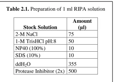
150-mM NaCl, 1% NP40, and 0.1% SDS and protease inhibitor (2× stock, 05 892 970 001, Roche)] was added for each 1 mg of tissue. This lysis buffer is suitable for target proteins that are membrane bound, nuclear or found in the whole cell. The recipe for the preparation of 1 ml RIPA buffer is found in Table 2.1.
Table 2.1. Preparation of 1 ml RIPA solution
Stock Solution Amount (μl) 2-M NaCl 75 1-M TrisHCl pH:8 50 NP40 (100%) 10 SDS (10%) 10 ddH2O 355 Protease Inhibitor (2x) 500
After tissues were suspended in the required amount of the RIPA buffer, they were homogenized with a 25-gauge, 2-mL syringe by passing through it 10-12 times on ice. Homogenates were incubated on ice for 30 minutes, during that period they were gently mixed three times. After the incubation, samples were centrifuged at 13,000 rpm for 20 minutes at 4°C, and the supernatant was collected and aliquoted. Following the determination of the soluble protein concentrations, protein samples were stored in -80°C.
2.4. Bradford Assay
To determine the concentration of the soluble proteins a Bradford assay was performed in a 96-well plate. Firstly, ddH2O for blanks, standards and unknown
24
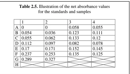
and 2.3. After that standards (bovine serum albumin (BSA, A7906, Sigma-Aldrich, St. Louis, MO, USA) ranging from 2-20 μg/ml) and unknown protein samples were loaded into the plate in duplicates, template designs is illustrated in Table 2.4. After ddH2O, BSA standards and protein samples were loaded, 250 μl of Bradford Reagent
(B6916, Sigma, St. Louis, MO, USA) was added into the wells, and a 96-well plate was mixed using the orbital plate shaker at 300 rpm at room temperature for 2 minutes. Then plate was incubated without shaking at room temperature for an additional 10 minutes. The plate was checked whether there were any air-bubbles in each well that could affect the absorbance values of the samples. In cases where there were air bubbles they were popped with a sterile syringe tip. Absorbance values were measured at 595 nm with a multi-plate reader (SpectraMax M5, Molecular Devices, Sunnyvale, CA, USA), and examples of the net absorbance values are found in Table 2.5. Blanks were assigned in the template design and the absorbance values of the blanks were subtracted from the absorbance values of the each well. Net absorbance of the standards and the concentrations were plotted, and a linear curve fit was applied to those data points and standard curve was created which was used for the concentration calculations of the unknown protein samples, which are shown in Figure 2.1.
25
Table 2.2. Preparations of the standards
BSA (μl) ddH2O (μl) Final Conc. (μg/ml) (1mg/1ml stock) Blank 0 5 0 Standard 1 0.5 4.5 2 Standard 2 1 4 4 Standard 3 2 3 8 Standard 4 3 2 12 Standard 5 4 1 16 Standard 6 5 0 20
Table 2.3. Preparation of the unknown protein samples
Protein (μl)
ddH2O (μl) Unknown Protein Samples 0.5 4.5
Table 2.4 Illustration of the template design for 96-well plate (loaded partially) 1 2 3 4 A STD0 STD0 f-tel-old f-tel-old B STD1 STD1 f-op-old f-op-old C STD2 STD2 f-cer-old f-cer-old D STD3 STD3 m-tel-old m-tel-old E STD4 STD4 m-op-old m-op-old F STD5 STD5 m-cer-old m-cer-old G STD6 STD6 H
26
Table 2.5. Illustration of the net absorbance values for the standards and samples
1 2 3 4 A 0 0 0.058 0.055 B 0.054 0.036 0.123 0.111 C 0.055 0.062 0.133 0.12 D 0.112 0.097 0.082 0.078 E 0.17 0.171 0.152 0.145 F 0.237 0.253 0.135 0.125 G 0.289 0.327 H
Figure 2.1. Illustration of the standard curve. Red and blue symbols are showing net absorbance values of the duplicates of the standards along with the corresponding concentration
27
2.5. Western Blot Protocol
Sodium dodecyl sulfate polyacrylamide gels (SDS-PAGE) were prepared for the gel electrophoresis, which was used to segregate the proteins according to their molecular weights. For the cohorts in which Postsynaptic density-95 (PSD-95) and the synaptophysin (SYP) levels were quantified, 10% resolving gels were prepared at a 1.0 mm thickness; 10% resolving gels were prepared at a 1.5 mm thickness for the cohorts which were used for the gepyhrin (GEP) quantification, the recipe of the 10% resolving gel is shown in Table 2.6. To eliminate the formation of the air bubbles in the resolving gel, 2-propanol was added onto the gels. After, resolving gels were polymerized, 2-propanol was discarded and the surface was rinsed with ddH2O and blotted with filter paper. Then 5% stacking gels were poured above the resolving gels, and the proper combs were placed carefully without inducing any air bubbles, the recipe of the 5% stacking gel is indicated in Table 2.7. Running buffer with a concentration of 10X (recipe is shown in Table 2.8) was diluted to a 1X concentration, gels were placed into the electrophoresis chamber, which was filled with ice-cold 1X running buffer, and then combs were removed from the wet surface to eliminate breakage in the stacking gel.
28
Table 2.6. 10% Resolving Gel Recipe
Materials Amount (ml) (for 10 ml) ddH2O 4.8 1.5-M Tris, pH 8.8 2.5 10% SDS 0.1 40% Bis-acrylamide 2.5 10% APS 0.1 TEMED 0.01
Table 2.7. 5% Stacking Gel Recipe
Materials Amount (ml) (for 8 ml) ddH2O 4.824 0.5-M Tris, pH 6.8 2 10% SDS 0.08 40% Bis-acrylamide 1 10% APS 0.08 TEMED 0.016
For the detection of the levels of PSD-95 and SYP 10 μg of proteins were used; and for the detection of GEP levels 40 μg of proteins was used. Each gel was run in cohorts that are indicated in the each Chapter in order to have reliable comparisons among the groups. After required concentrations were calculated, samples were mixed with 2X loading buffer (recipe is indicated in Table 2.9), they were incubated on dry bath at 95oC for 10 minutes, quick spanned afterwards; and waited on ice until the loading of the samples to the SDS-PAGE gels.
29
Table 2.8. 10X Running Buffer, pH 8.3
Materials Amount (For 1L) 250-mM Tris 30.285 g 1.9-M Glycine 144.134 g 10% SDS 100 ml ddH2O Up to 1 L
Table 2.9. 2X Loading Buffer
Stock Solution Amount
(ml)
10% SDS 4
20% Glycerol 2
4% BPB 0.1
0.125-M Tris, pH 6.8 2.5
Each time 10% DTT was added freshly
Samples were loaded with fine pipette tips without inducing air-bubbles into the wells, and as a marker pre-stained protein ladder (26616, Thermo Scientific Paisley, UK) with the size range of 10-180 kDa was used to determine the molecular weights of the selected proteins and loaded into each gel. A Mini-PROTEAN Tetra Cell electrophoresis system (BioRad, CA, USA) was used for the electrophoresis. Gels were run at 90 volts (V) for 30 minutes, after samples reached to the resolving gel they were run at 120V for approximately 100 more minutes for adequate separation.
30
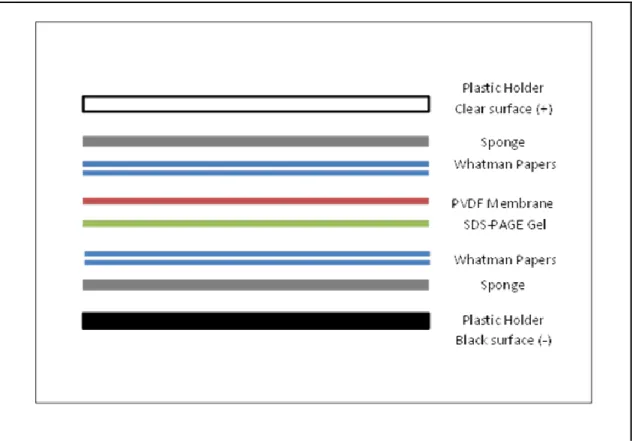
Gels were removed from electrophoresis chamber and glass holders, and put into the 4oC transfer buffer to equilibrate. Polyvinylidene difluoride (PVDF) membranes and the whatman filter papers were prepared in the required dimensions, which was 7.9x9.8 cm. PVDF membrane was activated with 100% Methanol (MeOH) for one minute, and then put into 4oC transfer buffer (recipe is shown in Table 2.10). Materials of the transfer system like sponges and whatman paper were soaked in the 4oC transfer buffer. They were placed into the Mini Trans-blot Electrophoretic Transfer Cell module (BioRad, CA, USA) as indicated in Figure 2.2. Proteins segregated in the SDS-PAGE gel were transferred into the PVDF membrane at 100V for 90 minutes in the cold room, which was at a temperature of 4oC; milliampere (mA) readings were checked during the transfer to eliminate increased resistance, which can occur due to content of the buffers or heating of the system during the transfer.
Table 2.10. Transfer Buffer
Materials Amount (For
2L)
25-mM Tris 6 g
195-mM Glycine 28 g
Methanol 400 ml
31
Figure 2.2. Illustration of the transfer assembly
Following the transfer, PVDF membranes were removed from the transfer chamber and the holders, and incubated with the blocking buffer for one hour at room temperature with rocking motion. The content of the blocking buffer was chosen according to the information sheet of the each antibody and the optimization experiments. For the cohorts in which the levels of PSD-95 and SYP were analyzed, non-fat milk powder was used as a blocking solution in the amount of 5% dissolved in Tris Buffered Saline with Tween 20 (TBS-T). TBS-T was prepared by diluting 10X TBS stock to 1X and adding 0.3% of Tween 20 to the diluted solution, the recipe for 10X TBS is indicated in Table 2.11. For the cohorts that were analyzed for the GEP levels, blocking solution was prepared with bovine serum albumin (BSA) (Sigma-Aldrich, St. Louis, MO, USA), at the amount of 5% dissolved in TBS-T.
32 Table 2.11. 10X TBS, pH 7.6 Materials Amount (For 1L) 100-mM Tris 12 g 1.35-M NaCl 88 g ddH2O Up to 1L
Following one hour of blocking at room temperature, membranes were quickly washed with TBS-T. Primary antibodies which were used in the Western blot experiments were anti-PSD-95 (abcam, Cambridge, UK: ab18258, 1:5000 dilution), anti-SYP (abcam, Cambridge, UK: ab32594, 1:20000 dilution) and anti-GEP (Santa Cruz Biotechnology, Santa Cruz, CA, USA: sc-6411, 1:1000 dilution). Anti-PSD-95 and anti-SYP were incubated simultaneously, and in this mix anti-β tubulin (Cell Signaling Technology, Danvers, AM, USA: #2146, 1:5000 dilution) was used as a housekeeping control. Anti-PSD-95, anti-SYP and anti-TUB primary antibodies mix were prepared with 5% non-fat milk powder in TBS-T. For anti-GEP incubations, different methods were used which are indicated in each Chapter; anti-GEP primary antibodies were prepared in 5% BSA in TBS-T. Membranes were incubated with primary antibodies for 18 hours at 4oC with rocking motion.
Membranes were taken from 4oC and the primary antibodies were collected. Membranes were washed with TBS-T for five times at room temperature with rocking motion for a duration of the following washing steps: 5, 5, 10, 5 and 5 minutes, respectively. After the washing steps were completed, membranes were incubated with appropriate secondary antibodies for 55 minutes at room temperature with rocking motion. Primary antibodies including anti-PSD-95, anti-SYP, anti-TUB and anti-ACTIN (abcam, Cambridge, UK: ab1608, 1:1000 dilution) were raised in
![Figure 3.2. Observed bands for the protein samples extracted from zebrafish brain lysate and mouse cortical lysate as positive control.[74] (adapted from Karoglu et al., 2017, Reprinted with permission from Elsevier)](https://thumb-eu.123doks.com/thumbv2/9libnet/5552306.108214/58.892.135.823.122.587/observed-extracted-zebrafish-cortical-karoglu-reprinted-permission-elsevier.webp)