onlay bone grafting to enable implant placement in
patients with atrophic jaw bones
Gokhan Gurler, DDS, PhD, Cagri Delilbasi, DDS PhD, Hasan Garip, DDS, PhD, Sukran Tufekcioglu, DDS.
ABSTRACT
ميعطتو
)ARS(ةيخنسلا تلاصيولحا ميسقت ةنراقلم :فادهلأا
يرومضلا كفلا ماظع يف
)AOBG(ةيتاذلا ماظعلا ةناطب
ةيزاولما ةساردلا هذه يف اضيرم
40ىلع ةساردلا تلمتشا :ةقيرطلا
ةعماج يف نانسلأا بطل لوبنطسا ةيلك يف تيرجأو ةيعاملجا
ةكامس سايق تم .م
2012-2015ينب ،ايكرت ،لوبنطسا ،لوبيديم
ناك.(
CBCT) بسولمحا يعطقلما ريوصتلا ةطساوب ةيلولأا ماظعلا
لكش
ساسأ ىلع
)n=23( AOGBو
)n=17(ىضرلما ةعومجم
ماظعلا مييقتل ةعباتم
CBCTتاسايقلا راركت
تم .ةفالحا كمسو
سايق تم .عرزي نم راطقأ ليجست تم .ةدايز دعب رهشأ 6–4 ةيقفلأا
ىلع بيسنتلا عرز دعب ةدحاو ةنس يف عورزلما مظعلا فاشترا
ءاقبلاو ةيحارلجا تافعاضلما مييقت تم .يمارونابلا يعاعشلا ريوصتلا
.ةايلحا ديق ىلع
ريثكب ىلعأ
AOBGةعومجم يف يئاهنلا ماظعلا ضرع ناك :جئاتنلا
عرز ةيلمع
44لاخدإ تم .(p=0.920)
ARSةعومجم يف كلذ نم
ةعومجم يف عرز
33لاخدإ تم ينح يف ،
AOBGةعومجم يف
ناكو .(p=0.920) عرزلا رطق يف يونعم قرف كانه نكي مل .
ARSو
ARSةعومجم يف
93.9%عرزلل ةايلحا ديق ىلع ءاقبلا لدعم
ةسرغ يف ماظعلا فاشترا ناكو .
AOBGةعومجم يف
93.1%هيلع تناك امم
AOBGةعومجم يف ىلعأ ةدحاو ةنس يف عرزلا
ةيحارج تافعاضم كانه تناك .(p=0.032)
ARSةعومجم يف
.حرلجا نادقفو ئيسلا ماسقنلاا كلذ يف ابم ،ةفيفط
ةينقتل ماظعلا عرزب ةطيلمحا ماظعلا فاشترا لدعم ناك :ةتمالخا
ةايلحا ديق ىلع ءاقبلا تلادعم نكلو ،
ARSةينقت نم ىلعأ
AOBG.ةهباشتم تناك اهعرزل
Objective: To compare alveolar ridge splitting )ARS( and autogenous onlay bone grafting )AOBG( in atrophic jaw bones.
Methods: Forty patients were included in this retrospective, parallel-group study conducted at the Istanbul Medipol University School of Dentistry, Istanbul, Turkey, between 2012-2015. The initial
bone thickness was measured by cone beam computed tomography )CBCT(. Patients were allocated into ARS )n=17( and AOGB )n=23( groups on the basis of ridge thickness and shape. Follow-up CBCT measurements to assess horizontal bone were repeated 4 to 6 months post augmentation. The diameters of the implants were recorded. Implant bone resorption was measured at one year post implant placement on panoramic radiography. Surgical complications and implant survival were evaluated.
Results: The final bone width in the AOBG group was significantly higher than that in the ARS group )p=0.029(. Forty-four implants were inserted in the AOGB group, whereas 33 implants were inserted in the ARS group. There was no significant difference regarding implant diameter )p=0.920(. Implant survival rate was 93.9% in the ARS group and 93.1% in the AOGB group. Peri-implant bone resorption at one year was higher in the AOBG group than in the ARS group )p=0.032(. There were minor surgical complications, including bad split and wound dehiscence.
Conclusion: The incidence of peri-implant bone resorption for the AOGB technique was higher than that for the ARS technique, but their implant survival rates were similar.
Saudi Med J 2017; Vol. 38 (12): 1207-1212 doi: 10.15537/smj.2017.12.21462 From the Department of Oral and Maxillofacial Surgery (Gurler, Delilbasi, Tufekcioglu), Istanbul Medipol University School of Dentistry and the Department of Oral and Maxillofacial Surgery (Garip), Marmara University School of Dentistry, Istanbul, Turkey.
Received 10th August 2017. Accepted 2nd October 2017.
Address correspondence and reprint request to: Dr. Gokhan Gurler, Department of Oral and Maxillofacial Surgery, Istanbul Medipol University School of Dentistry, Istanbul, Turkey. E-mail: [email protected]
T
he use of dental implants in oral rehabilitation of partially and totally edentulous jaws is common practice in oral surgery. To achieve ideal results in implant dentistry, there needs to be sufficient quantity and quality of both hard and soft tissues. There are a number of reconstruction techniques available to provide implant-supported restorations in patients with resorbed jaws, including the use of vascularized or nonvascularized grafts and tissue regeneration techniques. Trauma, periodontal disease, infection, neoplasms, malformation, and atrophy can cause bone loss.1,2 Depending on the type of atrophy, different surgical procedures can be used, such as onlay block graft, guided bone regeneration, sandwich osteotomy with inter-positional graft, alveolar ridge splitting, and sinus lifting.3 The autogenous onlay block graft technique was first described by Branemark et al4 in 1975 and has been commonly used in reconstruction of bone deficiency. Autogenous bone graft has osteogenic, osteoconductive, and osteoinductive properties for bone regeneration; therefore, it still remains the gold standard in reconstructive surgery.5,6 Donor sites for free autogenous bone graft techniques consist of extraoral )iliac crest, calvarial bone, rib, and tibia( and intraoral )symphysis or ramus mandible, zygomatic buttress, and tuberosity of maxilla( sites. Because of easy access, low morbidity, short healing periods, minimal graft resorption and high bone density, intraoral sites are preferred over extraoral sites.1,3 Alveolar ridge splitting )ARS( has been described to achieve buccal displacement of the vestibular wall. This technique augments the width of the alveolar bone and creates space for the dental implants. There needs to be a minimal cortical bone thickness of 3 mm together with trabecular bone between the cortical layers to perform ARS.7-9 There are some advantages of ARS compared with autogenous onlay bone grafting )AOBG(, including fewer surgical complications and lower morbidity, as well as a shorter healing time.7-9 Various surgical procedures have been described to augment deficit bone. Most research discusses the outcomes of these techniques, but there is still controversy in selecting the preferred technique because of a lack of comparative studies.10,11 The aim of this study was to compare surgical outcomes of the alveolar ridge splitting )ARS( versus AOBG to enable implant placement in patients with horizontally atrophic jaw bones.Methods. This retrospective, parallel-group study included 40 )21 female, 19 male( patients who were referred to the Department of Oral and Maxillofacial Surgery, Istanbul Medipol University School of Dentistry, Istanbul, Turkey for implant placement between 2012 and 2015. The study protocol was approved by the local ethical committee, and the adhered to the guidelines of the Helsinki Declaration. All patients underwent primary clinical and radiographic examinations. Those who were diagnosed as having inadequate bone volume for implant placement were further evaluated with CBCT to measure horizontal )alveolar ridge width( and vertical )envisaged implant height( recipient bone dimensions and distance to adjacent anatomical structures. All patients were informed in advance about the need for bone reconstruction prior to implant placement. Only healthy patients without chronic systemic diseases and those with only horizontal bone deficiency were included in the study. Patients with any maxillofacial syndrome or having history of facial trauma were excluded. Preoperative crestal bone thickness was measured from 4 mm below the alveolar crest by cone beam computed tomography )CBCT(. In patients in whom the width of the alveolar crest was ≥3 mm, the ARS procedure was performed )n=17(, and in patients in whom it was <3 mm, the AOBG procedure was performed )n=23(. All of the procedures were performed under intravenous sedation and local anesthesia by 2 oral and maxillofacial surgeons with similar surgical experience. Postoperative medication was identical for all patients, including antibiotics )875 mg amoxicillin + 125 clavulanic acid; Glaxo Smith Kline İlaçları San. ve Tic. A.Ş., İstanbul, Turkey(, an anti-inflammatory analgesic )100 mg flurpiprofen; Sanovel İlaç Sanayi ve Ticaret A.Ş, İstanbul, Turkey(, and a mouth rinse )0.2% chlorhexidine gluconate; Drogsan İlaçları Sanayi ve Ticaret A.Ş., Ankara, Turkey( for 5 days.
In the AOBG group, a mid-crestal horizontal incision, together with 2 vertical vestibular releasing incisions, was made in the recipient site and the mucoperiosteal flap was elevated accordingly. The mandibular symphysis was exposed through a 2-layer incision between the deepest part of the vestibule and the lip. Mental nerves were also exposed bilaterally to prevent neural damage. The corticocancellous block graft was harvested with a 4-mm-diameter trephine bur under copious sterile saline irrigation. Intensive care was taken not to damage the roots of the teeth, as well as lingual cortex and inferior border of the mandible. The symphysis cavity was filled with resorbable hemostatic sponges and the wound was primarily closed in layers with a 4-0 vicryl suture. The bone block was fixed to Disclosure. Authors have no conflict of interests, and the
recipient site with one or 2 titanium mini screws after performing decortication of the recipient bone. A layer of resorbable collagen membrane )Collagene AT, Centro di odontoiatria operative Srl, Padova, Italy( was used to cover the bone block. Periosteal releasing incisions were made where necessary to close the flap without tension. The flaps were repositioned and primarily closed with a 3-0 vicryl suture. In ARS group, a mid-crestal incision was made at the edentulous area and the mucoperiosteal flap was elevated. One horizontal and 2 vertical osteotomies were performed with a piezosurgery device under copious sterile saline irrigation, and then the bony fragment was extended buccally. The space between the 2 fragments was filled with corticocancellous xenograft bone chips )Hypro-Oss, Bioimplon GmbH, Giessen, Germany( and covered with a collagen membrane )Collagene AT, Centro di odontoiatria operative Srl, Padova, Italy(. Periosteal releasing incisions were made where necessary to close the flap without tension. The flap was primarily closed using 4-0 vicryl sutures.
Follow-up CBCT measurements were repeated to measure bone gain from 4 to 6 months post augmentation, prior to implant placement using the same reference point. The diameter of the implants placed was recorded. Panoramic radiography was performed one week postoperatively to measure the peri-implant bone height. The distance between the implant apex and the highest point of the mesial and distal peri-implant bone crests, respectively, was measured, and the mean was calculated. Alveolar bone resorption was measured on panoramic radiographs one year following implant placement using the same method and the difference was considered the amount of bone resorption.
Radiographic measurements were performed by 2 authors of the study who did not perform the operations. If there was any difference in opinion, a final agreement was reached after mutual consultation. Intraoperative and postoperative complications with regard to augmentation and implant placement )wound infection, exposure of graft, paresthesia, bad split, implant failure( were also evaluated.
The data were assessed using the IBM SPSS Statistics 22 program )IBM SPSS, Armonk NY, USA(. To compare the intergroup variables student’s t-test was used, and p≤0.05 was considered significant.
Results. The AOBG procedure was performed in only one site of 23 patients )14 male, 9 female, age range 23-53 years, mean age 31 years(. Seven sites were in the maxillary anterior region, 7 were in the maxillary posterior region, and 9 in the mandibular posterior
region. A total of 44 dental implants were placed in this group after 4 to 6 months following augmentation. The ARS procedure was performed in only one site of 17 patients )12 female, 5 male, age range 26-58 years, mean age 33 years(. Five sites were in the maxillary anterior region and 12 sites were in the mandibular posterior region.
The mean bone width measured prior to implantation was 6.3 mm in AOBG group and 5.85 mm in ARS group. The mean final bone width in the AOBG group was significantly higher than that of the ARS group )p=0.029(. The mean implant diameter inserted in AOBG group was 4.3 mm and 4.1 mm in the ARS group. There was no statistically significant difference regarding implant diameter inserted between the groups )p=0.920(. The amount of bone resorption measured on panoramic radiographs varied between 1.5 mm to 2 mm )mean 1.62 mm( in the AOBG group and 0.3 to 0.6 mm )mean 0.5 mm( in the ARS group. There was significantly more bone resorption in the AOBG group than the ARS group )p=0.032(.
Bad split, implant failure and wound dehiscence were complications encountered in both groups. In the ARS group, a bad split was seen in 2 patients. The segment was stabilized with a titanium mini screw and the operation was continued. Wound dehiscence was observed within one week following the surgery in one patient of the ARS group and 4 patients of the AOBG group. Secondary wound healing was achieved by local dressing using antimicrobial rinse and supportive systemic antibiotics. We did not observe severe infection, sensory disturbance, or bleeding in any of the patients.
A total of 33 dental implants were placed after 4 to 6 months following augmentation in this group. The mean follow-up for the patients following implant insertion was 3.1 years in ARS group and 3.3 years in AOBG group. Two implants failed in 2 patients in the ARS group )93.9% survival rate(, whereas 3 implants failed in 3 patients in the AOBG group )93.1% survival rate(. There was no significant difference in terms of implant survival rate between the groups )p=0.714(. The data of the study are shown in Table 1.
Discussion. Rehabilitation of the edentulous jaws with dental implants is a predictable and satisfactory option; however, there are some obstacles due to the quality and quantity of the bone. The alveolar bone undergoes irreversible and progressive resorption following tooth extraction. Horizontal bone loss occurs faster and to a greater extent than vertical bone loss.12 Bone contour and volume need to be restored for sufficient implant stability and proper positioning. The
success of an implant relies heavily on primary stability at the time of initial placement. This is primarily accomplished with a sufficient quantity and good quality of bone. Lack of bone may result in lingual position of the implant which may alter occlusal load distribution. Restoring vertical deficiency is more challenging; therefore, we only included horizontal deficiency in this study.
Common methods to restore horizontal deficiency include guided bone regeneration, ridge split, and block bone grafts.11 Guided bone regeneration is a commonly used technique to augment deficit bone by utilizing different graft materials. Among those materials, autogenous bone is considered as the gold standard owing to various advantages. Intraoral sites are preferred to extraoral donor sites to harvest autogenous bone graft due to easier access, lower morbidity and a lower complication rate.11,13,14
Autogenous onlay bone grafting is an advantageous technique for alveolar reconstruction because of its osteoinductive and osteoconductive properties, its ability to provide sufficient bone volume, and its biocompatibility. Nevertheless, the need for a donor site, surgical complications, and the need for delayed implant placement decrease the preference for this method. The common sites for intraoral block bone harvesting are the mandibular symphysis and ramus region. Clavero et al10 stated that access to symphysis region is easier than that to the ramus region; however, a greater amount of bone with a higher density of cortical bone can be obtained with the ramus graft. Moreover, lower morbidity and fewer complications are observed with ramus grafts. At our clinic, we prefer to harvest block bone mainly from the symphysis region as there is a lower risk of neural damage and better patient
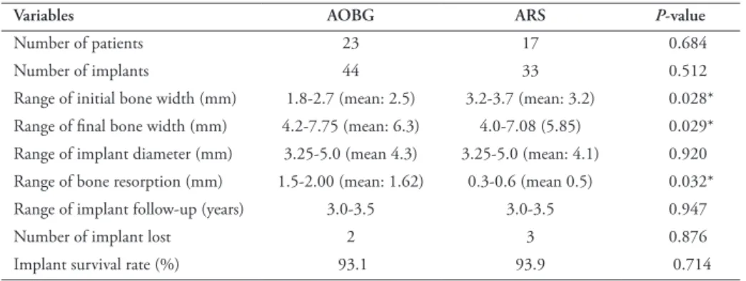
compliance. Ridge splitting provides bone gain by creating a green stick fracture and is considered a reliable and non-invasive technique. The main principle is the splitting and widening of the buccal plate anteriorly. The main advantage of this technique is a predictable amount of bone gain; rapid vascularization, leading to improved bone healing; and bone remodelling similar to that on fracture healing. However, it is important to note that there should be a layer of cancellous bone between the cortical plates to enable splitting.7-9 In a systematic review on ridge augmentation techniques, Milinkovic and Cordaro15 stated that the mean initial horizontal thickness where autogenous block grafts were used was 3.2 mm )range, 2.5 to 3.89 mm(. The linear bone gain was 4.3 mm )range 2.7 to 5.0 mm( after a healing time of approximately 5.6 months. The mean width of the reconstructed bone was 7.51 mm. The mean width of the alveolar bone where ridge splitting was performed was 3.37 mm )range 2.4 to 4.29 mm(. The bone dimension increased to a mean of 6.33 mm 2 to 6 months following augmentation. The bone gained was significantly higher in the AOBG group than in the ARS group, although the initial bone width was less in the former group in our study. However, we gained a sufficient amount of bone to place implants in both groups. The literature suggests that the use of bovine bone particles together with a resorbable membrane minimizes resorption of the autogenous block graft.12-14 One possible reason that there were higher levels of peri-implant bone resorption in AOBG group may be explained by lack of use of bovine particles to support the bone block. Further research may be conducted using different graft materials to evaluate their effects on bone gain and peri-implant bone resorption. Table 1 - Data of 40 )21 female, 19 male( patients included in the study.
Variables AOBG ARS P-value
Number of patients 23 17 0.684
Number of implants 44 33 0.512
Range of initial bone width )mm( 1.8-2.7 )mean: 2.5( 3.2-3.7 )mean: 3.2( 0.028* Range of final bone width )mm( 4.2-7.75 )mean: 6.3( 4.0-7.08 )5.85( 0.029* Range of implant diameter )mm( 3.25-5.0 )mean 4.3( 3.25-5.0 )mean: 4.1( 0.920 Range of bone resorption )mm( 1.5-2.00 )mean: 1.62( 0.3-0.6 )mean 0.5( 0.032* Range of implant follow-up )years( 3.0-3.5 3.0-3.5 0.947
Number of implant lost 2 3 0.876
Implant survival rate )%( 93.1 93.9 0.714
Implants can be placed during or after bone augmentation.16 Penarrocha-Diago et al17 conducted a study on 42 patients having 71 implants )33 delayed and 38 simultaneously inserted( following horizontal ridge augmentation. They concluded that both protocols resulted in high implant survival and success rates, but marginal bone loss was significantly higher in the simultaneous group. In this study, we inserted implants between 4-6 months following augmentation to achieve good primary stability and minimize additional surgical complications.
Implant survival rates vary among the research based on the augmentation technique preferred. For guided bone regeneration, the survival rate was approximately 96%; for onlay bone graft, it was approximately 91%; and for ridge expansion, it was approximately 94%.8 In the systematic review conducted by Aloy-Prosper et al11 the survival rate was between 97% and 100%. They indicated that there was no difference between onlay grafts and guided tissue regeneration in terms of implant survival rate, and this rate was similar to that of implants placed in the native bone. Chappuis et al13 stated that the symphysis graft was superior to the ramus graft, considering that graft resorption may be lower because of higher microvascularization in the symphysis graft. In this study, we only harvested a bone graft from the symphysis as it has some advantages over a ramus graft.
Surgical complication is an important factor to consider, in addition to several others, including the amount of bone required, type of bone )cortical or cancellous or both(, morphology of the recipient site, and resorption of the bone graft, during bone harvesting.10 Complications related to augmentation may originate from either donor or recipient site. Sensory disturbance is the most common complication whereas wound dehiscence, infection and graft loss are among other encountered complications. A review published by Milinkovic et al15 indicated that there is a tendency to use a ramus graft in the reconstruction of horizontal defects, with the possibility of obtaining a mean bone gain of 2.95 mm. The complication rate is low )6.8%(, the most common being a buccal bone fracture. Altiparmak et al7 also reported the buccal bone fracture as the most commonly seen complication, followed by temporary graft exposure. We observed several intraoperative and postoperative complications, all of which were managed uneventfully.
Study limitation. The small number of patients in the study groups and the use of different bone materials )xenograft and autograft( can be considered
as limitations of this study; however, the results give insights about outcomes of each technique and provide a comparison of both techniques.
In conclusion, AOBG and ARS are effective methods to gain bone in order to place implants in atrophic jaws. Surgical complications are rare, and the implant survival rate is high. However, there was a higher incidence of peri-implant bone loss in the AOBG group than in the ARS group. We recommend further investigation using different donor sites and graft materials with a larger patient sample to evaluate long-term bone gain.
References
1. Sakkas A, Schramm A, Karsten W, Gellrich NC, Wilde F. A clinical study of the outcomes and complications associated with zygomatic buttress block bone graft for limited preimplant augmentation procedures. J Craniomaxillofac Surg 2016; 44: 249-256.
2. Chiapasco M, Zaniboni M, Boisco M. Augmentation procedures for the rehabilitation of deficient edentulous ridges with oral implants. Clin Oral Implant Res 2006; 17: 136-159. 3. Restoy-Lozano A, Dominguez-Mompell JL, Infante-Cossio P, Lara-Chao J, Espin-Galvez F, Lopez-Pizarro V. Reconstruction of mandibular vertical defects for dental implants with autogenous bone block grafts using a tunnel approach: clinical study of 50 cases. Int J Oral Maxillofac Surg 2015; 44: 1416-1422. 4. Brånemark PI, Lindstrom J, Hallen O, Breine U, Jeppson PH,
Ohman A. Reconstruction of the defective mandible. Scand J
Plast Reconstr Surg 1975; 9: 116-128.
5. Lynch SE, Genco RJ, Marx R. Tissue engineering: applications in maxillofavial surgery and periodontics, 1st ed. Chicago: Quintessence Publishing; 1999. p. 83-98.
6. Misch CE, Scoretecci GM, Benner KU. Implants and restorative dentistry. London )UK(: M Duntz; 2001. p. 144-145. 7. Altiparmak N, Akdeniz SS, Bayram B, Gulsever S, Uckan S.
Alveolar ridge splitting versus autogenous onlay bone grafting: complications and implant survival rates. Implant Dent 2017; 26: 284-287.
8. Tarun Kumar AB, Triveni MG, Priyadharshini V, Mehta DS. Staged Ridge Split Procedure in the Management of Horizontal Ridge Deficiency Utilizing Piezosurgery. J Maxillofac Oral Surg 2016; 15: 542-546.
9. Bassetti MA, Bassetti RG, Bosshardt DD. The alveolar ridge splitting/expansion technique: a systematic review. Clin Oral
Implants Res 2016; 27: 310-324.
10. Clavero J, Lundgren S. Ramus or chin grafts for maxillary sinus inlay and local onlay augmentation: comparison of donor site morbidity and complications. Clin Implant Dent Relat Res 2003; 5: 154-160.
11 Aloy-Prósper A, Peñarrocha-Oltra D, Peñarrocha-Diago M, Peñarrocha-Diago M. The outcome of intraoral onlay block bone grafts on alveolar ridge augmentations: a systematic review. Med Oral Patol Oral Cir Bucal 2015; 20: 251-258. 12. Fu JH, Wang HL. Horizontal bone augmentation: the decision
tree. Int J Periodontics Restorative Dent 2011; 31: 429-436. 13. Chappuis V, Cavusoglu Y, Buser D, von Arx T. Lateral ridge
augmentation using autogenous block grafts and guided bone regeneration: A 10-year prospective case series study. Clin
14. von Arx T, Buser D. Horizontal ridge augmentation using autogenous block grafts and the guided bone regeneration technique with collagen membranes: a clinical study with 42 patients. Clin Oral Implants Res 2006; 17: 359-366.
15. Milinkovic I, Cordaro L. Are there specific indications for the different alveolar bone augmentation procedures for implant placement? A systematic review. Int J Oral Maxillofac Surg 2014; 43: 606-625.
16. Kuchler U, von Arx T. Horizontal ridge augmentation in conjunction with or prior to implant placement in the anterior maxilla: a systematic review. Int J Oral Maxillofac Implants 2014; 29: 14-24.
17. Peñarrocha-Diago M, Aloy-Prósper A, Peñarrocha-Oltra D, Guirado JL, Peñarrocha-Diago M. Localized lateral alveolar ridge augmentation with block bone grafts: simultaneous versus delayed implant placement: a clinical and radiographic retrospective study. Int J Oral Maxillofac Implants 2013; 28: 846-853.