Sinus surgery complicated by ventricular fibrillation in a young
patient: Inverted (reverse) Takotsubo cardiomyopathy
Genç bir hastada sinüs cerrahisi sonrası gelişen ventrikül fibrilasyonu:
Ters Takotsubo kardiyomyopatisi
Department of Cardiology, Medipol University Faculty of Medicine, İstanbul, Turkey Gültekin Günhan Demir, M.D., Gamze Babur Güler, M.D., Ekrem Güler, M.D.,
Hacı Murat Güneş, M.D., Filiz Kızılırmak, M.D.
Özet– Takotsubo kardiyomiyopatisi (TTK), apikal balonlaş-ma veya stres kardiyomiyopatisi olarak da bilinmektedir ve epikardiyal koroner arterlerde kritik darlığın eşlik etmediği geçici sol ventrikül fonksiyonu bozukluğu ile kendini göste-rir. En sık başvuru şekli akut substernal göğüs ağrısı, bazen ani nefes darlığı ve senkop ve nadiren de şok şeklinde olup tabloya ST-segment yükselmesi ve kardiyak biyobelirteçle-rin yükselmesi eşlik eder. Ters TTK ise çok daha nadir görü-lür ve bazal ve/veya midventriküler segmentlerin hipokinezi-si ile karakterizedir. Bu yazıda, kronik hipokinezi-sinüzit ve nazal polip nedeniyle nazal sinüs cerrahisi geçiren ve cerrahi sonrası ventrikül fibrilasyonu gelişen 27 yaşındaki bir kadın hasta sunuldu.
Summary– Takotsubo cardiomyopathy (TTC), also known as left ventricular apical ballooning syndrome or stress car-diomyopathy, is characterized by transient left ventricular systolic dysfunction and the absence of obstructive lesion in the epicardial coronary arteries. The most common pre-sentation is acute substernal chest pain, although occa-sionally dyspnea and syncope, and rarely shock with ST-segment elevation and elevated cardiac biomarkers have been observed. Inverted (reverse) TTC is a rare pattern characterized hypokinesis of the basal and midventricular segments. Presently described was case of a 27-year-old woman with ventricular fibrillation following endoscopic na-sal sinus surgery.
T
akotsubocardiomy-opathy (TTC) is char-acterized by transient left ventricular systolic
dys-function in the apical and mid segments, with the ab-sence of obstructive lesion in the epicardial coronary
arteries.[1] TTC predominantly occurs in
post-meno-pausal women (90%) and is most frequently
trig-gered by emotional or physical stress.[2] Also known
as left ventricular apical ballooning syndrome, in-verted (or reverse) TTC is a less common pattern of stress cardiomyopathy, involving the basal seg-ments of the left ventricle, and typically occurring in
younger patients.[3] Described in the present report
is the case of a young female patient who presented with reverse TTC after undergoing endoscopic sinus surgery.
CASE REPORT
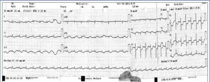
A 27-year-old woman with chronic sinusitis and nasal polyposis was admitted for endoscopic sinus surgery. She was otherwise healthy and had no risk factors for cardiovascular disease. Preoperative electrocar-diogram (ECG) and blood tests were unremarkable. Endoscopic sinus surgery was performed under gen-eral anesthesia induced with propofol. During the emergence period (recovery from general anesthesia), the patient developed ventricular fibrillation, and was successfully resuscitated. ECG obtained after defibril-lation revealed ST-segment elevation in DI and aVL leads, and ST depression in inferior and V3–6 leads (Figure 1). Echocardiography demonstrated hypoki-nesia in the basal inferior and posterior segments of the left ventricle, and estimated ejection fraction was Received:July 25, 2015 Accepted:November 27, 2015
Correspondence: Dr. Gültekin Günhan Demir. Birlik Mahallesi, Bahçeler Caddesi, No: 5, 34220 Esenler, İstanbul, Turkey.
Tel: +90 212 - 440 10 00 e-mail: [email protected] © 2016 Turkish Society of Cardiology
Abbreviations:
ECG Electrocardiogram TTC Takotsubo cardiomyopathy
40 percent (Figure 2). The patient was immediately transferred to the catheterization laboratory for nary angiography, which revealed normal left coro-nary system and osteal plaque formation in the right coronary artery.
The patient received appropriate treatment in the coronary care unit and was extubated the following day. Biochemical blood tests, including that for mag-nesium, were normal. Troponin I levels were mildly elevated (peak level: 0.058 ug/L; upper limit: 0.023 ug/L). Serial ECG follow-up showed complete reso-lution of ST-segment elevation. Strain and strain rate imaging analysis obtained prior to discharge revealed ongoing recovery, with the exception of posterobasal
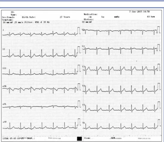
hypokinesia (Figure 3). Control echocardiographic examination showed improved left ventricular ejec-tion fracejec-tion, confirmed with cardiac magnetic reso-nance imaging. No cardiac symptoms were observed during postoperative course, and control ECG was completely normal (Figure 4).
DISCUSSION
Sato et. al described the first case of TTC in 1990,[4] and though it was initially considered to be a rare condition, recent retrospective data has demonstrat-ed that cases of TTC representdemonstrat-ed approximately 2% of all suspected cases of acute coronary syndrome, owing to increased awareness among cardiologists. [4] Though over a quarter-century has passed since its first description, pathophysiological mechanisms of TTC have yet to be clarified. Several mechanisms have been proposed, including coronary microvas-cular dysfunction, coronary vasospasm, induction of myocardial stunning by catecholamine excess and enhanced sympathomimetic activity, myocardial mi-croinfarction, and impaired cardiac fatty acid metabo-lism. Cardiotoxicity induced by catecholamine excess and coronary microvascular dysfunction are the most popular hypotheses thus far.[5]
Though no universally accepted definition exists, that most commonly used in daily practice and re-search incorporates Mayo Clinic diagnostic criteria, which were modified in 2008, and are as follows:
1) Transient hypokinesis, akinesis, or dyskinesis
Figure 1. Electrocardiogram obtained immediately after defibrillation showing ST elevation in DI and aVL leads, and ST depres-sion in inferior and V3–6 leads.
Figure 2. Transthoracic echocardiographic parasternal long-axis image showing hypokinesia in the basal septum and posterior segments (blue arrows). Hyperkinetic image in the remaining segments of the left ventricle.
of the left ventricular mid-segments with or without apical involvement. Regional wall motion abnormali-ties that extend beyond a single epicardial vascular distribution. A stress trigger is often, but not always present.
2) Absence of obstructive coronary disease or an-giographic evidence of acute plaque rupture.
3) Newly occurring electrocardiographic abnor-malities (ST-segment elevation and/or T-wave
inver-Figure 3. Measurement of left ventricle strain and strain rate, demonstrating ongoing left ven-tricular recovery, with the exception of posterobasal hypokinesia.
Figure 4. Electrocardiogram obtained 1 month after discharge showing normal sinus rhythm and no ST-T segment alteration.
sion) or modest elevation in cardiac troponins.
Ab-sence of pheochromocytoma or myocarditis.[6]
Regional wall motion abnormalities and electro-cardiographic alterations (usually ST-segment el-evation) that extend beyond a single coronary artery distribution, and documentation of normal or near-normal coronary arteries should be the key features in TTC recognition, as in the present case. Most cases of TTC described include ECG changes (usually ST-segment elevation), apical ballooning, and documen-tation of normal coronary arterial tree. Although a typically benign course has been recognized, TTC is also known to rarely cause life-threatening ventricu-lar arrhythmias and sudden cardiac death. Syed et al. reported ventricular fibrillation prevalence of 1.8% (15 of 816 cases) in a review in which arrhythmia
occurrence with TTC was investigated.[7] This rate
was reported as 4.2% (4 of 105 cases) elsewhere.[8]
Presentation of TTC with arrhythmia may challenge the clinician during differential diagnosis, as in the present case. Requisition of defibrillation may stun left ventricle functions and motions. However, echo-cardiographic images of the present patient were ob-tained after exposure to defibrillation, and it would have been impossible to discriminate the stunning of the left ventricle from the pathophysiologic process of TTC leading wall motion abnormality and ventricular fibrillation.
TTC has 4 subtypes: classical, reverse (or in-verted), midventricular, and localized. Reverse TTC is characterized by transient systolic dysfunction of the basal segments of the left ventricle. Unlike the profile of classical TTC, which includes post-meno-pausal women, reverse TTC has been significantly associated with younger age and mental or physical stress, as opposed to catecholaminergic states.[9] Re-verse TTC has also been associated with diRe-verse con-ditions and diseases, general anesthesia, and surgery.
[10] Reverse TTC can present with hemodynamic
de-terioration during any phase of anesthesia or surgery. Insufficient anesthesia and severe abrupt surgical pain stimulus are considered responsible for the catechol-amine surge.[11] In addition, reverse TTC can be trig-gered during recovery from general anesthesia, as in the present case.[12,13]
TTC presents with chest pain, electrocardio-graphic and echocardioelectrocardio-graphic abnormalities, and increased cardiac biomarkers, mimicking acute
coronary syndrome and mandating prompt differen-tiation. Coronary angiography is usually essential for diagnostic confirmation, and most patients have an-giographically normal epicardial coronary arteries or mild atherosclerosis, as the typical patient profile is a post-menopausal woman. Furthermore, it was recent-ly suggested in a report by Napp et al. that TTC and coronary artery disease can coexist, and that detection of obstructive coronary artery disease should not ex-clude diagnosis of TTC in all cases.[14] Similarly, re-gional wall motion abnormality beyond a single epi-cardial artery territory is a typical echocardiographic finding in patients with TTC. A similar debate regard-ing association of myocarditis and TTC is ongoregard-ing.
[15] Myocardial injury biomarkers may be normal or
mildly elevated, different from the pattern observed in acute coronary syndromes.
Presently described was a case of reverse TTC following endoscopic sinus surgery in a young fe-male patient. In spite of good prognosis and outcome, prompt recognition and treatment is of primary im-portance in this uncommon pattern of an uncommon disease.
Conflict-of-interest issues regarding the authorship or article: None declared.
REFERENCES
1. Horowitz JD, Frenneaux MP. Tako-Tsubo cardiomyopathy: new insights into pathogenesis. In: Bonow RO, Mann DL, Zipes DP, Libby P, editors. Braunwald’s Heart Disease: A Textbook of Cardiovascular Medicine. Philadelphia, PA: El-sevier; 9th ed. 2012. p. 1565.
2. Roshanzamir S, Showkathali R. Takotsubo cardiomyopathy a short review. Curr Cardiol Rev 2013;9:191–6. Crossref
3. Song BG, Chun WJ, Park YH, Kang GH, Oh J, Lee SC, et al. The clinical characteristics, laboratory parameters, electro-cardiographic, and echocardiographic findings of reverse or inverted takotsubo cardiomyopathy: comparison with mid or apical variant. Clin Cardiol 2011;34:693–9. Crossref
4. Sato H, Taiteishi H, Uchida T. Takotsubo-type cardiomyopa-thy due to multivessel spasm. In: Clinical aspect of myocar-dial injury: From ischemia to heart failure, Kodama K, Haze K, Hon M (Eds), Kagakuhyouronsha Tokyo 1990. p. 56. 5. Komamura K, Fukui M, Iwasaku T, Hirotani S, Masuyama T.
Takotsubo cardiomyopathy: Pathophysiology, diagnosis and treatment. World J Cardiol 2014;6:602–9. Crossref
6. Scantlebury DC, Prasad A. Diagnosis of Takotsubo cardiomy-opathy. Circ J 2014;78:2129–39. Crossref
with takotsubo cardiomyopathy: a literature review. Europace 2011;13:780–8. Crossref
8. Song BG, Chung SM, Kim SH, Kim HJ, Kang GH, Park YH, et al. The QT prolongation and clinical features in patients with takotsubo cardiomyopathy: Experiences of two tertiary cardiovascular centers. Anadolu Kardiyol Derg 2014;14:162– 9. Crossref
9. Ramaraj R, Movahed MR. Reverse or inverted takotsubo car-diomyopathy (reverse left ventricular apical ballooning syn-drome) presents at a younger age compared with the mid or apical variant and is always associated with triggering stress. Congest Heart Fail 2010;16:284–6. Crossref
10. Cvorovic V, Stankovic I, Panic M, Stipac AV, Zivkovic A, Neskovic AN, et al. Establishing the diagnosis of inverted stress cardiomyopathy in a patient with cardiac arrest dur-ing general anesthesia: a potential role of myocardial strain? Echocardiography 2013;30:E161–3. Crossref
11. Gołębiewska J, Stopczyńska I, Dębska-Ślizień A, Bohdan M, Gruchała M, Rutkowski B. Tako-tsubo cardiomyopathy on the first day after renal transplantation - case report and
litera-ture review. Transplant Proc 2014;46:2920–2. Crossref
12. de Boer HD, Booij LH. Takotsubo cardiomyopathy and an-aesthesia: case report and review of the literature. Rev Esp Anestesiol Reanim 2014;61:284–9. Crossref
13. Bortnik M, Verdoia M, Schaffer A, Occhetta E, Marino P. Ventricular fibrillation as primary presentation of takotsubo cardiomyopathy after complicated cesarean delivery. World J Cardiol 2012;4:214–7. Crossref
14. Napp LC, Ghadri JR, Bauersachs J, Templin C. Acute coro-nary syndrome or Takotsubo cardiomyopathy: The suspect may not always be the culprit. Int JCardiol 2015;187:116–9. 15. Y-Hassan S. Myocarditis is an essential feature rather than an
exclusion criterion for takotsubo syndrome: Case report. Int J Cardiol 2015;187:304–6. Crossref
Keywords: Inverted Takotsubo cardiomyopathy; sinus surgery; ven-tricular fibrillation.
Anahtar sözcükler: Ters Takotsubo kardiyomiyopatisi; sinüs cerra-hisi; ventriküler fibrilasyon.