ABSTRACT
Pericardial decompression syndrome (PDS) is an infrequent but mostly fatal complication after successful pericardiocentesis. We present a case of a 27-year-old female patient with suspected malignancy and cardiac tamponade treated with pericardiocentesis, followed by acute pulmonary edema and cardiac arrest 2 hours later. We also reviewed possible mechanisms involved in PDS pathophysiology in the context of literature data.
Key Words: Pericardiocentesis; decompression; tamponade
Başarılı Perikardiyosentez Sonrası Nadir ve Tehlikeli Bir Komplikasyon: Perikardiyal Dekompresyon Sendromu
ÖZET
Perikardiyal dekompresyon sendromu (PDS), başarılı perikardiyosentez sonrası görülebilecek nadir ancak ölümcül bir tablodur. Yirmi yedi yaşında malignite sebebiyle takip edilen, kardiyak tamponad sebebiyle perikardiyosentez yapılan ve sonraki ikinci saatte dispne, akut pulmoner ödem ve kardiyak arrest gelişen olguyu sunduk. PDS patofizyolojisinde etkili mekanizmalar ve olası tedavi literatür bağlamında gözden geçirildi. Anahtar Kelimeler: Perikardiyosentez; dekompresyon; tamponad
Ekrem Güler E-mail: [email protected] Submitted: 16.09.2015 Accepted: 08.10.2015
© Copyright 2017 by Koşuyolu Heart Journal. Available on-line at
www.kosuyoluheartjournal.com
253
Koşuyolu Heart J 2017;20(3):253-255 • DOI: 10.5578/khj.10324 ● CASE REPORT
Correspondence
INTRODUCTION
Pericardiocentesis is a life-saving procedure commonly performed in cardiology practice. Injuries of the cardiac wall, coronary arteries, and lungs are well-known complications; pericardial decompression syndrome (PDS) is a lesser-known but serious complication of pericardiocentesis(1). We present a case of PDS following pericardiocentesis in a patient with
suspected malignancy and cardiac tamponade and also a review of the current literature. CASE REPORT
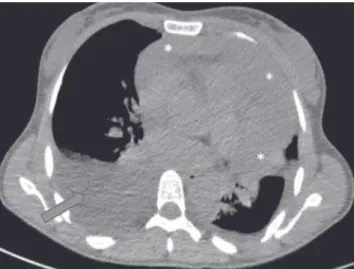
A 27-year-old female patient with an ovarian mass was evaluated for malignancy and presented with progressive shortness of breath and palpitations. The patient had orthopnea; jugular venous pressure was increased; and the heart sounds were muffled on auscultation. On physical examination, blood pressure was 100/60 mmHg, heart rate was 106 bpm, respiratory rate was 25/min, and saturated oxygen was 94%. Chest X-Ray and computed tomography showed an enlarged cardiac silhouette and bilateral pleural effusion (Figure 1,2). Echocardiographic examination revealed pericardial effusion surrounding the entire heart. Signs of cardiac tamponade were detected, and pericardiocentesis was scheduled. After obtaining informed consent and using local anesthesia, initially 500 mL and then 480 mL of serous fluid was drained via the subxyphoid route. The symptoms of the patient improved right after the procedure; however, 2 hours later, new-onset dyspnea, tachycardia, and hypotension developed. The patient was intubated for progressive respiratory failure, and control echocardiography showed no pericardial effusion. However, left ventricular ejection fraction, which was normal before the index procedure, was 20% with global hypokinesia. The patient was followed up with inotropic support; however, shortly afterwards, bradycardia and cardiac arrest occurred, and cardiopulmonary resuscitation was undertaken. The patient died despite the supportive treatment.
Gamze Babur Güler, Ekrem Güler, Gültekin Günhan Demir, Hacı Murat Güneş Medipol University Faculty of Medicine, Department of Cardilogy, İstanbul, Turkey
Pericardial Decompression Syndrome: an
Uncommon But Dangerous Complication
After Successful Pericardiocentesis
254
Koşuyolu Heart J 2017;20(3):253-255 ● Pericardial Decompression After PericardiocentesisDISCUSSION
We described a patient with PDS presenting with pulmonary edema and cardiogenic shock following pericardiocentesis. In the literature, various definitions have been used for PDS(2).
The estimated prevalence of this mortal complication was reported to be 4.8%(3). In the analysis of a case series of 35
patients with PDS, it was reported that the onset of PDS after the procedure ranged from seconds to 48 hours. Mortality was seen in 10 patients (29%), which was only associated with surgical drainage. Various clinical presentations of PDS were recorded as follows: 10 (29%) with cardiogenic pulmonary edema without shock, 14 (40%) with left ventricular failure, 3 (9%) with right ventricular failure, 7 (20%) with biventricular failure, and 1 (3%) with non-cardiogenic pulmonary edema(4). In our case,
pulmonary edema and subsequent cardiogenic shock occurred 2 hours after the procedure.
The mechanism underlying PDS remains unclear; however, autonomic dysfunction, hemodynamic overload, and ischemia hypotheses were postulated. According to the sympathetic overdrive hypothesis, transient tachycardia and inotropic effect caused by excessive catecholamine release diminishes following pericardial drainage. Altered sympathetic tone uncovers masked left ventricular dysfunction(5).
Increased coronary vascular resistance caused by increased pericardial pressure results in a decline in coronary blood flow(6).
It is assumed that mechanical pressure of increased pericardial pressure on coronary arteries leads to myocardial stunning and hibernation, which ends up as transient systolic dysfunction. Moreover, malignant myocardial infiltration and chemotherapy suppress systolic functions, particularly in PDS cases associated with malignancy(2). Basmaji et al. performed nuclear perfusion
imaging in their PDS case and detected fixed perfusion defects without any inducible ischemia. Coronary angiography revealed no significant stenosis, and EF was 57% in magnetic resonance imaging(7). In our case, it was not possible to investigate ischemia
and myocardial disease due to failed resuscitation.
A hemodynamic hypothesis suggests pulmonary edema as a consequence of preload-afterload mismatch. The rapid drainage of pericardial effusion causes a sudden increase in venous return and overloaded left ventricle(8). Besides, increased right cardiac
output, compared with left cardiac output, may have caused ventricular dysfunction(9).
PDS treatment includes supportive therapy for pulmonary edema and cardiogenic shock. Gradual drainage was suggested for the prevention of PDS. However, there is no data regarding the efficacy and safety of the amount of drainage fluid. Current hypotheses suggest impaired cardiac functions even in the absence of PDS symptoms. Further investigation of the optimal drainage rate of pericardial fluid and its impact on cardiac functions with new echocardiographic or invasive imaging methods will help better the understanding of PDS.
CONCLUSION
The utilization of new echocardiographic imaging modalities may help to prevent PDS by determining the optimal rate and amount of drainage fluid.
REFERENCES
1. Karamichalis JM, Gursky A, Valaulikar G, Pate JW, Weiman DS. Acute pulmonary edema after pericardial drainage for cardiac tamponade. Ann Thorac Surg 2009;88:675-7.
2. Angouras DC, Dosios T. Pericardial decompression syndrome: a term for a well-defined but rather underreported complication of pericardial drainage. Ann Thorac Surg 2010;89:1702-3.
3. Dosios T, Theakos N, Angouras D, Asimacopoulos P. Risk factors affecting the survival of patients with pericardial effusion submitted to subxiphoid pericardiostomy. Chest 2003;124:242-6.
Figure 2. CT image showing pericardial (*) and pleural effusions (arrow). Figure 1. Chest X-Ray of the antero-posterior view shows a cardio thoracic index > 0.5 and bilateral pleural effusion (arrow).
255
● Koşuyolu Heart J 2017;20(3):253-255 Güler GB, Güler E, Demir GG, Güneş HM.
4. Pradhan R, Okabe T, Yoshida K, Angouras DC, DeCaro MV, Marhefka GD. Patient characteristics and predictors of mortality associated with pericardial decompression syndrome: a comprehensive analysis of published cases. Eur Heart J Acute Cardiovasc Care 2015;4:113-20.
5. Wolfe MW, Edelman ER. Transient systolic dysfunction after relief of cardiac tamponade. Ann Intern Med 1993;119:42-4.
6. Skalidis EI, Kochiadakis GE, Chrysostomakis SI, Igoumenidis NE, Manios EG, Vardas PE. Effect of pericardial pressure on human coronary circulation. Chest 2000;117:910-2.
7. Basmaji SG, Peretz-Larochelle M, Bernier ML. Pericardial decompression syndrome: A rare and potentially dramatic complication of pericardiocentesis. Int J Cardiol 2015;178:297-8.
8. Vandyke Jr WH, Cure J, Chakko CS, Gheorghiade M. Pulmonary edema after pericardiocentesis for cardiac tamponade. N Engl J Med 1983;309:595-6.
9. Konstam MA, Levine HJ. Pulmonary edema after pericardiocentesis. N Engl J Med 1984;310:391.