Case Report
Sarcoidosis presenting as penile mass
Hüseyin Semiz, Senol Kobak1Department of Internal Medicine, Ege University, Faculty of Medicine, Izmi, 1Department of Rheumatology, Istinye University, Faculty of Medicine, LIV Hospital, Istanbul, Turkey
ABSTRACT
Sarcoidosis is an inflammatory disease with unknown cause characterized by noncaseating granuloma formations. It may present with bilateral hilar lymphadenopathy, skin lesions, eye, and musculoskeletal system involvement. Rare involvement of the genital organs (prostate, testis, epididymis) has also been reported. However, penile involvement is observed quite rare. In this paper, we report a patient with penile mass who was diagnosed with sarcoidosis on the basis of the laboratory, radiological, and pathological investigations.
KEY WORDS: Penile mass, presentation, sarcoidosis
Address for correspondence: Prof. Senol Kobak, Department of Rheumatology, Istinye University, Faculty of Medicine, LIV Hospital, Istanbul, Turkey. E‑mail: [email protected]
INTRODUCTION
Sarcoidosis is an inflammatory disease with unknown cause characterized by noncaseating granuloma formation.[1] Although the pathogenesis of disease is not clear yet, it is thought that there is a Th1 cell‑mediated immune response against unknown antigens. Environmental, genetic and infectious agents are discussed in the pathogenesis of sarcoidosis. Noncaseating granuloma formation in the biopsy specimen material may be used as diagnostic tool.[2] It may present with bilateral hilar lymphadenopathy, skin lesions, the involvement of eye and symptoms on the locomotor system. Rare involvement of the genital organs (prostate, testis, epididymis) has also been reported.[3,4] However, penile involvement is observed quite rare. In this paper, we mentioned about a patient with penile mass who was diagnosed with sarcoidosis according to histopathological examination.
CASE REPORT
A 47‑year‑old male patient admitted to a urology physician because of the swelling and mass in the penis for nearly
Access this article online Quick Response Code:
Website: www.lungindia.com
DOI:
10.4103/0970-2113.205326
© 2017 Indian Chest Society | Published by Wolters Kluwer - Medknow
6 months. Soft tissue biopsy from the penile mass was performed, and noncaseating granuloma formation was reported on histological examination. According to this result, the patient was admitted to our rheumatology clinic for advanced investigations. There is no known feature in his past medical and family history. On physical examination, sensitivity, and reduced range of motion in both shoulders joints and sensitivity, reduced range of motion, and swelling in the both ankle joints were detected. In his systemic examination, rough lung sounds were determined during auscultation. In laboratory tests; fasting blood glucose, liver, and renal functions were normal. Complete blood count, thyroid function tests, and routine urinalysis were normal. In serological tests, negative results for rheumatoid factor, anti‑nuclear antibodies, anti‑cyclic citrullinated peptide,
anti‑neutrophil cytoplasmic antibodies, and
anti‑double‑stranded DNA were determined. Hepatitis markers (hepatitis B virus, hepatitis C virus) and HIV were normal. Serum angiotensin converting enzyme (ACE) level was high (111 U/L; normal range: 8–52 U/L) [Table 1]. Chest X‑ray revealed bilateral hilar enlargement. Thorax computed tomography was performed; multiple mediastinal and hilar lymph nodes
This is an open access article distributed under the terms of the Creative Commons Attribution- NonCommercial-ShareAlike 3.0 License, which allows others to remix, tweak, and build upon the work non-commercially, as long as the author is credited and the new creations are licensed under the identical terms.
For reprints contact: [email protected]
How to cite this article: Semiz H, Kobak S. Sarcoidosis presenting as penile mass. Lung India 2017;34:275-7.
Semiz and Kobak: Sarcoidosis presenting as penile mass Table 1: Laboratory findings in patient with sarcoidosis
Hemoglobin 13,7 gr/dl 10,8‑14,9 gr/dl
WBC 6300/uL 3040‑9640/uL
Platelet count 269000/uL 150000‑400000/uL
BUN 18 mg/dl 7‑20 mg/dl
Creatinine 0,83 mg/dl 0,5‑0,9 mg/dl
Uric acid 6,8 mg/dl 3,4‑7 mg/dl
ALT 27 U/L 0‑31 U/L
AST 29 U/L 0‑32 U/L
Calcium 10,1 mg/dl 8,6‑10,2 mg/dl
Total protein 7,5 g/dl 6,4‑8,3 g/dl
Albumin 4,6 g/dl 3,4‑4,8 g/dl
TSH 1,36 ng/dl 0,27‑4,2 ng/dl
CRP 0,39 mg/dl 0‑0,5 mg/dl
ESR 2 mm/sa 0‑20 mm/sa
RF 7,9 iu/ml 0‑14 iu/ml
ACE 111 U/L 8‑52 U/L
WBC: White blood count, ALT: Alanine Aminotransferase, AST: Aspartate Aminotransferase, TSH: Thyroid-Stimulating Hormone, RF: Rheumatoid factor, CRP: C-reactive protein, ACE: Angiotensin- converting enzyme
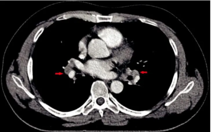
with the greatest dimension of 16 mm and parenchymal infiltration like reticular opacities and frosted glass in the right upper lobe were reported (sarcoidosis Stage 2) [Figure 1]. Abdominal ultrasonography was normal. The ankle ultrasonography showed periarticular soft tissue swelling. After the clinical, radiological, histopathological and laboratory tests, diagnosis of sarcoidosis was considered. Hydroxychloroquine 200 mg/day and corticosteroid 40 mg/gün were initiated. In his follow‑up examination after 3 months, a significant regression in his clinical symptoms was observed. Six months later, thorax CT was performed; mediastinal and hilar lymphadenopathy, as well as the findings in the lung, were completely regressed. Corticosteroids were reduced gradually and then discontinued, and the patient was just followed up. Three months later after stopping treatment, he was admitted again with complaints of a dry cough and exertional dyspnea, bilateral ankle arthritis. On thorax CT sarcoidosis Stage 2 findings were reported. After the clinical, laboratory, and radiological tests, disease relapse was considered. Corticosteroid 40 mg/day, methotrexate 15 mg/week, folic acid 5 mg/week were initiated. In his follow‑up examination after 3 months, a significant regression in her clinical symptoms was observed, and there was a regression in his control chest X‑ray. Outpatient follow‑up is still in progress for the patient with representing overall good condition.
DISCUSSION
Sarcoidosis is an inflammatory disease with unknown cause characterized by noncaseating granuloma formations. It has been suggested that the disease develops in genetically susceptible individuals after exposure to certain environmental and infectious agents affecting inflammatory responses and granuloma formation.[5] There is no gold standard diagnostic test for sarcoidosis. Although serum ACE level is high in the sarcoidosis patients, it is not specific for sarcoidosis. In general, the biopsy sample with noncaseating epithelioid granulomas is typical for the diagnosis of sarcoidosis.[6] However,
Figure 1: Torax computed tomography showed bilateral hilar lymphadenopathy
these histopathological involvement can also be seen in tuberculosis, leprosy, Sjögren’s syndrome, Behcet’s disease, and berylliosis. At the same, there are shown several organ involvements in autopsy studies. Sarcoidosis primarily affects the skin, lungs, eyes, bones, and lymph nodes. Genital organ involvement is rare, and epididymis, prostate, and testicular involvement have been reported.[3,4] However, seminal vesicles, scrotum, and spermatic cord involvement are very rare. A patient with scrotum swelling was diagnosed for sarcoidosis by Khan et al. as a result of the inguinal lymph node biopsy.[7] There is not so much information about penile sarcoidosis at the literature. Penile involvement which is characterized by skin lesions and develops in the scars area of the penis was more often reported than penile mass involvement like our case.[8,9] Whittaker et al. reported a patient who admitted with soft tissue swelling in the penis diagnosed with sarcoidosis as a result of the biopsy.[10] They reported that the disease was not responded to corticosteroids and disease was regressed after partial penectomy and radiotherapy. Genital involvement of the sarcoidosis is seen in the second stage of the disease. These lesions get better with fibrosis and hyalinization between 6 months and 3 years. Penile mass involvement in sarcoidosis is not a common situation. Usually, subcutaneous nodule involvement is more frequent.[11] Subcutaneous sarcoidosis is fairly uniform clinical condition, usually occurs at the early stage of the disease and may be useful in the diagnosis of systemic sarcoidosis.[12] Usually, it is not associated with severe systemic sarcoidosis and chronic fibrotic disease.[13]
CONCLUSION
The case with sarcoidosis presented as penile mass was reported. The diagnosis was made as a result of the biopsy and the other investigations. In patients with penile and genital lesions, sarcoidosis should be kept in mind, and biopsy should be considered for true diagnosis.
Financial support and sponsorship
Nil.
Semiz and Kobak: Sarcoidosis presenting as penile mass
Conflicts of interest
There are no conflicts of interest.
REFERENCES
1. Newman LS, Rose CS, Maier LA. Sarcoidosis. N Engl J Med
1997;336:1224‑34.
2. Chen ES, Moller DR. Etiology of sarcoidosis. Clin Chest Med
2008;29:365‑77, vii.
3. Ryan DM, Lesser BA, Crumley LA, Cartwright HA, Peron S, Haas
GP, et al. Epididymal sarcoidosis. J Urol 1993;149:134‑6. 4. Hausfeld KF. Primary sarcoidosis of the scrotum: Case report. J
Urol 1961;86:269‑72.
5. Smith G, Brownell I, Sanchez M, Prystowsky S. Advances in the
genetics of sarcoidosis. Clin Genet 2008;73:401‑12. 6. Grosser M, Dittert DD, Luther T. Re: Molecular detection of
M. tuberculosis DNA in tuberculosis and sarcoidosis. Diagn Mol
Pathol 2001;10:66‑8.
7. Khan SA, Richenberg JL, Fletcher MS. Scrotal swelling in
sarcoidosis. J R Soc Med 2004;97:487‑8.
8. Mahmood N, Afzal N, Joyce A. Sarcoidosis of the penis. Br J Urol
1997;80:155.
9. Vitenson JH, Wilson JM. Sarcoid of the glans penis. J Urol 1972;108:284‑9.
10. Whittaker M, Anderson CK, Clark PB. Sarcoidosis of the penis treated by radiotherapy. Br J Urol 1975;47:325‑30.
11. Mahajan VK, Sharma NL, Sharma RC, Sharma VC. Cutaneous sarcoidosis:
Clinical profile of 23 Indian patients. Indian J Dermatol Venereol Leprol 2007;73:16‑21.
12. Rubinstein I, Baum GL, Hiss Y. Subcutaneous sarcoidosis– clinicopathological study of 10 cases. J Urol 1986;135:1016‑7. 13. Su O, Onsun N, Topukçu B, Ozçelik HK, Cakiter AU, Büyükpinarbasili N.
Disseminated scar sarcoidosis may predict pulmonary involvement in sarcoidosis. Acta Dermatovenerol Alp Pannonica Adriat 2013;22:71‑4.