Tarihi / Received: 21.08.2009, Kabul Tarihi / Accepted: 05.10.2009
Yazışma Adresi /Correspondence: Assist. Prof.Mehmet BULUT, MD Dicle University, Medical Faculty ORIGINAL RESEARCH / ÖZGÜN ARAŞTIRMA
Outcomes of the distal intraarticular humeral fractures treated by
olecranon osteotomy
Distal humerus eklem içi kırıklarının olekranon osteotomisi ile tedavi
sonuçları
Erhan Yılmaz1, Mehmet Bulut2
1
Firat University, Medical Faculty, Department of Orthopaedics & Traumatology, Elaziğ - Turkey
2
Dicle University, Medical Faculty, Department of Orthopaedics & Traumatology, Diyarbakır-Turkey
ABSTRACT
Objectives: To evaluate the management and outcome
of intraarticular fractures of the distal humerus treated by open reduction and internal fixation using the olecranon osteotomy technique.
Materials and methods: Twenty-one patients with
in-traarticular fractures of the distal humerus were treated by open reduction and internal fixation. The mean age of the patients was 41.6 years and the mean follow-up pe-riod was 25.3 months. All fractures were type C accord-ing to the AO/ASIF fracture classification system. Chev-ron type olecranon osteotomy was performed within 12-24 h after the injury in all cases but 4 of them. Physical and radiological examination of patients with the appro-priate range checks were made.
Results: All fractures united within average duration of
3.2 months. Excellent or good results were found in pa-tients less than 50 years-old, in women, when passing time from injury to surgery was within 12 hours and when early mobilization was achieved. The complica-tions were seen as 2 (9.6%) transient neuropraxia of the ulnar nerve, 2 (9.6%) failure of fixation, 1 (4.8%) het-erotopic ossification and 1 (4.8%) olecranon non-union. Fracture type (C1) and time from occurrence of injury to surgery (<12 hours) are the main prognostic factors for achieving the excellent/ good functional results.
Conclusions: The critical factors for a successful
out-come of intraarticular fractures of the distal humerus in-cluded meticulous surgical technique, stable internal fix-ation, surgical experimentation and early controlled postoperative mobilization.
Key words: Distal humerus, intraarticular fracture,
sur-gical treatment, olecranon osteotomy, early mobilization.
ÖZET
Amaç: Distal humerusun eklem içi kırıklarında olekranon
osteotomisi yöntemiyle açık redüksiyon ve internal tesbit yapılan hastalarda tedavi sonuçlarının değerlendirilmesi.
Gereç ve yöntem: Distal humerusta eklem içi kırığı olan
21 hastaya olekranon osteotomisi yapılarak açık redük-siyon ve internal tesbit uygulandı. Hastaların ortalama yaşı 41.6 yıl ortalama takip süresi 25.3 ay idi. Kırıkların tamamı AO/ASIF kırık sınıflandırma sistemine göre tip-C idi. Chevron tipi olekranon osteotomisi yapılan hastaların dördü hariç diğerleri yaralanmadan sonraki ilk 12-24 saat içerisinde ameliyata alındı. Uygun aralıklarla hastaların fizik muayene ve radyolojik kontrolleri yapıldı.
Bulgular: Kırıkların tamamı ortalama 3.2 ayda kaynadı.
Bayanlarda, 50 yaşın altında, yaralanmayla ameliyata alınma arasındaki süre 12 saatin altında olan ve erken hareket başlanan hastalarda mükemmel ve iyi sonuçlar elde edildi. Komplikasyon olarak 2 (9.6%) hastada geçici unlar sinir nöropatisi, 2 (9.6%) hastada tesbit yetersizliği, 1 (4.8%) hastada heterotopik ossifikasyon, 1 (4.8%) has-tada da olekranonda kaynamama görüldü. Kırığın tipi (C1) ve yaralanmayla ameliyata alınma arasında geçen zaman (12 saat) mükemmel ve iyi fonksiyonel sonuçlar elde etmede ana belirleyici faktörlerdi.
Sonuçlar: Dikkatli ve titiz cerrahi teknik, stabil internal
tesbit, cerrahi tecrübe ve ameliyat sonrası erken kontrol-lü hareket distal humerusun eklem içi kırıklarının başarılı sonuçları için kritik faktörlerdir.
Anahtar kelimeler: Distal humerus, eklem içi kırık,
cerahi tedavi, olekranon osteotomisi, erken hareket
INTRODUCTION
Distal humerus fractures are difficult to manage successfully because of the local anatomic con-straints, the frequent presence of communication, displacement and osteopenia1-6. Standard treatment
and protocols for these fractures have not been de-veloped. Although reasonable results were reported after conservative treatment in the past, it usually results in loss of elbow movement and permanent disability1,7,8. Moreover, accurate reconstruction of the articular surface is not always possible by
closed manipulation1. The recent trend for dis-placed, intra-articular fractures of the distal hume-rus is open reduction and stable osteosynthesis with early rehabilitation2,9,10,11,12,5,13.
Adequate exposure is critical for visualisation of the fracture fragments during reduction and fixation, and it is generally agreed that the best ex-posure of both columns of the distal part of the humerus and articular surface is achieved through a posterior approach14,9,11,15. Various approaches that mobilize the triceps tendon (triceps-sparing approaches) have been described, but have limita-tions in exposure and extensibility. The triceps split has been used as a standard approach for dis-tal diaphyseal fractures. Its use for periarticular fractures has not been well described6. The most recent studies have showed that the articular sur-face can be exposed via an olecranon osteotomy more than the other approaches16,17,9,3,18,19.
The purpose of this study was to retrospec-tively review the effect of fracture type, sex, age, time from injury to surgery and immobilization pe-riod on the results of distal intra-articular humerus fractures treated with stable internal fixation fol-lowed by early active movement.
PATIENTS AND METHODS
A total of 21 patients with comminuted intra-articular fractures of the distal humerus were treated by open reduction and internal fixation. There were 13 men and 8 women with an average age of 41.6 years (range, 17-62). Left elbow in-volvement was found in 12 and right in 9. Average follow-up duration was 25.3 months (range 9-48).
By using the classification system of AO/ASIF, six fractures were type C1, 11 were type C2, and four were type C3. Two cases had grade 1 open fracture (case numbers 7 and 20). The cause of the fractures were traffic accident in 11, fall in 9, and sport injury in one.
Seventeen fractures were treated early within 24 hours. Four fractures (surgery was postponed until swelling had subsided in three cases and the severity of associated injuries made early operation impossible in one case) had delayed treatment (>24 hrs).
One of the patients had multiple fractures else-where in the body. Two patients had associated fractures in the forearm area and one required
fas-ciotomy of the forearm due to compartment syn-drome.
Operative Techniques
The patient was either placed supine with the af-fected arm placed across the chest or prone with the involved extremity flexed and hanging off the operating table. The pneumatic tourniquet was used. A straight posterior incision with radial de-viation across the tip of the olecranon was made. The ulnar nerve was then identified and carefully protected (at the end of the procedure, the nerve was found in place in 8 patients and transposed an-teriorly in 13 patients). Intra-articular chevron os-teotomy was performed approximately two centi-metres from the tip of the olecranon with a high-speed micro-oscillating saw to cut up to the sub-chondral bone. The osteotomy was completed with an osteotome used as a lever to crack through the articular surface. The proximal part of the ole-cranon was elevated with the triceps, which pro-vides excellent exposure as far as seven centime-tres proximal to the joint line before the radial nerve is threatened. The elbow capsule was incised and the fracture fragments were identified by care-fully dissecting soft tissue and muscular attach-ments, as necessary. The articular fragments were inspected, and then carefully irrigated, and clots are removed with gentle curettage, taking care not to remove any bone.
The first step in the osteosynthesis was to re-duction of the condyles and reconstruction of the joint surface. Medial and lateral condyles were fixed together with a cancellous lag screw. The next step was to anatomically reattach the condyles to the humeral shaft. Stable fixation was achieved by using two plates in 10 cases. Implants should not be placed in the coronoid or olecranon fossa. To avoid fixation failure before bone healing, a cancellous bone graft was used for bone defects and comminution. At the end of the procedure, the olecranon was reduced and then fixed with two longitudinal 2.0 mm K-wires and an 18-gauge ten-sion band wire or 6.5 mm. cancellous screw.
The tourniquet was deflated, and hemostasis was obtained. A hemovac drain was placed and a meticulous wound closure was completed.
Usually by the second postoperative day, ac-tive or acac-tive-assisted range of elbow motion exer-cises as pain permitted was started in patients with good bone quality and rigid osteosynthesis. Longer
immobilization (>3 weeks) was used when the bone quality was poor and the stability of the os-teosynthesis was questionable. No continuous pas-sive motion machines were used. After the postop-erative 6th week, resisted exercises were started and normal daily activities resumed. Strenuous physical exercise was only allowed after radiologi-cal evidence of union. For prophylaxis of het-erotopic bone formation, patients received a six-week course of indomethacin (25 mg three times a day) beginning within twenty-four hours after sur-gery.
Pearson-Spearman correlation analysis was performed for statistical analyses. SPSS vs. 11.5 was used for statistical analyses. P less than 0.05 was accepted significant.
RESULTS
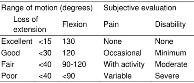
All patients treated in our series had anatomic re-constitution of the joint surface in the early postop-erative radiograph. All fractures united in average 3.2 months (range 7-19 weeks). Non-union was not determined in humerus. The clinical results were evaluated according to the criteria of Jupiter9 (Ta-ble 1). These criteria consist of range of elbow mo-tion (ROM) and subjective findings. When the
re-sults were graded according to the range of motion, excellent postoperative results were observed in 7 patients (33%) and good results were observed in 7 patients (33%) at final follow-up. Four patients (19%) had fair results, and three patients (14%) had poor results (Table 2).
Backing of one K-wire in olecranon were ob-served one patient (4.8%) (Case number: 5), but this did not make severe loss of elbow function and olecranon osteotomy united without complication.
Fixation failure depends on screw loosening were determined in two case (9.6%) (Case number: 10-18) with type C1 and C2 fracture. In these cas-es, fracture side was strengthened with lateral plate by using lateral longitudinal humeral exposure. Table 1. Criteria for grading results12.
Range of motion (degrees) Subjective evaluation Loss of
extension Flexion Pain Disability
Excellent <15 130 None None
Good <30 120 Occasional Minimum Fair <40 90-120 With activity Moderate Poor <40 <90 Variable Severe
Table 2. Overview of clinical data Case
Age/ sex
Inj. limb
A.O. class. Time from injury to surgery (hrs) Immobilization (weeks) Length of follow-up (mos.) Range of motion Subjective evaluation Postop. Com-plications 1 58-F L C1 16 < 3 27 Excellent Excellent 2 38-F R C3 5 < 3 32 Good Good
3 56-M L C2 4 > 3 14 Fair Good Paraesthesias
4 19-M R C2 7 days < 3 42 Poor Fair
5 29-M L C1 14 > 3 22 Good Excellent K-wire backing
6 22-M R C2 4 < 3 17 Excellent Good
7 43-M L C3 7 < 3 31 Fair Good
8 42-M L C2 8 < 3 25 Excellent Excellent Paraesthesias
9 56-M L C2 6 days > 3 29 Fair Fair
10 27-F L C1 8 < 3 39 Excellent Excellent Fix.failure
11 24-M L C2 2 days < 3 28 Fair Poor
12 17-M R C1 12 > 3 19 Good Good
13 43-M L C2 9 < 3 26 Excellent Excellent
14 47-F R C2 14 < 3 20 Good Excellent Olecranon
nonunion
15 59-M R C3 11 days > 3 44 Poor Fair Heterotopic
ossification
16 62-F L C2 5 > 3 48 Good Excellent
Table 3. Excellent and good results rate according
to the fracture types, sex, age, and time from injury to surgery and immobilization period
Characteristic Excellent or Good rating P Fracture type C1 (n=6) 6 (100 %) C2 (n=11) 6 (55 %) C3 (n=4) 2 (50 %) 0.017 Sex Male (n=13) 7 (54 %) Female (n= 8) 7 (88 %) NS Age <50 y (n= 13) 10 (77 %) >50 y (n=8) 4 (50 %) NS
Time from injury to surgery
<24 hrs (n=17) 14 (82 %) >24 hrs (n=4) 0 (0 %) 0.004 Immobilization period <3 wk (n=13) 10 (77 %) >3 wk (n=8) 4 (50 %) NS NS: not significant
Primary bone grafting was performed in two cases (9.6%) (Case number: 15-20). Both of them were type C3 fracture. One patient had only pain with activity, but a “poor” result due to elbow stiffness.
Heterotopic ossification was seen in only one patient (4.8%) (Case number: 15) whose operation had been delayed for 11 days because of multiple injuries. This patient required a second procedure to excise it. A fair over-all result was achieved.
Transient neuropraxia of the ulnar nerve due to its mobilization before the olecranon osteotomy was found in two cases (9.6%) (Case number: 3-8) and recovered completely with only observation.
According to the functional results; 6 patients (100 %) in the C1 fracture type group, 6 (55 %) of 11 patients in C2 fracture type group and 2 (50 %) of 4 patients in C3 fracture type group had excel-lent and good results (p=0.017). Seven (54 %) of 13 men and 7 (82 %) of 8 women had excellent and good results (p=0.378). Ten (77%) of 13 pa-tients with early mobilization (<3 weeks; average 11.9 days; SD, 4.2 days) of the elbow had excellent or good results, whereas only 4 (50 %) of 8 pa-tients who were mobilized later (>3 weeks;
aver-age 26.1 days; SD, 2.8 days) achieved the same excellent and good level (p=0.097).
According to the subjective criterions, the re-sults were found to be excellent in 8 (38%), good in 8 (38%), fair in 3 (14%) and poor in 2 (10%) (Table II). There were no statistical correlation be-tween the age, sex, immobilization period and sub-jective results.
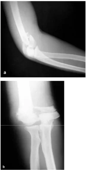
We used two plates for rigid fixation of frac-ture and continuity the fracfrac-ture alignment in 6 cas-es with type C2 fracture and in 4 cascas-es with type C3 fracture. Usually, one was lateral condyl plate and other was reconstruction plate (Scherman plate) (Fig 1-2-3).
Figure 1 a - b. Preoperative roentgenogram of the
Figure 2 a - b. Six weeks after the operation.
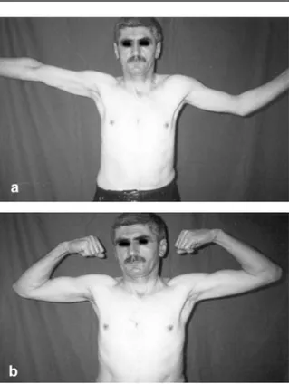
Figure 3 a - b. Functional result at 14 months DISCUSSION
Fractures of the distal humerus are difficult to treat both by the nature of injury and most sur-geons do not have a great deal of experience with
them20,1,5. Most intra-articular fractures of the distal
humerus are displaced, and the successful treat-ment of any articular fracture demands an anat-omic reduction, stable fixation and the ability to al-low early elbow motion21,17,12.
There are numerous surgical exposures of the elbow joint14,22,4. The posterior approach through an olecranon osteotomy is the most widely
used16,9,3,4. This approach provides excellent
visu-alization, particularly of the distal articular frag-ments and excellent exposure for plate applica-tion9,4. An anatomic comparison in cadavers dem-onstrated only 35% of the articular surface is ade-quately exposed with a standard triceps splitting approach In comparison, a triceps reflecting ap-proach allowed access to approximately 46% of the articular surface Employing the most extensive exposure, the olecranon osteotomy, only 57% of the articular anatomy is fully exposed23,19.
The elbow joint tolerates immobilization poor-ly and the functional outcome after surgical treat-ment is unavoidably worsened by prolonged
im-mobilization5. Therefore, early mobilization of the elbow has been recommended, but this requires stable fixation of the fracture1,24,18,13. However, pa-tient-related factors, such as poor bone quality or extensive comminution of the fracture, may neces-sitate prolonged immobilization in order to avoid non-union of the fracture. In our series, 10 of 13 patients in early active elbow mobilization group showed excellent or good postoperative results, 4 of 8 patients in late mobilization group had excel-lent and good results.
Fair and poor results were reported in ad-vanced-age and male cases25,7,18. In our study, we found fair and poor functional results especially in > 50 years-old cases and in male cases. This may be possibly because of inferior bone quality and longer immobilization in older patients and high C2 – C3 fracture frequency in male cases.
There is a correlation between the late surgery and heterotopic ossification17. We found one het-erotopic ossification in a case with poor functional result and fair subjective evaluation that had oper-ated after 11 days from trauma.
Dual plates are necessary to maintain the anat-omic alignment of the fracture, while buttressing the fragments and preventing loss of reduction un-der torsion or bending loads. This is especially true in type C3 fractures26. It should be noted that dou-ble plating appears to be superior to single plating or to the use of screws and K-wires alone26,9,4,8. We used two plates in 10 cases with type C2 and C3 fracture for rigid fixation. Usually, one plate was lateral condyl plate and other was medial recon-struction plate.
Three different types of olecranon osteotomy can be used, intra-articular tranverse, extra-articular oblique and intraextra-articular chevron2. We believe that chevron osteotomy was enhanced sta-bility and union, for this reason, we used chevron osteotomy. Non-union of the olecranon osteotomy has been reported to occur between 2-5 %20,26,17,9. In this study, 1 (4.8%) olecranon non-union was observed and reosteosynthesis with 6.5 mm long cancelleos screw and tension band wiring was per-formed.
Ulnar nerve is at obvious risk both from the injury and subsequent surgery to the distal hume-rus. Ulnar nerve injury has been reported as
%5-2020,1,27. Two (9.6%) transient ulnar nerve
paraes-thesias in cases with not anteriorly transposed were
encountered in the present study. We agree with Jupiter et al.9 that, anterior transposition of the ul-nar nerve should be done whenever the nerve is re-traction or when a metal implant is likely to cause mechanical irritation.
Mechanical failure of fixation is common in patients with severe comminution and displace-ment (type C2, C3 according to AO/ASIF classifi-cation)8. Henley2 reported 5 cases of implant brea-kage in a group of 33 patients. Sodergard et al.5 re-ported 29.5% mechanical failure rate in 18 cases and advised a nonsurgical approach when the bone is osteoporotic. We observed 2 (9.6%) fixation failures due to the screw loosening. Our rate was lower than the literature.
Pajarinen and Bjorkenheim18 did not find any correlation between the type of fracture and post-operative result. In this study, C1 fracture type had excellent and good functional result than other fracture types.
In conclusion, the successful treatment of comminuted intra-articular distal humerus fractures demands stable osteosynthesis with anatomic re-duction of the joint surface and early mobilisation. REFERENCES
1. Gupta R. Intercondylar fractures of the distal humerus in adults. Injury 1996; 27: 569-572.
2. Henley MB. Intra-articular distal humeral fractures in adults. Orthop Clin North Am 1987;18:11-23.
3. Kinik H, Atalar H, Mergen E. Management of distal hume-rus fractures in adults. Arch Orthop Trauma Surg 1999; 119: 467-469.
4. Ring D, Jupiter JB. Fractures of the distal humerus. Orthop Clin North Am 2000;31:103-113.
5. Sodergard J, Sandelin J, Bostman O. Postoperative compli-cations of distal humeral fractures. Acta Orthop Scand 1992; 63: 85-89.
6. Ziran BH, Smith WR, Balk ML, Manning CM, Agudelo JF. A true triceps-splitting approach for treatment of distal humerus fractures: a preliminary report. J Trauma 2005; 58:70-75.
7. Kundel K, Braun W, Wieberneit J, Ruter A. Intra-articular distal humerus fractures. Factors affecting functional out-come. Clin Orthop 1996;332:200-208.
8. Zhao J, Wang X, Zhang Q. Surgical treatment of commin-uted intra-articular fractures of the distal humerus with double tension band osteosynthesis. Orthopedics 2000;23:449-452.
9. Jupiter JB, Neff U, Holzach P, Allgower M. Intercondylar fractures of the humerus. An operative approach. J Bone Joint Surg 1985;67:226-239.
10. Letsch R, Chmit-Neuerburg KP, Sturmer KM, Walz M. Intra-articular fractures of the distal humerus. Surgical treatment and results. Clin Orthop 1989;241:238-244.
11. McKee MD, Wilson TL, Winston L, Schemitsch EH, Ri-chards RR. Functional outcome following surgical treat-ment of intra-articular distal humeral fractures through a posterior approach. J Bone Joint Surg 2000;82:1701-1707. 12. Pollock JW, Faber KJ, Athwal GS. Distal humerus
frac-tures. Orthop Clin North Am 2008;39:187-200.
13. Zagorski JB, Jennings JJ, Burkhalter WE, Uribe JW. Comminuted intra-articular fractures of the distal humeral condyles. Surgical vs. nonsurgical treatment. Clin Orthop 1986;202:197-204.
14. Ebraheim NA, Andreshak TG, Yeasting RA, Saunders RC, Jackson WT. Posterior extensile approach to the elbow joint and distal humerus. Orthop Rev 1993;22:578-582. 15. Ring D, Gulotta L, Chin K, Jupiter JB. Olecranon
osteot-omy for exposure of fractures and nonunions of the distal humerus. J Orthop Trauma 2004;18:446-449.
16. Eralp L, Kocaoglu M, Sar C, Atalar AC. Surgical treat-ment of distal intra-articular humeral fractures in adults. Int Orthop 2001;25:46-50.
17. Helfet DL, Schmeling GJ. Bicondylar intra-articular frac-tures of the distal humerus in adults. Clin Orthop 1993;292:26-36.
18. Pajarinen J, Bjorkenheim JM. Operative treatment of type C intercondylar fractures of the distal humerus: Results af-ter a mean follow-up of 2 years in a series of 18 patients. J Shoulder Elbow Surg 2002;11:48-52.
19. Wilkinson JM, Stanley D. Posterior surgical approaches to the elbow: a comparative anatomic study. J Shoulder El-bow Surg 2001;10:380-382.
20. Aitken GK, Rorabeck CH. Distal humeral fractures in the adult. Clin Orthop 1986;207:191-197.
21. Anglen J. Distal humerus fractures. J Am Acad Orthop Surg 2005;13:291-297.
22. O’Driscoll SW. The triceps-reflecting anconeus pedicle (TRAP) approach for distal humeral fractures and nonun-ions. Orthop Clin North Am 2000;31:91-101.
23. Archdeacon MT. Combined olecranon osteotomy and pos-terior triceps splitting approach for complex fractures of the distal humerus. J Orthop Trauma 2003;17:368-373. 24. Holdsworth BJ, Mossad MM. Fractures of the adult distal
humerus elbow function after internal fixation. J Bone Joint Surg 1990; 72-B: 362-365.
25. Caja VL, Moroni A, Vendemia V, Sabato C, Zinghi G. Surgical treatment of bicondylar fractures of the distal humerus. Injury 1994; 25: 433-438
26. Gabel GT, Hanson G, Bennett JB, Noble PC, Tullos HS. Intra-articular fractures of the distal humerus in the adult. Clin Orthop 1987; 216: 99-108.
27. Sodergard J, Sandelin J, Bostman O. Mechanical failures of internal fixation in T and Y fractures of the distal hume-rus. J Trauma 1992; 33: 687-690.