DOI: 10.1501/commuc_0000000222 ISSN 1303-6025 E-ISSN 2651-3749
http://communications.science.ankara.edu.tr/index.php?series=C
Received by the editors: December 10, 2018; Accepted: December 14, 2018.
Key word and phrases. Bryophyte, Moss, Bartramiaceae, SEM, spores morphology, capsule structure Submitted via II. Aerobiology and Palynology Symposium 07-10 October 2018 (APAS 2018)
© 2018 Ankara University Communications Faculty of Sciences University of Ankara Series C: Biology SPORE MORPHOLOGY OF SOME BARTRAMIACEAE SPECIES
(BRYOPHYTA) IN TURKEY
TALIP CETER, MERVE CAN GOZCU, GURAY UYAR Abstract. In this study, spore morphology of Bartramia pomiformis Hedw., B. halleriana Hedw., Philonotis calcarea (Bruch & Schimp.) Schimp. and Plagiopus oederianus (Sw.) H.A.Crum & L.E.Anderson were examined by light microscopy (LM) and scanning electron microscopy (SEM). All spores are small, the length of the polar axis (P) is between 12.36 µm and 19.14 µm, equatorial diameter (E) is between 19.59 µm and 26.23 µm. The smallest spores of them are Philonotis calcarea and the biggest spores of them are Plagiopus oederianus. The shapes of the spores are determined as suboblate for Bartramia pomiformis, B. halleriana, and oblate for Philonotis calcarea, Plagiopus oederianus. Verrucate-granulate, gemmate-microecinate, perforate, gemmate-pilate and verrucate ornamentation types were observed on the distal pole. In addition to spore morphology, capsule structures were also examined and were photographed with SEM. As a result, the spore size, shapes, ornamentation types and the capsule structures show some differences among these species and these differences can be used as distinctive characters in the identifications of them.
1. Introduction
Bartramiaceae Schwägr. is a large and cosmopolitan family with almost 400 species. The family members, in general, have globose to oblong-cylindric capsules, with the neck absent or inconspicuous, and they have dense, often reddish tomentum on their stems [1]. They generally spread on soil and rocks in moist habitats [2]. The family are represented by 18 taxa of Turkey. The taxa belong to genera Anacolia Schimp. (A. menziesii (Turner) Paris, A. webbii (Month.) Schimp.), Bartramia Hedw. (B. aprica Mull. Hal, B. halleriana Hedw., B.
ithyphylla Brid., B. pomiformis Hedw.), Philonotis Brid. (P. caespitosa Jur., P. calcarea var. calcarea (Bruch & Schimp.) Schimp., P. calcarea var. orthophylla
Schiffner, P. calcarea var. seriatifolia Schiffner, P. capillaris Lindb., P. fontana (Hedw.) Brid., P. hastata (Duby) Wijk & Margad., P. marchica (Hedw.) Brid., P.
rigida Brid., P. seriata Mitt., P. tomentella Molendo) and Plagiopus Brid. (P. oederianus (Sw.) H.A.Crum & L.E.Anderson) [3]. The division of Bartramiaceae
and separation of the genera has been made according to gametophytic and sporophytic characters. Some of the characters used were leaf cells, leaf shape, capsule structure and spore ornamentations [4]. The spore morphology is an important taxonomic character for this family. Some recent papers have shown that the spore external morphology is useful to characterize moss taxa at the generic and specific levels [5-12]. However, the bryophyte spores in Turkey are not fully known.
For this reason, in this study, the detailed spore morphological structures of some Turkish Bartramiaceae species were studied with light microscope (LM) and scanning electron microscope (SEM), in order to contribute to the taxonomy and palynology works. In addition to spore morphology, capsule structures were examined and photographed with SEM.
2. Material And Methods
The spore and capsule materials were obtained from the bryophyte herbarium of Ankara Hacı Bayram Veli University Polatlı Science and Arts Faculty. List of spores examined are given in the Table 1. The external surfaces of the spores were observed using light microscopy (LM) and scanning electron microscopy (SEM). The spores were prepared by the Wodehouse [13] method for LM photographs. Measurements of the diameters in the polar axis (P) and in the equatorial view (E) were taken in 20 randomly selected spores. The Simpson and Roe graphical test was used for graphical calculations [14]. The capsules were observed using stereo microscopy and scanning electron microscopy. The mouth diameter, length and width in 20 randomly selected capsules were measured. Olympus SZX7 model light microscope, BX47 model stereo microscope and SC 100 Model image analysis system were used to photograph and measurements the spores and the capsules.
For scanning electron microscopy, the spores and capsules were directly placed onto stubs which have double-sided carbon band. The stubs were coated with gold-palladium alloy at voltage of 40 mV for 60 seconds in a vacuum evaporator and examined with Quanta Feg 250 scanning electron microscopy in Kastamonu University Central Research Laboratory.
The terminology for spore morphology was proposed by Erdtman [15], Boros and Járai-Komlódi [16], Blackmore and Barnes [17], Punt et al. [18] and Kapp et al. [19].
TABLE 1. The details of specimens and localities.
Species Localities
Bartramia pomiformis
Sakarya (Akyazı); on soil, Fagus orientalis Lipsky, Carpinus betulus L. and Castenea sativa Mill forest, 232 m. alt., 40°39'20''N, 30°39'20''E, 12.05.2018. Bartramia
halleriana
Bursa (İznik); on rock, Fagus orientalis Lipsky and Carpinus betulus L. forest, 775 m. alt., 40°32'29''N, 29°51'59''E, 11.05.2018.
Philonotis calcarea Sakarya (Akyazı); on rock, Pinus sylvestris L., Fagus orientalis Lipsky and Carpinus betulus L. forest, 1283 m. alt., 40°30'49''N, 30°39'47''E, 12.05.2018. Plagiopus
oederianus
Bursa (İznik); on rock, Fagus orientalis Lipsky and Carpinus betulus L. forest, 775 m. alt., 40°32'29''N, 29°51'59''E, 11.05.2018.
TABLE 2. The spore morphological parameters (values in μm).
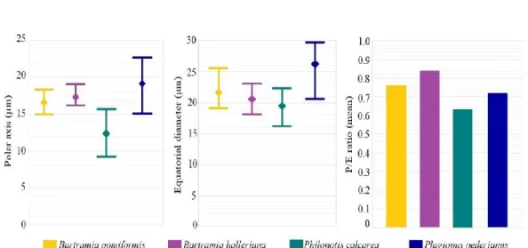
FIGURE 1. Graphical comparison of the length of polar axis (P), equatorial diameter (E) and P/E rotations (minimum, maximum and mean values).
Species P E (mean)P/E Shape Ornamentations
min max mean min max mean
B.pomiformis 15.0 18.48 16.63 19.10 25.6 21.67 0.76 Sub-oblate Verrucate-granulate B.halleriana 16.21 19.09 17.29 18.18 23.18 20.64 0.84 Sub-oblate Gemmate-microechinate
P.calcarea 9.39 15.75 12.36 16.21 22.42 19.59 0.63 Oblate Gemmate-pilate
3. Results
Bartramia pomiformis: Spores are small, suboblate, heteropolar and katalept. The
length of the polar axis is average 16.63 µm, equatorial diameter is average 21.67 µm in diameter and the ratio of polar axis to equatorial diameter is ~0.76 (Table 2, Figure 1). The sclerine surface is ornamented with big verrucae. The top of the verrucae shows granulate ornamentations in the distal face. The elements are irregular in shape and size, between 1.0-3.5 µm long. The wall structure is thinner on the proximal face. The aperture consists of monolete leptoma. Leptoma shape is ellipsoidal. The surface of leptoma is ornamented by sparse verrucate and granulate elements (Figure 2, 3). Capsules are inclined, globose to ovoid, asymmetrical, furrowed, average 1.8 mm long and 0.9 mm wide, ~2 times as long as wide. Peristome is double, teeth are lanceolate, average 300 µm, reddish-brown, striate, finely papillose. Endostome segments are present, finely seriate-papillose. Cilia available but less developed (Figure 4, 5).
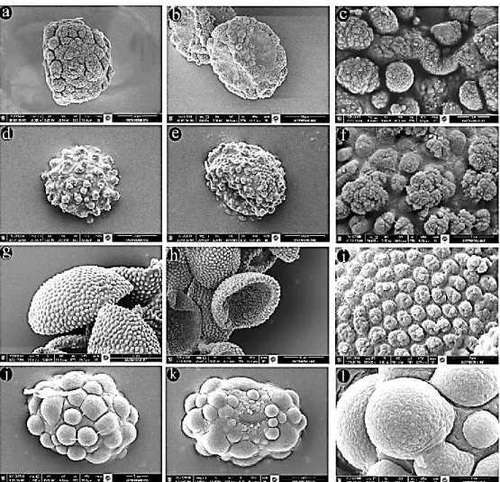
FIGURE 2. LM spore microphotograph. a, b: Bartramia pomiformis; c, d: B. halleriana; e, f: Philonotis calcarea; g, h: Plagiopus oederianus (a, h: equatorial view; b, d, f: proximal view; c, e, g: distal view).
Bartramia halleriana: Spores are small, suboblate, heteropolar and katalept. The
length of the polar axis is average 17.29 µm, equatorial diameter is average 20.64 µm in diameter and the ratio of polar axis to equatorial diameter is ~0.84 (Table 2, Figure 1). The sclerine surface is ornamented by gemmate with microechinate elements. It was seen perforate ornamentation among the gemmae. The length of the elements is between 0.5-2.2 µm. The aperture consists of irregular concave leptoma (Figure 2, 3). Capsules are subglobose to ovoid, furrowed, average 2.0 mm
wide and 0.9 mm long, ~2 times as long as wide. Operculum is conic and convex. Peristome is double, teeth are lanceolate, average 455 µm, brownish-red, strongly striate, papillose. Endostome segments are present, seriate-papillose. Cilia available but less developed (Figure 4, 5).
Philonotis calcarea: Spores are small, oblate, heteropolar and katalept. The length
of the polar axis is average 12.36 µm, equatorial diameter is average 19.59 µm in diameter and the ratio of polar axis to equatorial diameter is ~0.63 (Table 2, Figure 1). The sclerine surface is ornamented by gemmate-pilate elements. The ornamentations are dense in the distal pole, diluted in the proximal pole. The length of the elements is between 0.6-1.0 µm. The aperture consists of concave leptoma (Figure 2, 3). Capsules are inclined, globose, asymmetrical, furrowed, average 3.0 mm long and 1.3 mm wide, ~2.2 times as long as wide. Peristome is present, teeth are lanceolate, average 345 µm, yellowish-red, striate, papillose (Figure 4, 5).
Plagiopus oederianus: Spores are small, oblate, heteropolar and katalept. The
length of the polar axis is average 19.14 µm, equatorial diameter is average 26.23 µm in diameter and the ratio of polar axis to equatorial diameter is ~0.72 (Table 2, Figure 1). The sclerine surface is ornamented by big densely verrucate elements. The length of the elements is between 2.5-5.5 µm. The aperture consists of slightly concave leptoma. The ornamentations were diluted and shrunken on the leptoma (Figure 2, 3). Capsules are suberect, subglobose, asymmetrical, furrowed, average 1.7 mm long and 1.1 mm wide, ~1.5 times as long as wide. Peristome is double, teeth are lanceolate, average 172 µm, reddish-brown, striate, finely papillose (Figure 4, 5).
4. Discussion
The spore morphology of Turkish Bartramia pomiformis, B. halleriana, Philonotis
calcarea and Plagiopus oederianus studied by LM and SEM. Among them, the
spores of Bartramia pomiformis have previously been photographed with SEM and LM; Bartramia halleriana, Philonotis calcarea and Plagiopus oederianus with only LM by Boros et al. [20]. In the present study, SEM photographs of these taxa were given in detail and the ornamentation structures were compared with the referred study.
FIGURE 3. SEM spore photograph. a-c: Bartramia pomiformis; d-f: B. halleriana; g-i:
Philonotis calcarea; j-l: Plagiopus oederianus (a, d, g, j: distal view; b, e, h, k: proximal
view; c, f, i, l: spore ornamentation).
All spores are small, the length of the polar axis is between 12.36 µm and 19.14 µm, equatorial diameter is between 19.59 µm and 26.23 µm. The smallest spores of them are Philonotis calcarea and the biggest spores of them are Plagiopus
oederianus. All spores are katalept and heteropolar. Apertures are concave leptoma
on proximal pole. The shapes of the spores are determined as suboblate for
oederianus. Spore wall is thick on distal pole, tapers to proximal pole and forms
leptoma. Ornamentations are differing on leptoma, near leptoma and on distal pole. Verrucate-granulate, gemmate-microecinate, perforate, gemmate-pilate and verrucate ornamentation types were observed on the distal pole. The results for
Bartramia pomiformis and Plagiopus oederianus presented here are in accordance
with Boros et al. [20]. However, gemmate-microechinate ornamentation types were observed in Batramia halleriana, and gemmate-pilate ornamentation types in
Philonotis calcarea in this study, while verrucate ornamentation types were
reported in Batramia halleriana, and clavate or baculate ornamentation types in
Philonotis calcarea in the study of Boros et al. [20].
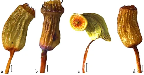
FIGURE 4. Stereo microscope capsule photograph. a: Bartramia pomiformis; b: B.
halleriana; c: Philonotis calcarea; d: Plagiopus oederianus (Scale bar: a, b, d: 0.25 mm; c:
0.50 mm).
The shapes and sizes of the capsules have some differences. The widths of the capsules are between 0.9 mm and 1.3 mm, length of capsules are between 1.7 mm and 3.0 mm. The smallest capsules are in Plagiopus oederianus. All capsules are asymmetrical and furrowed. All species have a developed peristome.
FIGURE 5. SEM capsule photograph a, b: Bartramia pomiformis; c: B. halleriana; d, e:
Philonotis calcarea; f, g: Plagiopus oederianus.
Consequently, the spore morphology and capsule structures in examined Bartramia
pomiformis, B. halleriana, Philonotis calcarea and Plagiopus oederianus show
distinguish characters which are important for taxonomic studies. References
[1] V. Virtanen, Phylogeny of the Bartramiaceae (Bryopsida) Based on Morphology and on rbcL, rps4, and trnL-trnF Sequence Data. The
Bryologist, 106 (2), (2003) 280-296.
[2] K. Dierβen, Distribution, ecological amplitude and phytosociological characterization of European bryophytes. Bryophytorum Bibliotheca, Band
56, (2001) Stuttgart.
[3] A. Erdag and H. Kurscher, Türkiye Bitkileri Listesi (Karayosunları). Ali
[4] V. Virtanen, Taxonomic Studies of the Bartramiaceae, Bryopsida. Helsingin
yliopiston kasvitieteen julkaisuja Publications in Botany from the University of Helsinki, Helsinki, 31, (2000).
[5] I. Potoglu Erkara and F. Savaroglu, Spore morphology of some Brachytheciaceae Schimp. species (Bryophyta) from Turkey. Nordic Journal
of Botany, 25, (2007) 194-198.
[6] F. Savaroglu, I. Potoglu Erkara, C. Baycu and M. Alkan, Spore morphology of some Bryaceae Schwägr. species (Bryophyta) from Turkey. International
Journal of Natural and Engineering Sciences 1 (2), (2007) 49-54.
[7] F. Savaroglu and I. Potoglu Erkara, Observations of spore morphology of some Pottiaceae Schimp. species (Bryophyta) in Turkey. Plant Systematics
and Evolution, 271, (2008) 93-99.
[8] N. Medina, B. Estebanez, F. Lara and V. Mazimpaka, On the presence of dimorphic spores in Orthotrichum affine (Bryopsida, Orthotrichaceae).
Journal of Bryology, 31, (2009) 127-129.
[9] B. Ascı, T. Ceter , N. Pınar, H. Colgeçen and B. Cetin, Spore morphology of some Turkish Tortula and Syntrichia species (Pottiaceae Schimp., Bryophyta. The Herb Journal of Systematic Botany, 17 (2), (2010) 165-180. [10] I. Caldeira, A. Luizi-Ponzo and V. Esteves, Palynology of selected species
of Fissidens (Hedw.). Plant Systematics and Evolution, 299, (2013) 187-195. [11] F. Savaroglu, Spore morphology of some Orthotrichaceae Arn. species
(Bryophyta) from Turkey. Bangladesh Journal of Botany, 44 (4), (2015) 499-506.
[12] T. Ceter and K. Canlı, Türkiyede yayılış gösteren bazı Grimmia (Grimmiaceae, Bryophyta) turlerin spor morfolojisinin incelenmesi. III. Aerobiyoloji, Palinoloji ve Alerjik Hastalıklarda Son Yenilikler Sempozyumu (APAS 2016), 5-7 Kasım 2016, Kastamonu/Türkiye, (2016). [13] R. Wodehouse, Pollen grains. Mc. Grew Hill, (1935) New York.
[14] A. Van der Pluym and M. Hideux, Application d’une mèthodologie quantitative á la palynologie d’Eryngium maritimum (Umbelliferae). Plant
Systematics and Evolution, 127, (1977) 55-85.
[15] G. Erdtman, Pollen and spore morphology/plant taxonomy; Gymnospermae, Pteridophyta, Bryophyta. Almquist and Wiksell, (1957) Stockholm.
[16] A. Boros and M. Járai-Komlódi, An atlas of recent European bryophyte spore. Akademiai Kiado, (1975) Budapest.
[17] S. Blackmore and S. Barnes, Pollen and Spores. Patterns of Diversification. The Systematics Association. Special Vol. No. 44. Clarendon Press, (1991) Oxford.
[18] W. Punt, S. Blackmore, S. Nilsson and A. Le Thomas, Glossary of pollen and spore terminology, contributions series No: 1. LPP foundation, (1994) Netherlands.
[19] R. Kapp, O. Davis and J. King, Pollen and spores, the American association of stratigraphic palynologists foundation. Texas A&M University, (2000) USA.
[20] A. Boros, M. Járai-Komlódi, Z. Töth and S. Nilson, An atlas of recent European Bryophyte spores. Akademiai Kiado, (1993) Budapest.
Current Address: TALIP CETER: Kastamonu University, Faculty of Science and Arts,
Department of Biology, Kastamonu, Turkey.
E-mail: [email protected]
ORCID: https://orcid.org/0000-0003-3626-1758
Current Address: MERVE CAN GOZCU: Hacı Bayram Veli University, Polatlı Faculty of
Science and Arts, Department of Biology, Ankara, Turkey.
E-mail: [email protected]
ORCID: https://orcid.org/0000-0001-7935-6314
Current Address: GURAY UYAR: Hacı Bayram Veli University, Polatlı Faculty of
Science and Arts, Department of Biology, Ankara, Turkey.
E-mail: [email protected]