DOI: 10.26650/IstanbulJPharm.2020.0001
Original Article
Effect of gel formulation obtained from Fomes
fomentarius on bleeding and clotting time:
A pilot study
Gülşah Gedik
1, Hülya Asan
2, Anıl Özyurt
2, Hakan Allı
3, Ahmet Asan
4, Hakan Nazlı
5,
Önder Sarp
51Trakya University, Faculty of Pharmacy, Department of Pharmaceutical Technology, Edirne, Turkey 2Trakya University, Faculty of Dentistry, Edirne, Turkey
3Mugla Sitki Kocman University, Faculty of Arts and Science, Department of Biology, Mugla, Turkey 4Trakya University, Faculty of Science, Department of Biology, Edirne, Turkey,
5Trakya University, Faculty of Pharmacy, Department of Pharmaceutical Technology, Edirne, Turkey
ORCID IDs of the authors: G.G. 0000-0003-4147-6729; H.A. 0000-0002-9650-9498; A.Ö. 0000-0002-3243-3156; H.A. 0000-0002-1238-3227; A.A. 0000-0002-4132-3848; H.N. 0000-0001-5763-1450; Ö.S. 0000-0003-1127-9273
Cite this article as: Gedik , G., Asan, H., Ozyurt, A., Alli, H., Asan, M., Nazli, H., & Sarp, O. (2020). Effect of gel formulation obtained from Fomes fomentarius on bleeding and clotting time: A pilot study. Istanbul Journal of Pharmacy, 50(3), 216-223.
ABSTRACT
Background and Aims: Fomes fomentarius (L.) Fr. (jar fungus) has been used extensively in the past by surgeons, barbers, dentists, and is therefore called surgeon's fungus. The hemostatic properties of F. fomentarius extracts are important in medicine. The aim of this study was to investigate the effect of the gel formulation obtained from Fomes Fomentarius on bleeding and clotting time in rat models.
Methods: The gel was made for this purpose and a cross-linked polymer of acrylic acid was used as a gelling agent. Results: The outcomes of the characterization analysis indicate the development of a successful gel formulation with optimum characteristics. Data from the antimicrobial study showed a good inhibition zone, with the strongest values against
Klebsi-ella pneumoniae, Candida krusei, C. tropicalis, C. guilliermondii. The gel administration shortened the hemostasis time after
bleeding from skin incisions by 63.9% and tail incisions by 55.63% from the original time. For coagulation time, these results are determined as 71.56% from the original time. The gel administration shortened the coagulation time after bleeding from the tail incisions by 69.9 % from the original time.
Conclusion: Our preliminary study showed that the gel formulation obtained from F. fomentarius is a potent hemostatic agent in a rat-bleeding model.
Keywords: Fomes fomentarius, bleeding time, coagulation time
Address for Correspondence:
Gülşah GEDİK, e-mail: [email protected]
This work is licensed under a Creative Commons Attribution 4.0 International License.
Submitted: 03.02.2020 Revision Requested: 18.05.2020 Last Revision Received: 19.07.2020 Accepted: 22.07.2020 INTRODUCTION
F. fomentarius, beech tree (Fagus sylvatica L.) and other deciduous species are developing as parasites or saprophytes, large wide variety of woody, perennial fungus with various sizes (Vetrovsky, Vorísková, & Snajdr, 2011). F. fomentarius is a white root fungus. Fruit body of between 5 and 50 cm across, 3 and 25 cm wide and 2 and 25 cm thick, which attaches broadly to the tree on which the fungus is growing. While typically shaped like a horse’s hoof, it can also be more bracket-like with an umbonate attachment to the substrate. The species typically has broad, concentric ridges, with a blunt and rounded margin. The flesh is hard and fibrous, and a cinnamon brown colour. The pores are circular, and there are 2–3 per millimeter. Odor pleasantly fungoid, taste rather bitter.
d · t; UNIVERSITY
•, .. s-s• - - P R E S S
A saprophyte or weak parasite on Fagus (beech), also on other hardwoods and rarely on conifers (Pacioni, 1985; Ellis & Ellis, 1990). Spores is oblong-ellipsoid, smooth, hyaline, 18-19x5-6 μ. It is known as “kav mantarı” in Turkey. The inside was a fungus used to light fire (Allı, Işıloğlu, & Solak, 2007).
F. fomentarius has been used extensively in the past by sur-geons, barbers and dentists, and is therefore called surgeon’s fungus. People used it as a medicine in Germany. In addition, F. fomentarius extract has been used in Europe, West Siberia and Indian folk medicine for dressing with iodine for external wounds and burns. Mushrooms are called “Wundschwamm” or “Chirurgenschwamm” in the alpine region of Switzerland. This preparation was sold in pharmacies in bandage form as an anti-bleeding agent and was used by Austrian farmers until the 19th century. Fomitin, the active ingredient of the F. fomentarius, is also used against dysmenorrhea, hemorrhoids and bladder diseases. There is information about the use of F. fomentarius for esophagus, stomach and uterine cancer in the literature (Grienke, Zöll, & Peintner, 2014). Also F. fomentarius has been used in traditional Chinese medicine for the treat-ment of oral ulcers, gastroenteritis, inflammations and various cancers (Chen, Zhao, & Li, 2011). In some studies, F. fomentarius has been reported to have hypoglycemic, anti-nociceptive, anti-inflammatory, anti-infective and anti-tumor activities (Se-niuk et al., 2011; Park et al., 2004). A study demonstrated that F. fomentarius ethanol extract inhibits cell motility and growth and induces apoptosis by inhibiting the phosphoinositide 3- kinase /AKT pathway and caspase activation (Seon-Ok, Min-Ho, & Kyung-Ran, 2019).
Several classes of metabolites were identified: Primary me-tabolites polysaccharides (glucans), polysaccharide–protein complexes, and secondary metabolites such as triterpene gly-cosides, esters and lactones (fungisterollinoleate, betulin 28-O-acetate), alcohols (7 ergostenol, sitosterol), aldehydes and ketones (protocatechualdehyde), organic acids, benzofurans (paulownin), coumarins (daphnetin), and volatile components (Grienke et al., 2014). The most important compounds with clinically beneficial activity are glucans. In vitro studies have suggested that large molecular weight or particular glucans can directly activate leukocytes, stimulating their phagocytic, cytotoxic and antimicrobial activities, including the production of reactive oxygen and nitrogen intermediates (Akramiene, Kondrotas, & Didziapetriene, 2007). The presence of polysac-charides in the polar extracts was considered to be very impor-tant because a previous study had shown that a water-soluble melanin-glucan complex (containing 80% melanins and 20 % β-glucans) completely inhibited the growth of C. albicans (Seniuk et al., 2011). The antifungal activities of F. fomentarius against Aspergillus flavus, A. fumigatus, Absidia orchidis and Can-dida krusei were shown in a study (Dresh et al., 2015).Hemor-rhagic properties of F. fomentarius extracts are important in medicine and dentistry. This feature of F. fomentarius, we want-ed to make human life safe by using these extracts to save lives of patients in traffic accidents, injuries, allergies and respiratory diseases, genetic disorders, hemophilia and bleeding and clot-ting disorders and to improve the quality of life.
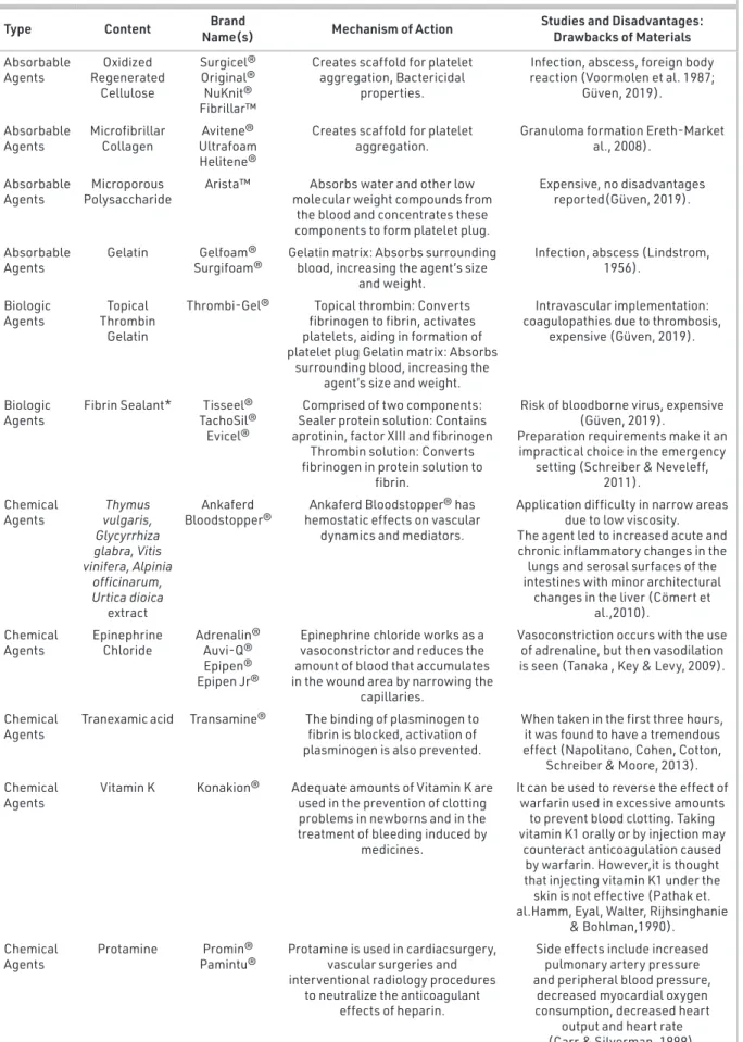
The characteristics of an ideal topical hemostat should include various properties such as, stopping arterial and venous bleed-ing in 2 minutes, no need special storage conditions, durable and light insensitive, long shelf live (>2 years), no need special training for its use, easy to use in difficult whether and war con-ditions, easy to be self applicable, effective on complex, tour-niqetless and hypothermic wounds, effective in patients using antiaggregants or anticoagulants, do not delay would healing, easily cleaned and bioabsorbed from the application area and provide an antimicrobial envirement, no local or systemic side effects and cost effective (Kheirabadi, 2011; Pusateri et al., 2003). Some hemostat agents types, mechanism of action, studies and disadvantages are given in Table1. Although very impor-tant studies have been carried out over the last years, a prod-uct with all the characteristics of the ideal topical hemostat, even with clinical trials and animal experiments, has not yet been developed. Our study was planned to demonstrate the bleeding-inhibiting properties of F. fomentarius.
MATERIALS AND METHODS Reagents and chemicals
Carbomer 940 powder (Lubrizol) and Triethanolamine (Sigma-Aldrich) was chosen for the gel formulation. Double-distilled water was used throughout the study.
Mushroom material
The aerial parts (fruit bodies) of F. fomentarius were collected by Hakan Allı on March 25, 2014, on Liquidambar orientalis Mill. in Köycegiz in the Toparlar region of Mugla, Turkey. A sample of the fungus was authenticated and a voucher specimen (No: 5379) was deposited in the herbarium of the Mugla University Faculty of Sciences. The fungus parts were ground to a powder using a porcelain mortar and pestle.
Preparation of gel formulation
The aqueous extract for formulation was obtained via extrac-tion of 5 g fungus powder with 100 mL distilled water at 80°C for 30 min using a water bath (Daihan Scientific, WB-22, Korea). The extract was filtered through Grade 1 Whatman paper. Car-bomer 940 powder (2%) was added to the filtrate. Triethanol-amine was then added and mixed with it enough to gel. The formulation was kept at 25±1°C for 48 hours to see a possible phase separation.
Characterization of gel formulation
Gel was evaluated as for its clarity, pH and viscosity. The experi-ments were repeated four times.
Clarity of formulation
The clarity of gels was determined by optical check under dark color background, and it was scaled as follows: turbid, +; clear, ++; and very clear, +++ (Okur, Yoltaş, &Yozgatli, 2016).
Measurement of pH
The pH of the formulation was detected by a digital pH-meter (Mettler Toledo S 220, Switzerland). Measurements were per-formed four times and an average of these measurements was accepted as the pH of the in-situ gels.
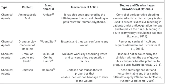
Table 1. Type, content, brand name, mechanism of action, studies and disadvantages of some hemostatic agents.
Type Content Name(s) Brand Mechanism of Action Studies and Disadvantages: Drawbacks of Materials Absorbable
Agents Regenerated Oxidized Cellulose
Surgicel® Original® NuKnit® Fibrillar™
Creates scaffold for platelet aggregation, Bactericidal
properties.
Infection, abscess, foreign body reaction (Voormolen et al. 1987;
Güven, 2019). Absorbable
Agents Microfibrillar Collagen UltrafoamAvitene® Helitene®
Creates scaffold for platelet
aggregation. Granuloma formation Ereth-Market al., 2008). Absorbable
Agents PolysaccharideMicroporous Arista™ molecular weight compounds from Absorbs water and other low the blood and concentrates these components to form platelet plug.
Expensive, no disadvantages reported(Güven, 2019).
Absorbable
Agents Gelatin Surgifoam® Gelfoam® Gelatin matrix: Absorbs surrounding blood, increasing the agent’s size and weight.
Infection, abscess (Lindstrom, 1956).
Biologic
Agents Thrombin Topical Gelatin
Thrombi-Gel® Topical thrombin: Converts fibrinogen to fibrin, activates platelets, aiding in formation of platelet plug Gelatin matrix: Absorbs
surrounding blood, increasing the agent’s size and weight.
Intravascular implementation: coagulopathies due to thrombosis,
expensive (Güven, 2019).
Biologic
Agents Fibrin Sealant* TachoSil® Tisseel® Evicel®
Comprised of two components: Sealer protein solution: Contains aprotinin, factor XIII and fibrinogen
Thrombin solution: Converts fibrinogen in protein solution to
fibrin.
Risk of bloodborne virus, expensive (Güven, 2019).
Preparation requirements make it an impractical choice in the emergency
setting (Schreiber & Neveleff, 2011).
Chemical
Agents vulgaris, Thymus Glycyrrhiza glabra, Vitis vinifera, Alpinia officinarum, Urtica dioica extract Ankaferd
Bloodstopper® hemostatic effects on vascular Ankaferd Bloodstopper® has dynamics and mediators.
Application difficulty in narrow areas due to low viscosity. The agent led to increased acute and chronic inflammatory changes in the lungs and serosal surfaces of the intestines with minor architectural
changes in the liver (Cömert et al.,2010).
Chemical
Agents Epinephrine Chloride Adrenalin®Auvi-Q® Epipen® Epipen Jr®
Epinephrine chloride works as a vasoconstrictor and reduces the amount of blood that accumulates in the wound area by narrowing the
capillaries.
Vasoconstriction occurs with the use of adrenaline, but then vasodilation is seen (Tanaka , Key & Levy, 2009).
Chemical
Agents Tranexamic acid Transamine® The binding of plasminogen to fibrin is blocked, activation of plasminogen is also prevented.
When taken in the first three hours, it was found to have a tremendous effect (Napolitano, Cohen, Cotton,
Schreiber & Moore, 2013). Chemical
Agents Vitamin K Konakion® Adequate amounts of Vitamin K are used in the prevention of clotting problems in newborns and in the treatment of bleeding induced by
medicines.
It can be used to reverse the effect of warfarin used in excessive amounts
to prevent blood clotting. Taking vitamin K1 orally or by injection may
counteract anticoagulation caused by warfarin. However,it is thought that injecting vitamin K1 under the skin is not effective (Pathak et. al.Hamm, Eyal, Walter, Rijhsinghanie
& Bohlman,1990). Chemical
Agents Protamine Pamintu®Promin® Protamine is used in cardiacsurgery, vascular surgeries and interventional radiology procedures
to neutralize the anticoagulant effects of heparin.
Side effects include increased pulmonary artery pressure and peripheral blood pressure,
decreased myocardial oxygen consumption, decreased heart
output and heart rate (Carr & Silverman, 1999).
Determination of viscosity
The viscosity of the in-situ gel formulations was performed with a vibro viscometer (AND, SV-10, Japan). The formulations were performed with 50 Hz at 32±2°C.
Spreadability studies of formulation
The spreadability of the gel was measured by spreading of 0.5 g of the gel on a circle of 2 cm diameter premarked on a glass plate and then a second glass plate was employed. Half kilo-gram of weight was permitted to rest on the upper glass plate for 5 min. The diameter of the circle after spreading of the gel was determined (Shinde, Pokharkar, & Modani, 2012).
Screening antimicrobial activity
All test microorganisms, including Klebsiella pneumoniae, Aci-netobacter baumannii, Staphylococcus aureus, vancomycin-re-sistant enterococci (VRE)+, Escherichia coli, Candida albicans (C.P. Robin) C. tropicalis (Castell.) C. krusei (Kudryavtsev), C. guillier-mondii (Kurtzman and Suzuki) and C. glabrata (H.W. Anderson) were obtained from the Duzce University Research Hospital (Duzce, Turkey). The microorganisms were stored in a refrigera-tor at 4°C prior to the study.
The disk diffusion method for antimicrobial susceptibility testing was carried out according to the Clinical and Labora-tory Standards Institute (CLSI) technique to assess the antimi-crobial activities of the gel formulation. The formulation was performed under sterile conditions in duplicate and repeated three times (Gedik et al., 2019).
In-vivo experimental design
Three healthy female rats were supplied by Trakya University’s Experimental Research Center Animal Laboratory for bleeding and clotting time tests. The study was approved by the Animal Experiments Local Ethics Committee of Trakya University (TU-HADYEK-2016/36). Experimental animals were maintained in
the same room in a clean environment with adequate ventila-tion under the following condiventila-tions: 1 atm pressure, 250C tem-perature, and 12-hour light-and-dark cycle. During the study, the rats were fed dry pellets and water ad libitum.
Bleeding assay
One of the rats was used as a control for determining normal bleeding and clotting time. The others were for testing 5% F. fomentarius gel formulation effect on bleeding and clotting time. On behalf of bleeding, two wound models were created in two different area of the rat body:
1) 5 mm diameter biopsy punch wound on nape, 2) 5 mm diameter tail cut wound
5% formula dose was prepared as separated 0.1 g sufficient applicable packages to fully-cover wound surface areas. All surgical procedures were performed under sterile conditions. The animals were anesthetized by an intramuscular injection of ketamine hydrochloride 50 mg/kg and xylazine hydrochlo-ride 10 mg/kg. All test procedures were performed according to Duke’s Method and Slide Method (Nilsson, Magnusson & Borchgrevink 1963; Waghmare & Muniyappanavar, 2018). Before creating a skin wound on the nape, the neck hair was shaved then cleaned with 70% alcohol. A 5 mm diameter skin was incised using a biopsy punch with a 2 mm depth. Bleed-ing time was counted with a stopwatch (refers to incision to end of bleeding). Three drops of blood were carried on a glass plate to observe the clotting time with another stopwatch si-multaneously.
A 5 mm diameter tail cut wound was created using a scalpel after the skin test. The cut tail was left on the ground at the same level as the heart. The same measurement procedures for bleeding and clotting time were conducted. All wounds
Table 1. Continued.
Type Content Name(s) Brand Mechanism of Action Studies and Disadvantages: Drawbacks of Materials Chemical
Agents Aminocaproic Acid Amicar® FDA to prevent recurrent bleeding in It has also been approved by the patients with traumatic hyphema.
Control of perioperative bleeding associated with cardiac surgery is also
used to prevent excessive bleeding in patients under anticoagulation therapy
and to reduce the risk of bleeding in acute promyelocytic leukemia patients
(Lu et al., 2015). Chemical
Agents Granular clay made out of smectite
WoundStat® It swells and thus can conform to any
wound. requires debridement (Schreiber et Removing can be difficult and al., 2011).
Chemical
Agents zeolite and Granular kaolin
QuikClot Combat Gauze®
QuikClot works by absorbing water and concentrating coagulation
factors.
It should not be touched by the clinician without the use of gloves. This substance has the potential to produce burns (Schreiber et al., 2011). Chemical
Agents Chitosan HemCon® Chitosan has mucoadhesive properties that enable the HemCon bandage to stick
to the wound.
These dressings are stiff and nonconformable and thus can be difficult to apply (Wedmore, McManus,
were sutured and cleaned with iodized antiseptic after the procedure was completed. The rats were placed in their cages and allowed to wake up.
Statistical analysis
Statistical analyses were conducted using SPSS, version 20.0 (SPSS, Inc., Chicago, IL). The mean and standard deviation were calculated for each group. All data were expressed as means and 95% confidence intervals (CIs) p<0.05 was considered sta-tistically significant.
RESULTS
Preparation and characterization of gel formulations
The physicochemical characterization parameters of gels are reported in Table 2. The clarity, viscosity and spreadability of all the formulations were found to be satisfactory. The pH of the developed gel ranged between 5.49 and 5.55. The pH of the gels was appropriate for the dermal application. The spread-ability of the gel was considered high by having, only two sec-onds. The therapeutic efficacy of gels depends on their spread.
Screening antimicrobial activity
The antimicrobial activity of gel formulation was performed against Klebsiella pneumoniae, Acinetobacter baumannii, Staph-ylococcus aureus, vancomycin-resistant enterococci (VRE)+,
Escherichia coli, Candida albicans, C. tropicalis, C. krusei, C. guillier-mondii and C. glabrata. The antimicrobial results of formulations are reported in Table 3. Agar well diffusion test results revealed that gel formulation showed a good inhibition zone with the strongest value against Klebsiella pneumoniae (10.3±0.79 mm), Candida krusei (11±0.26 mm), C. tropicalis (9±0.17 mm), C. guil-liermondii (10.2 ±0.4 mm) (Gedik et al., 2019)
Bleeding assay
5 mm diameter biopsy punch wound on nape:
Results obtained from the experiments were summarized in the Table 4. After confirming continuous bleeding from the full-thickness skin wound, the application of the gel resulted in rapid hemostasis. The gel administration shortened the he-mostasis time after bleeding from the skin incisions by 3:32:57 minutes or 63.9% from the original time of 9:48:39. For coag-ulation time these results are determined as follows: 2:31:20 minutes or 71.56 % from the original time of 8:51:50.
5 mm diameter tail cut wound:
Results obtained from the experiments were summarized in the Table 5. After confirming continuous bleeding from the tail wound, the application of the gel resulted in rapid hemostasis. The gel administration shortened the hemostasis time after bleeding from the tail incisions by 5:27:05 minutes or 55.63%
Table 2. Clarity, pH, viscosity, spreadability of gel formulation.
Clarity ++ pH 5.52 ± 0.01
Viscosity (P) 34.1P ± 0.22 (29,4 oC) Spreadability (cm) 1.72 ± 0.17 (≤ 2 seconds)
The data are presented as the mean ± standard deviation (SD).
Table 3. Antibacterial and antifungal activity of gel formulation.
Test Microorganisms Inhibition zones (mm)*
Gel Formulation E GN AM AMB FLU KTC
Klebsiella pneumoniae 10.3 ± 0.79 NT - 7 NT NT NT Acinetobacter baumannii - NT 9 10 NT NT NT Staphylococcus aureus 10.5 ± 0.43 14 NT 11 NT NT NT **VRE+ - 10 12 9 NT NT NT Escherichia coli 7.0 ± 0.01 11 NT - NT NT NT Candida krusei 11 ± 0.26 NT NT NT 9 - -C. albicans 7 ±0.45 NT NT NT 9 8 9 C. tropicalis 9 ±0.17 NT NT NT - - -C. guilliermondii 10.2 ±0.4 NT NT NT 8 - -C. glabrata 10 ±0.62 NT NT NT - - 15
*GN: Gentamicin 30 μg; AM: Ampicillin 10 μg; AMB: Amphotericin B 100 μg; FLU: Fluconazole 25 μg; KTC: Ketoconazole 10 μg; NT: Not tried. **Vancomycin Resistant Enterococci.
Table 4. Bleeding Assay with Biopsy Punch Wound on Nape.
Control Gel Application Bleeding
Time Coagulation Time Bleeding Time Coagulation Time
9:48:39 (588sn) (n=1) 8:51:50±0.07 (531sn) (n=3) 3:32:57±0.09 (212sn) (n=2) 2:31:20±0.06 (151sn) (n=6) (p=0.002)
from the original time of 12:17:06. For coagulation time these results are determined as follows: 2:01:69 minutes or 69.9% from the original time of 6:42:48.
DISCUSSION
This study evaluated the hemostatic effects of 5% F. fo-mentarius gel formulation in vivo in rat skin–tail bleeding models. Today, in addition to conventional anti-bleeding methods, the use of extracts from various plants and marine organisms has become popular. The aim of this study was to investigate the effect of the F. fomentarius which is a com-mon fungal species on bleeding time and clotting time. Gel formulation provides better application property and stabil-ity in comparison to cream and ointment. Topical gel drug administration is a localized drug delivery system anywhere in the body (Kaur & Guleri, 2013). The gel was made for this purpose and cross-linked polymer of acrylic acid was used as a gelling agent. Carbomer 940 polymer is a white pow-der, crosslinked polyacrylic acid polymer. Its short flow, non-drip properties are ideal for applications such as clear gels, hydroalcoholic gels, and creams. This gels are optimum vis-cosity at 25°C and physiological temperature, making them optimum gelling agent for dermal applications (https:// www.lubrizol.com/en/Personal-Care/Products/Product-Finder/Products-Data/Carbopol-940-polymer).
The gel formulation characterized based on its pH, clarity, and viscosity. Physicochemical characterization of in situ gel lations is an important subject to be considered in the formu-lation part, especially those intended for dermal application. The gel has a clear appearance on visual inspection. The pH of the developed gel ranged between 5.49 and 5.55. Ideally, dermal formulations should possess pH in the range of 5-6, for minimizing the discomfort of patient or skin irritation due to acidic pH and microbial growth on the skin because of basic pH (Okur, Çağlar, & Arpa, 2017). The outcomes of the charac-terization analysis indicate the development of successful gel formulation with optimum characteristics.
The spreadability of the gel was considered high by having a low spread of time. The gel spreading helps in the uniform ap-plication of the gel to the skin, so the prepared gels must have a good spreadability and satisfy the ideal quality in topical ap-plication. Furthermore, this is considered an important factor in patient compliance with treatment. This gels viscosity prop-erties with possible pseudoplastic behavior observed in the formulation, confirms the characteristic of high spreadability due to the decrease in viscosity when applying certain force,
and at the same time has the property of remaining at the ap-plication site without drain (Carvalho et al., 2010).
Data from the antimicrobial study showed a good inhibition zone with the strongest value against Klebsiella pneumoniae, Candida krusei, C. tropicalis, C. guilliermondii. Similar to the our study, Kolundžić et al. (2016) tested the antimicrobial activity of F. fomentarius extracts of different polarity, especially against Gram-negative and Gram-positive bacteria that are commonly related to dermal and hospital infections (Staphylococcus aure-us Rosenbach, S. epidermidis (Winslow and Winslow), Micrococ-cus luteus (Schroeter), Bacillus subtilis (Ehrenberg), EnterococMicrococ-cus faecalis (Andrewes and Horder), Escherichia coli (Migula) Castel-lani and Chalmers, Klebsiella pneumoniae (Schroeter) Trevisan and Pseudomonas aeruginosa (Schröter). They indicated that their F. fomentarius extracts (C-cyclohexane, D-dichlorometh-ane, M-methanol and A-aqueous) displayed strong crobial activity. Hereby, it can be concluded that the antimi-crobial efficiency of the fungi gel formulation could provide a convenient surrounding for hemostatic efficacy by preventing wound infections caused by bleeding.
Pyranose is a collective term for saccharides that have a chemi-cal structure that includes a six-membered ring consisting of five carbon atoms and one oxygen atom. This saccharide’s pKa is also 11.8 in strong acid character. These electrolyte struc-tures are biological environments force them to load cationic. (http://foodb.ca/compounds/FDB015634).
It has an anti-hemorrhagic effect by positively charged poly-saccharide and polypoly-saccharide-protein complex structure by binding chemical and mechanical effects with negatively charged erythrocytes, activating platelets and forming an ad-hesive structure in the tissue and forming a physical barrier around the injured vessel.
In our study, the gel administration shortened the hemostasis time after bleeding from the skin incisions by 63.9% and the tail incisions by 55.63% from the original time. For coagulation time these results are determined as follows 71.56% from the original time in skin incisions model and 69.9% from the origi-nal time in the tail incisions model.
CONCLUSION
Our preliminary study showed that the gel formulation ob-tained from F. fomentarius is a potent hemostatic agent for cutaneous bleeding resulting from surgical skin defects in a rat-bleeding model. The use of the gel in especially in patients with hemostatic abnormalities, those undergoing anticoagu-lant or antiaggregant therapy, pregnant patients, or people with cancer may be an alternative to other hemostatic agents for achieving effective bleeding control. Future controlled clinical trials are needed to evaluate the efficacy of the plant extract in the control of hemorrhaging during surgery. In particular, the search for hemostatic agent of natural origin is progressing rapidly, which points to the need for further studies exploring the utilization of the therapeutic agents from F. fomentarius.
Table 5. Bleeding Assay with Tail Cut Wound.
Control Gel Application Bleeding
Time Coagulation Time Bleeding Time Coagulation Time
12:17:06 (737sn) (n=1) 6:42:48±0.11 (402sn) (n=3) 5:27:05±0.07 (327sn) (n=2) 2:01:69±0.02 (121sn) (n=6) (p=0.006)
I
Ethics Committee Approval: The study were approved by animal experiments local ethics committee of Trakya University (TUHADY-EK-2016/36) and indicated in the method section.
Peer-review: Externally peer-reviewed.
Author Contributions: Conception/Design of Study- G.G., A.Ö., H.A.; Data Acquisition- G.G., A.Ö.; Data Analysis/Interpretation- G.G., H.N., Ö.S.; Drafting Manuscript- G.G., A.Ö., H.A., H.A., A.A.; Critical Revision of Manuscript- A.A., G.G.; Final Approval and Accountability- G.G., H.A., A.Ö., H.A., A.A., H.N., Ö.S.; Technical or Material Support- H.A., G.G.; Su-pervision- A.A., G.G.
Conflict of Interest: The authors have no conflict of interest to de-clare.
Financial Disclosure: Authors declared no financial support.
REFERENCES
• Akramiene, D., Kondrotas, A., Didziapetriene, J. & Kevelaitis, E. (2007). Effects of beta-glucans on the immune system. Medicina,
43(8), 597–606. https://doi.org/10.3390/medicina43080076
• Allı, H., Işıloğlu, M. & Solak, M. H. (2007). Macrofungi of Aydın Prov-ince. Mycotaxon, 99(1), 163–165. Retrieved from https://www.re-searchgate.net/publication/282726082_Macrofungi_of_Aydin_ Province_Turkey_65/link/5c8966b5a6fdcc3817526251. • Carr, J. A., Silverman, N. (1999). The heparin-protamine interaction:
A review. The Journal of Cardiovascular Surgery, 40(5), 659–666. Re-trieved from https://europepmc.org/article/med/10596998. • Carvalho, F. C., Barb, M. S., Sarmento, V. H. V., Chiavacci, L. A., Netto,
F. M. & Gremiao, M. P. D. (2010). Surfactant systems for nasal zid-ovudine delivery structural, rheological and mucoadhesive prop-erties. Journal of Pharmacy Pharmacology, 62(4), 430–439. https:// doi.org/10.1211/jpp.62.04.0004.
• Chen, W, Zhao Z, Li Y. (2011). Simultaneous increase of mycelial biomass and intracellular polysaccharide from Fomes fomentarius and its biological function of gastric cancer intervention.
Carbo-hydrate Polymers, 85(2), 369–375.
https://doi.org/10.1016/j.carb-pol.2011.02.035.
• Cömert, M., Karakaya, K, Barut, F., Çakmak, K. G., Uçan, H. B.,
Gül-tekin, F. A., Emre, A. U., Taşçılar, Ö., Irkörücü, O., Ankaralı, H. (2010).
Does intraabdominal use of Ankaferd Blood Stopper cause in-creased intraperitoneal adhesions. National Trauma and
Emer-gency of Surgery Journal, 16(5), 383–389. Retrieved from https://
pubmed.ncbi.nlm.nih.gov/21038113.
• Dresch, P., D´Aguanno, M. N., Rosam, K., Grienke, U., Rollinger, J. M., Ursula Peintner, U. (2015). Fungal strain matters: colony growth and bioactivity of the European medicinal polypores Fomes
fo-mentarius, Fomitopsis pinicola and Piptoporus betulinus. AMB Express, 5(4). https://doi.org/10.1186/s13568-014-0093-0.
• Ellis, M. B. & Ellis, J. P. (1990). Fungi without gills (Hymenomycetes
and Gasteromycetes). An Identification Handbook. XI, 329 S., 543
Abb. Chapman and Hall, London, New York, Tokyo, Melbourne, Madras, 1990. ISBN 0‐412‐36970‐2
• Ereth-Mark, H., Schaft, M., Ericson, E., Wetgen, N., Nuttal, G., Oliver, W. C. (2008). Comparative safety and efficacy of topical hemostat-ic agents in a rat neurosurghemostat-ical model. Neurosurgery, 63, 369–372. https://doi.org/10.1227/01.neu.0000327031.98098.dd.
• Gedik, G., Dülger, G., Asan, H., Özyurt, A., Allı, H. & Asan A. (2019). The antimicrobial effect of various formulations obtained from
Fomes fomentarius against hospital isolates. Mantar Dergisi- The Journal of Fungus, 10(2), 103–109. https://doi.org/
10.30708/man-tar.535994.
• Grienke, U., Zöll, M., Peintner, U. & Rollinger, J. M. (2014). European medicinal polypores–A modern view on traditional uses. Journal
of Ethnopharmacology, 154(3), 564–583. https://doi.org/10.1016/j.
jep.2014.04.030.
• Güven, R. (2019). An experimental study investigating the effect of local hemostatic agents on hemostatic, histopathological, anti-mullerian hormone and postoperative intraabdominal adhesion in a laceration model of complicated ovarian hyperstimulation (Exper-tise thesis in medicine). Available from tez.yok.gov.tr. (No. 535721). • Kaur, L. P., Guleri, T. K. (2013). Topical Gel: A Recent Approach for
Novel Drug delivery. Asian Journal of Biomedical and
Pharmaceuti-cal Sciences, 3(17), 1–5. https://doi.org/10.15272/AJBPS.V3I17.183.
• Kheirabadi, B. S. (2011). Evaluation of topical hemostatic agents for combat wound treatment. The United States Army Medical
De-partment Journal, 2, 25–38. Retrieved from https://pubmed.ncbi.
nlm.nih.gov/21607904.
• Kolundžić, M., Grozdanić, N. D., Dodevska, M., Milenković, M., Sisto, F., Miani, A. & Kundaković, T. (2016). Antibacterial and cytotoxic activities of wild mushroom Fomes fomentarius (L.) Fr.
Polypo-raceae. Industrial Crops and Products, 79, 110–115. https://doi.
org/10.1016/j.indcrop.2015.10.030.
• Lindstrom, P. A. (1956). Complications from the use of absorb-able hemostatic sponges. American Medical Association
Ar-chive of Surgery, 73(1), 133–141.
https://doi.org/10.1001/arch-surg.1956.01280010135018.
• Lu, J., Meng, H., Meng, Z., Sun, Y., Pribis, J. P., Zhu, C., Li, Q. (2015). Ep-silon aminocaproic acid reduces blood transfusion and improves the coagulation test after pediatric open-heart surgery: A meta-analysis of 5 clinical trials. International Journal of Clinical and
Experimental Pathology, 8(7), 7978–7987. Retrieved from https://
pubmed.ncbi.nlm.nih.gov/26339364.
• Napolitano, L. M., Cohen, M. J., Cotton, B. A., Schreiber, M. A., Moore, E. E. (2013). Tranexamic acid in trauma: how should we use it ? Journal of Trauma and Acute Care Surgery, 74(6), 1575–86. https://doi.org/10.1097/ta.0b013e318292cc54.
• Nilsson, I. M., Magnusson, S., Borchgrevink, C. (1963).The Duke and Ivy methods for determination of the bleeding time.
Throm-bosis et Diathesis Haemorrhagica 10.02: 223–234. Retrieved from
https://europepmc.org/article/med/14081283
• Okur, N. Ü., Çağlar, E. Ş., Arpa, M. D. & Karasulu, H. Y. (2017). Preparation and evaluation of novel microemulsion-based hydrogels for dermal delivery of benzocaine. Pharmaceutical Development and Technology,
22(4), 500–510. https://doi.org/10.3109/10837450.2015.1131716.
• Okur, N. Ü., Yoltaş, A. & Yozgatli, V. (2016). Development and char-acterization of voriconazole loaded in situ gel formulations for ophthalmic application. Turkish Journal of Pharmaceutical
Scienc-es,13(3): 311–317. https://doi.org/10.4274/tjps.2016.05
• Pacioni, G. (1985). Mac Ency Mushrooms Toadstools (Macdonald encyclopedias). London.
• Park, Y. M., Kim, I. T., Park, H. J., Choi, J. W., Park, K. Y., Lee, J. D., Nam, B. H., Kim, D. G., Lee, J. Y. & Lee, K. T. (2004). Anti-inflammatory and anti-nociceptive effects of the methanol extract of Fomes
fomen-tarius. Biological & Pharmaceutical Bulletin, 27(10), 1588–1593.
https://doi.org/10.1248/bpb.27.1588.
• Pathak, A., Hamm, C. R., Eyal, F. G., Walter, K., Rijhsinghani, A., Bohl-man, M. (1990). Maternal Vitamin K administration for preven-tion of intraventricular hemorrhage in preterm infants. Pediatric
Research, 27, 219. Retrieved from https://www.ncbi.nlm.nih.gov/
pmc/articles/PMC7043360.
• Pusateri, A. E., McCarthy, S. J., Gregory, K. W., Harris, R. A., Cardenas, L., McManus, A. T., & Goodwin Jr, C. W. (2003). Effect of a chitosan-based hemostatic dressing on blood loss and survival in a model of severe venous hemorrhage and hepatic injury in swine.
Jour-nal of Trauma and Acute Care Surgery, 54(1), 177–182. https://doi.
• Schreiber, M. A, Neveleff J. D. (2011). Achieving hemostasis with topical hemostats: Making clinically and economically appropri-ate decisions in the surgical and travma settings. The Association of Perioperative Registered Nurses Journal, 94(5), 1–20. https:// doi.org/10.1016/j.aorn.2011.09.018.
• Seniuk, O. F., Gorovoj, L. F., Beketova, G. V., Savichuk, N. O., Rytik, P. G., Kucherov, I. & Prilutsky A. (2011). Anti-infective properties of the melanin-glucan complex obtained from medicinal tinder bracket mushroom, Fomes fomentarius (L.: Fr.) Fr.
(Aphyllophoro-mycetideae). International Journal of Medicinal Mushrooms, 13(1),
7–18. https://doi.org/10.1615/intjmedmushr.v13.i1.20.
• Seon-OK, L., Min-Ho, L., Kyung-Ran, L., Eun-Ok, L., Hyo-Jeong, L. (2019). Fomes fomentarius Ethanol Extract Exerts Inhibition of Cell Growth and Motility Induction of Apoptosis via Targeting AKT in Human Breast Cancer MDA-MB-231 Cells. International
Journal of Molecular Science, 20(5), 1147. https://doi.org/10.3390/
ijms20051147.
• Shinde, U., Pokharkar, S. & Modani, S. (2012). Design and evalu-ation of microemulsion gel system of nadifloxacin. Indian
Jour-nal of Pharmaceutical Sciences, 74(3), 237–247. https://dx.doi.
org/10.4103%2F0250-474X.106066.
• Tanaka, A. K., Key, N. S., Levy, J. H. (2009). Blood coagulation: He-mostasis and thrombin regulation. Anesthesia & Analgesia, 108(5), 1433–1446. http://doi.org/10.1213/ane.0b013e31819bcc9c.
• Vetrovsky, T., Vorísková, J., Snajdr, J., Gabriel, J. & Baldrian, P. (2011). Ecology of coarse wood decomposition by the saprotrophic fun-gus Fomes fomentarius. Biodegradation, 22(4), 709–718. https:// doi.org/10.1007/s10532-010-9390-8.
• Voormolen, J. H. C., Ringers, J., Bots, G. A. M., Van Der Heide, A., Hermans, J. (1987). Hemostatic agents: Brain tissue reaction and effectiveness. Neurosurgery, 20(5), 702–709. https://doi. org/10.1227/00006123-198705000-00005.
• Waghmare, R. V., Muniyappanavar, N. S. (2018). Influence of blood groups on bleeding and clotting time. International Physiology
6(3), 200–204. https://dx.doi.org/10.21088/ip.2347.1506.6318.6.
• Wedmore, I., McManus, J. G., Pusateri, A. E., Holcomb, J. B. (2006). A special report on the chitosan-based hemostatic dressing: ex-perience in current combat operations. Journal of Trauma, 60(3), 655–658. https://doi.org/10.1097/01.ta.0000199392.91772.44. • FOODB. (2019, November 19) Retrieved from http://foodb.ca/
compounds/FDB01563.
• Lubrizol. (2019, December 28) Retrieved from https://www.lubr- izol.com/en/Personal-Care/Products/Product-Finder/Products-Data/Carbopol-940-polymer.