temporal and masseter muscles secondary to bruxism in
Turkish patients
Hasan Garip, DDS, PhD, Sukran Tufekcioglu, DDS, PhD, Emre Kaya, MD, PhD.
ABSTRACT
ةغضالما و ةيغدصلا ةلضعلا ةماخض ينب ةقلاعلا ليلتح :فادهلأا
يننرلا مادختساب يغدصلا يكفلا لصفملل يلصفلما صرقلا قلازناو
.يقابطلاا ززضلا ىضرم دنع يسيطانغلما
100دنع يغدص يكف لصفم
100ةساردلا هده نمضتت :ةقيرطلا
ةحارج مسق يف ةلجاعملل ينلولمحا و يقابطلاا ززضلاب صخشم ضيرم
,لوبيديم لوبناطسا ةعماج و ةرمرم ةعماج يف ينكفلا و هجولا و مفلا
.م
2016لولاا نوناك نوناك ىتح و
م2015يناثلا نوناك خيرات نم
مادختساب يسيطانغم يننرلاب ةيعاعش ةروصل ىضرلما عيمج عضخ
جردتلا ةداعتسا ىدص ةينقتب ليلكلاا ريوصتلا تم .لاست
1,5زاهج
مفلل قلاغلاا و حتفلا عضو يف ةلئام ةيمهس عطاقم ىلع لوصلحاو
دخأ و ةغضالما و ةيغدصلا ةلضعلل يروحم عطقم يرجأو . دنع
.قلاغلاا عضو يف اهنم لك سايق
ىدم(
mm 1.11±0.24تناك يلصفلما صرقلل ةناخث لقأ :جئاتنلا
ةكامس تناك ,ةناخث رثكلأا عضولما يف .)
0.6-1.7ينب حوارتي
)
20 - 9,2لدعبم(
mm 13.65±2.19حوارتت ةغضالما ةلضعلا
8.7-لدعبم(
mm 12.98±2.4تناك ةيغدصلا ةلضعلا امنيب
,يعيبط لكشب َاعضوتم ناك
24% ,يلصفلما صرقلل ةبسنلاب .)
21ودبت .يفلخ عضوت وذ ناك
2%,يمامأ ع ّضوت يف ناك
74%يعيبطلا عضوتلا ىضرم دنع نخثأ حضاو لكشب ةيغدصلا ةلضعلا
يلصفلما صرقلل يماملأا عضوتلا ىضرم عم ةنراقلماب يلصفلما صرقلل
.)
p=0.035(
ووذ ىضرلما يف نخثأ حضاو لكشب ةيغدصلا ةلضعلا تدب :ةتمالخا
صرقلل يماملاا عضوتلا عم ةنراقلماب يلصفلما صرقلل يعيبطلا عضوتلا
ةقلاعلا مييقتل ةيفاضا تاسارد ءارجاب حصني .)
p=0.035( يلصفلما
يف يلصفلما صرقلا عضوت و ةطيلمحاو ةغضالما تلاضعلا عيمج ينب
.يقابطلاا ززضلا ىضرم
Objectives: To analyze the relationships between temporalis and masseter muscle hypertrophy and temporomandibular joint (TMJ) disc displacement in patients with severe bruxism using magnetic resonance imaging (MRI).
Methods: This retrospective study included 100 patients with severe bruxism, referred to the Department of Oral and Maxillofacial Surgery, University of Marmara and Istanbul Medipol University, Istanbul, Turkey, between January 2015 and December 2016. Patients underwent TMJ MRI with a 1.5-T system in open and closed mouth positions. The masseter and temporalis muscles were measured in the axial plane when the patient’s mouth was closed.
Results: At its thinnest, the disc averaged was 1.11±0.24 mm. At their thickest, the masseter averaged was 13.65±2.19 mm and temporalis muscles was 12.98±2.4 mm. Of the discs, 24% were positioned normally, 74% were positioned anteriorly, and 2% were positioned posteriorly. The temporalis muscle was significantly thicker in patients with normally positioned discs than in those with anteriorly positioned discs (p=0.035).
Conclusions: The temporalis muscle was significantly thicker in patients with normally positioned discs than in those with anteriorly positioned discs (p=0.035). Additional studies should be conducted to evaluate the relationships between all masticatory and surrounding muscles and disc movements in patients with bruxism.
Saudi Med J 2018; Vol. 39 (1): 81-85 doi: 10.15537/smj.2018.1.20873 From the From the Department of Oral and Maxillofacial Surgery (Garip), Faculty of Dentistry, Marmara University; from the Department of Oral and Maxillofacial Surgery (Tufekcioglu), School of Dentistry, İstanbul Medipol University; from the Department of Radiology (Kaya), Goztepe Training and Research Hospital, Istanbul Medeniyet University, Istanbul, Turkey.
Received 6th August 2017. Accepted 15th November 2017.
Address correspondence and reprint request to: Dr. Hasan Garip, Department of Oral and Maxillofacial Surgery, Faculty of Dentistry, Marmara University, Istanbul, Turkey. E-mail: [email protected] ORCID ID: orcid.org/0000-0001-5580-6640
T
he temporomandibular joint (TMJ) is a special joint that forms the synovial articulation between the temporal bone and lower mandible bilaterally.1,2A biconcave fibrocartilage articular disc divides the TMJ into superior and inferior compartments.3 The
articular disc in the TMJ is postulated to behave as a shock absorber and to distribute the joint load.4,5
However, the effects of certain support of the articular disc on the mechanics of the TMJ are not understood clearly.3,5 Temporomandibular disorders (TMDs)
affect the stomatognathic system. The masticatory muscles and TMJ are the most affected structures.6,7
Temporomandibular disorders may be associated with neuromuscular, anatomical, and psychological factors. Limitation of physiological activity, joint sounds such as clicking, muscle and joint sensitivity or pain, and limitation and deviation of mandibular movements are the main signs and symptoms of TMDs. Headaches and neck and ear symptoms may also be seen in these patients.8,9 Disc displacement is a change in disc location
from its normal position on top of the mandibular condyle in the TMJ. Abnormal positioning may cause TMJ clicking and pain and may limit jaw function.10
Temporomandibular joint disc displacement is the main clinical sign of internal derangement of the TMJ, which progresses from reducible to non-reducible. The jaw elevator muscles, such as the temporal and masseter muscles, are commonly affected in TMD.11
The masseter is a muscle that elevates the mandible. It also plays an important role in the lower facial shape. It originates from the zygomatic arch and inserts into the angle of the mandible.12 The temporalis, another
masticatory muscle, enables elevation and retraction of the mandible. It originates from the temporal fossa up to the inferior temporal line and the inner surface of the temporal fascia and inserts into the coronoid process of the mandible.13 Bruxism is associated with numerous
negative outcomes, such as chronic local muscular contracture, inflammation, and localized muscular hypertrophy, which may in turn cause myofascial pain.14 In addition, significant associations have
been found among unilateral chewing, possible sleep bruxism, and TMDs.15 Sleep bruxism also negatively
affects masticatory muscle function. Magnetic resonance imaging is accepted as the gold standard for TMJ imaging. It enables visualization of the
position and morphology of the disc and masticatory muscles without exposing the patient to radiation.1,16
The present study aimed to evaluate the relationships between temporalis and masseter muscle hypertrophy and TMJ disc displacement with MRI in patients with severe bruxism.
Methods. This retrospective study included 100 TMJs from 100 randomly selected patients with severe bruxism, referred to the Departments of Oral & Maxillofacial Surgery (OMFS), University of Marmara and Istanbul Medipol University, Istanbul, Turkey, between January 2015 and December 2016.
Patients underwent TMJ MRI with a 1.5-T system (8-channel radiofrequency head coil, Philips Initial Ingenia, Best, Holland, 2013). After coronal T1-weighted (T1W) spin echo (SE) and axial-coronal T2-weighted (T2W) SE acquisitions, sagittal oblique images were repeated in open and closed mouth positions with T2-weighted SE sequences. The gradient pulse sequences and parameters were as follows: for T2W images, field of view (FOV) 160, slice thickness 3 mm, repetition time (TR) 345 ms, echo time (TE) 11 ms, flip angle 30, matrix 256×256, and acquisition mode 2-dimensional (2D); and for T1W images, FOV 160, slice thickness 3 mm, TR 400 ms, TE 20 ms, FA 90, matrix 256×256, and acquisition mode 2D.
The TMJ MRI protocol at the Radiology Department of Istanbul Medipol University consists of T1W sequences in the coronal and sagittal planes, T2W sequences in the axial plane, and proton-density-weighted (PDW) sequences in the coronal plane. In addition, 2 PDW images are obtained in the sagittal plane while the patient’s mouth is open and closed. The masseter and temporalis muscles were measured in the axial plane at their thickest points while the patient’s mouth was closed.
This study was carried out in agreement with International and Turkish laws on clinical experimentation, and in accordance with the Declaration of Helsinki. The research protocol was approved by the Ethics Committee of Istanbul Medipol University. The study was conducted at the Departments of OMFS, University of Marmara and Istanbul Medipol University, Istanbul, Turkey.
The inclusion criteria were the presence of TMJ clicking or locking, limitation of mouth opening, and/ or TMJ pain. The exclusion criteria were the presence of condylar hyperplasia, rheumatoid arthritis, congenital craniofacial syndrome, benign or malignant parotid disease, rhabdomyoma, and lymphangioma.
Disclosure. Authors have no conflict of interests, and the work was not supported or funded by any drug company.
Statistical analysis. The data were evaluated using SPSS Statistics 22.0 (IBM SPSS, Turkey). The conformity of parameters to a normal distribution was evaluated using the Shapiro-Wilks test. Fisher’s exact test was used for comparing qualitative data. Pearson correlation analysis was used for determining relationships between parameters. The level of significance was set to p<0.05.
Results. The 100 patients enrolled consisted of 18 men and 82 women, with a mean age of 33.44±14.45 years (Table 1). Masseteric and temporal muscle hypertrophy related to bruxism was identified in all patients. There were no significant differences between males and females in terms of disc location, average thinnest diameter of the disc (p=0.078), or thickness of the masseteric muscle (p=0.086) or temporal muscle (p=0.674). There were also no significant differences
in disc location (p=0.096) or thickness (p=0.343), or thickness of the masseteric muscle (p=0.717) or temporal muscle (p=0.941) between the left and right sides.
Moreover, Pearson correlation analysis revealed no significant relationship between the thinnest diameter of the disc and the thickest points of the masseteric muscle (p=0.253) and temporal muscle (p=0.900). There was also no significant difference in masseteric muscle thickness between discs in the normal and anterior positions (p=0.539). The temporal muscle was significantly thicker in patients whose discs were in the normal position than in patients with discs in an anterior position (p=0.035).
Discussion. It is unclear whether masticatory muscle hypertrophy is reactive or idiopathic. Masticatory muscle hyperactivity or parafunctions cannot be verified in all instances of hypertrophy. Masticatory muscle hyperactivity has been described as a possible cause. Compensatory and stress hypertrophy has been assumed in most cases.17 The signs and
symptoms of bruxism include tooth grinding, abnormal tooth wear, an indented tongue, muscle hypertrophy, jaw locking, morning headache, facial pain or fatigue, and reports of tooth grinding or jaw clenching sounds during sleep.18 This study evaluated the temporal
and masseteric muscle thickness and disc position in patients with severe bruxism. Bruxism was diagnosed based on self-reporting, awareness of sounds during sleep by a roommate, and clinical examination. In our cohort of patients with bruxism, all of the patients had temporal and masseteric muscle hypertrophy. The mean maximum thickness of the masseteric muscle was 13.65±2.19 mm. There were no significant differences between the gender or between the left and right sides. In addition, the mean maximum thickness of the temporalis was 12.98±2.4 mm and there were also no significant differences between the gender or the left and right sides.
In a systematic review of bruxism and TMDs18 and
in the study conducted by Jiménez-Silva et al,14 bruxism
was associated with myofascial pain, arthralgia, and joint pathology such as disc displacement and joint noises. Jussila et al19 investigated the prevalence of TMDs in
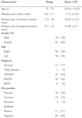
1,962 subjects (1,050 women, 912 men) undergoing medical and dental examinations and found that the most common TMD diagnosis was reducible disc displacement and that TMD was diagnosed in females more often than in males. In our cohort, we also found that the most common TMD diagnosis was reducible disc displacement (43%). The disc was positioned Table 1 - Descriptive results of 100 temporomandibular joint (TMJ)
from 100 randomly selected patients with severe bruxism.
Characteristic Range Mean ± SD
Age (y) 15 - 79 33.44 ± 14.45
Thinnest part of disc (mm) 0.6 - 1.7 1.11 ± 0.24 Thickest part of masseter muscle
(mm) 9.2 - 20 13.65 ± 2.19
Thickest part of temporal muscle
(mm) 8.7 ± 21 12.98 ± 2.4 Gender (%) Male 18 (18) Female 82 (82) Side Right 50 (50) Left 50 (50) Diagnosis Normal 17 (17) TMJ arthrosis 2 (2) NRADD 32 (32) RADD 43 (43) RDD 6 (6) Disc position Normal 24 (24) Anterior 74 (74) Posterior 2 (2) Arthritis Positive 17 (17) Negative 83 (83)
NRADD - non-reducible anterior disc displacement, RADD - reducible anterior disc displacement, RDD - reducible disc displacement,
normally in 24%, posteriorly in 2%, and anteriorly in 74%. The disc was positioned anteriorly more often in males (94.4%) than in females (69.5%).
In a clinical study of 49 patients, Dergin et al1 found
no correlation between the type of muscle attachment and disc displacement, articular surface degeneration, or disc degeneration. We also found no significant difference between the masseteric muscle thickness for discs positioned normally and anteriorly, while the temporal muscle was significantly thicker in patients in whom the disc was positioned normally (p=0.035). Articular surface degeneration was seen only in 2 patients and was not a significant finding.
Liu et al20 reported that disc perforation commonly
occurred in the bilaminar zone and the lateral part of the articular disc. In addition, medial disc perforation developed more frequently in patients with non-reducible displacement. Abnormal relationships between the intra-articular disc and the condyle and condylar fossa were the main cause of disc perforation. The authors pointed out that articular disc perforation occurred most commonly at the posterolateral attachment of the disc to the retrodiscal tissue.21
In our cohort, we did not see any disc perforation. Stratmann et al22 found a significant gradient of
decreasing disc thickness in the mediolateral axis of the part with intermediate density.22 The disc ranged
from 6 to 1.7 mm at its thinnest point, averaging 1.11±0.24 mm. There was no significant relationship between the location where the disc was thinnest and the thickness of the masseteric and temporal muscles (p>0.05).
Previous studies have not examined relationships between the masticatory muscles and disc displacement in Turkish patients with severe bruxism. The present study revealed no significant association between the thinnest point of the disc and the maximum thickness of the masseteric and temporal muscles, and we also did not find a significant difference in masseteric muscle thickness between the cases in which the discs were positioned normally or anteriorly. However, the temporal muscle was significantly thicker in patients in whom the disc was positioned normally than in patients in whom it was positioned anteriorly (p=0.035).
Sleep bruxism negatively affected masticatory muscle function. The masseter muscle pulls the mandible upward, whereas the temporalis muscle unclenches and clenches the jaw, controls retraction and elevation of the mandible, and makes fine mandibular adjustments.23
Our findings indicate that a strong, thick temporalis muscle stabilizes mandibular movements and probably helps to keep the disc in a normal position.
A limitation of our study is its retrospective nature. This study was conducted solely on the bruxist Turkish patients, and result of the research cannot be generalized to all masticatory muscle hypertrophies.
In conclusion, TME function affects disc movements. The present study demonstrated that the temporalis muscle directly alters disc function. Additionally, anterior disc displacement occurs in significantly fewer patients with thicker temporalis muscles. This study can guide further clinical studies regarding the relationship between the disc and the masticatory muscles in patients with bruxism.
References
1. Dergin G, Kılıc C, Gozneli R, Yıldırım D, Garip H, Moroglu S. Evaluating the correlation between the lateral pterygoid muscle attachment type and internal derangement of the temporomandibular joint with an emphasis on MR imaging findings. J Craniomaxillofac Surg 2012; 40: 459-463. 2. Kuroda S, Tanimoto K, Izawa T, Fujihara S, Koolstra JH,
Tanaka E. Biomechanical and biochemical characteristics of the mandibular condylar cartilage. Osteoarthritis Cartilage 2009; 17: 1408-1415.
3. Nickel J, Spilker R, Iwasaki L, Gonzalez Y, McCall WD, Ohrbach R, et al. Static and dynamic mechanics of the temporomandibular joint: plowing forces, joint load and tissue stress. Orthod Craniofac Res 2009; 12: 159-167.
4. Scapino RP, Canham PB, Finlay HM, Mills DK. The behaviour of collagen fibres in stress relaxation and stress distribution in the jaw-joint disc of rabbits. Arch Oral Biol 1996; 41: 1039-1052. 5. Yildirim D, Dergin G, Tamam C, Moroglu S, Gurses B. Indirect measurement of the temporomandibular joint disc elasticity with magnetic resonance imaging. Dentomaxillofac
Radiol 2011; 40: 422-428.
6. McNeill C. Management of temporomandibular disorders: concepts and controversies. J Prosthet Dent 1997; 77: 510-522. 7. Suvinen TI, Kemppainen P. Review of clinical EMG studies related to muscle and occlusal factors in healthy and TMD subjects. J Oral Rehabil 2007; 34: 631-644.
8. Ciancaglini R, Testa M, Radaelli G. Association of neck pain with symptoms of temporomandibular dysfunction in the general adult population. Scand J Rehabil Med 1999; 31: 17-22.
9. Parker MW. A dynamic model of etiology in temporomandibular disorders. J Am Dent Assoc 1990; 120: 283-290.
10. Dolwick MF, Katzberg RW, Helms CA. Internal derangements of the temporoman- dibular joint: fact or fiction. J Prosthet
Dent 1983; 49: 415-418.
11. Chandu A, Suvinen TI, Reade PC, Borromeo GL. Electromyographic activity of frontalis and sternocleidomastoid muscles in patients with temporomandibular disorders. J Oral
Rehabil 2005; 32: 571-576.
12. Drake R, Vogl AW, Mitchell A. Gray’s Anatomy for Students. 2nd edition. Philadelphia (PA): Churchill and Livingstone; 2009. p. 927.
13. Lee JY, Kim JN, Kim SH, Choi HG, Hu KS, Kim HJ, Song WC, Koh KS. Anatomical verification and designation of the superficial layer of the temporalis muscle. Clin Anat 2012; 25: 176-181.
14. Jiménez-Silva A, Peña-Durán C, Tobar-Reyes J, Frugone-Zambra R. Sleep and awake bruxism in adults and its relationship with temporomandibular disorders: A systematic review from 2003 to 2014. Acta Odontol Scand 2017; 75: 36-58.
15. Yalçın YD, Yılmaz N, Koraltan M, Aydın E. A survey on the potential relationships between TMD, possible sleep bruxism, unilateral chewing, and occlusal factors in Turkish university students. Cranio 2017; 35: 308-314.
16. Kaya K, Dulgeroglu D, Unsal-Delialioglu S, Babadag M, Tacal T, Barlak A, et al. Diagnostic value of ultrasonography in the evaluation of the temporomandibular joint anterior disc displacement. J Craniomaxillofac Surg 2010; 38: 391-395, 17. Graziano P, Dell’Aversana OG, Astarita F, Ponzo LM, Nunziata
R, Salzano G, et al. Bilateral hypertrophy of masseteric and temporalis muscles, our fifteen patients and review of literature.
Eur Rev Med Pharmacol Sci 2016; 20: 7-11.
18. Bertazzo-Silveira E, Stuginski-Barbosa J, Porporatti AL, Dick B, Flores-Mir C, Manfredini D, et al. Association between signs and symptoms of bruxism and presence of tori: a systematic review. Clin Oral Investig 2017; 21: 2789-2799.
19. Jussila P, Kiviahde H, Näpänkangas R, Päkkilä J, Pesonen P, Sipilä K, et al. Prevalence of Temporomandibular Disorders in the Northern Finland Birth Cohort 1966. J Oral Facial Pain
Headache 2017; 31: 159-164.
20. Liu XM, Zhang SY, Yang C, Chen MJ, Cai XY, Haddad MS, et al. Correlation between disc displacements and locations of disc perforation in the temporomandibular joint. Dentomaxillofac
Radiol 2010; 39: 149-156.
21. Holmlund A, Hellsing G. Arthroscopy of the temporomandibular joint. A comparative study of arthroscopic and tomographic findings. Int J Oral Maxillofac Surg 1988; 17: 128-133. 22. Stratmann U, Schaarschmidt K, Santamaria P. Morphometric
investigation of condylar cartilage and disc thickness in the human temporomandibular joint: significance for the definition of osteoarthrotic changes. J Oral Pathol Med 1996; 25: 200-205.
23. Palinkas M, Bataglion C, Luca CG, Machado CN, Theodoro GT. Impact of sleep bruxism on masseter and temporalis muscles and bite force. Cranio 2016; 34: 309-315.
Authorship entitlement
Excerpts from the Uniform Requirements for Manuscripts Submitted to Biomedical Journals updated November 2003.
Available from www.icmje.org
The international Committee of Medical Journal Editors has recommended the following criteria for authorship; these criteria are still appropriate for those journals that distinguish authors from other contributors.
Authorship credit should be based on 1) substantial contributions to conception and design, or acquisition of data, or analysis and interpretation of data; 2) intellectual content; and 3) final approval of the version to be published. Authors should meet conditions 1, 2, and 3.
Acquisition of funding, collection of data, or general supervision of the research group, alone, does not justify authorship.