Corresponding author/İletişim kurulacak yazar: [email protected]
Submitted/Başvuru: 22.05.2019 • Accepted/Kabul: 12.07.2019 • Published Online/Online Yayın: 26.08.2019 ©Telif Hakkı 2019 J Ist Faculty Med - Makale metnine jmed.istanbul.edu.tr web sayfasından ulaşılabilir. ©Copyright 2019 by J Ist Faculty Med - Available online at jmed.istanbul.edu.tr
LASER-ASSISTED-INDOCYANINE-GREEN-ANGIOGRAPHY
VERSUS CONVENTIONAL ASSESSMENT TO PREDICT OR LOCATE
NECROTIC AREAS ON MASTECTOMY FLAPS: A PROSPECTIVE
CLINICAL TRIAL
MASTEKTOMİ FLEBİ NEKROZUNU TAHMİN YA DA LOKALİZE EDEBİLEN LAZER
YARDIMLI İNDOSİYANİN YEŞİLİ ANJİOGRAFİNİN KONVANSİYONEL YÖNTEMLE
KIYASLANMASI: PROSPEKTİF KLİNİK ÇALIŞMA
Fatih Levent BALCI1 , Cihan URAS2
1Medipol University Mega Hospital, Department of General Surgery, Istanbul, Turkey 2Acibadem University, Department of General Surgery, Istanbul, Turkey
ORCID IDs of the authors: F.L.B. 0000-0001-8460-9355; C.U. 0000-0002-6838-2311
Cite this article as: Balci FL, Uras C. Laser-assisted-indocyanine-green-angiography versus conventional assessment to predict or locate necrotic areas on mastectomy flaps: A prospective clinical trial. J Ist Faculty Med 2019;82(4):193-8. doi: 10.26650/IUITFD.2019.0040
ÖZET
Amaç: Bu çalışmada meme-başı koruyucu mastektomi (MBKM) fleplerindeki nekrozu yada nekroz lokalizasyonunu, lazer yardımlı indosiyanin yeşilli angiografinin (LYIYA) tahmin edip edemeyece-ğini konvansiyonel gözlem ile kıyaslayarak saptamaktı.
Yöntem: Meme kanseri nedeniyle 21 hastaya MBKM ve eşzamalı silikon implant rekonstrüksiyon yapıldı. Ondokuz hastada flep üze-rindeki hipoperfuze alanların lazer absorpsiyon derecesini anla-mak için LYIYA sayıları kullanıldı. Elde edilen sayılar konvansiyonel gözlem ile kıyaslanarak LYIYA’nın nekroz prediktivitesi saptandı. Bulgular: Bu 19 mastektomi flebinin 3’ünde (15,8%) parsiyel cilt nekrozu saptanmış ve LYIYA sayısı ≤7 olarak saptanmıştır. LYIYA’nın duyarlılığı, özgüllüğü, yalancı pozitifliği ve doğruluğu sırasıyla 43%, 100%, 57%, ve 79% olarak bulunmuştur. LYIYA ≤7 olan hastalarda daha çok nekroz saptanmış, 60 yaş üstü, sigara öyküsü, BMI >30 veya intraoperatif tumescent solusyonu kullanı-lanlarda LYIYA ≤7 uyumsuz bulunmuş, nekroz tahmininde yanılgı-ya sebep olmuştur.
Sonuç: LYIYA sayısı ≤7 bulgusu, mastektomi flep nekrozunu tah-min edebilmede kullanılabilecek tek anlamlı parametredir. LYIYA aynı anda nekrozun nerde lokalize olduğunu ve sınırlarını da gös-terebilmektedir.
Anahtar Kelimeler: SPY, lazer yardımlı indosiyanin yeşilli angi-ografi, meme başı koruyucu mastektomi, meme kanseri
ABSTRACT
Objective: The aim of this study was to determine whether la-ser-assisted-indocyanine-green-angiography (LA-ICGA) could ac-curately predict flap necrosis in comparison to conventional clini-cal assessment and visually identify its location during immediate reconstruction following a nipple-sparing mastectomy (NSM). Methods: Twenty-one patients with breast cancer were prospec-tively enrolled to undergo NSM with immediate implant recon-struction. In 19 cases LA-ICGA numbers were used to show the level of laser absorption of hypo-perfused areas on the mastec-tomy flaps. Those numbers were compared to conventional as-sessment to assess the predictive value of LA-ICGA.
Results: Of the 19 mastectomy flaps, 3 (15.8%) examples of partial skin flap necrosis with an LA-ICGA value of ≤7 was observed. The sensitivity, specificity, false-positive rate, and accuracy of LA-IC-GA were 43%, 100%, 57%, and 79%, respectively. Patients with an LA-ICGA value of ≤7 were found more likely to develop mastecto-my flap necrosis, whereas patients aged >60 or, a history of smok-ing, a BMI >30, or intraoperative use of tumescence solution con-taining epinephrine were more likely to have an LA-ICGA score ≤7 which is not clinically reliable in predicting necrosis.
Conclusion: Our results indicate that a low LA-ICGA score ≤7 is the only significant factor in predicting mastectomy flap ne-crosis. LA-ICGA could accurately show the location of nene-crosis. Keywords: SPY, intraoperative angiography, nipple-sparing mastectomy, breast Cancer
INTRODUCTION
Immediate reconstructive procedures have been made more feasible through surgical approaches that pre-serve the natural skin envelope, including skin-sparing and the nipple-sparing mastectomy. Critical issues in im-mediate reconstructive procedures include coverage of the implant, the use of an appropriate tissue expander or silicone, and the quality and viability of tissue flaps. The quality and viability of mastectomy flaps is a central challenge in reconstructive surgery. A leading cause of early complications following reconstructive procedures is insufficient perfusion in the tissue flaps (1-4). For this reason, surgeons have evaluated and employed many methods to assess tissue perfusion in the intraoperative setting. An ideal technique would allow for the accurate identification of perforating vessels and their perfusion zones, assessment of tissue perfusion, and identification of tissue at risk for necrosis. Clinical judgment is the most widely used method for evaluating blood supply and tis-sue viability (5). However, some evidence suggests that clinical assessment alone is not entirely reliable for as-sessing tissue perfusion (6,7).
To reduce the occurrence of mastectomy necrosis, in-traoperative laser-assisted indocyanine green angi-ography, has been increasingly advertised as a useful preventative measure (8,9). Laser-assisted indocyanine green angiography (LA-ICGA) provides live imaging of fluorescent indocyanine green injected into the pa-tient’s bloodstream, which then permeates the papa-tient’s tissue. When such a patient’s mastectomy flap is viewed under the scanner, well-perfused tissue appears fluores-cent and ischemic tissue appears dark. This technolo-gy allows the immediate assessment of the viability of the mastectomy flap and facilitates intraoperative de-cision-making concerning how much tissue to excise so that no ischemic tissue is left behind that will result in mastectomy necrosis (10).
The fluorescence agent ICG has a short half-life, binds strongly to plasma proteins, and has an excellent safe-ty profile, allowing for rapid clearance from tissues and the performance of repeat evaluations during the same surgical procedure (11). Recent work suggests that the LA-ICGA also provides greater accuracy compared to fluorescein or clinical judgment for the prediction of mastectomy flap necrosis (8). This study aims to deter-mine whether LA-ICGA can accurately predict skin flap necrosis and visually identifies its location during nipple or skin-sparing mastectomies with immediate reconstruc-tion at our institureconstruc-tion.
METHODS
Twenty-one consecutive patients diagnosed with invasive breast cancer who underwent nipple-sparing
mastecto-mies (NSM) with immediate silicone gel implant recon-struction were prospectively enrolled in the study. Those skin flaps or edges, which were predicted to be ischemic by LA-ICGA, but physically assessed as normal by the surgeon, were not excised intraoperatively to assess the accuracy of SPY-Q numbers (represent quantitative laser absorption of hypoperfused areas) on flap necrosis. The SPY-Q numbers equal to or fewer than seven were con-sidered as predictors for tissue necrosis (12). Exclusion criteria included skin-reducing NSMs, delayed recon-struction, iodine allergy or intolerance to ICG dye. The study was approved by the Acibadem University Ethics Committee (#2016-11/12), and informed consent was ob-tained from all the enrolled patients.
Surgical technique
Since periareolar or medial/lateral extension incisions result in a slightly higher rate of nipple necrosis after NSMs followed by immediate reconstruction, lateral in-cisions which start at least 2 cm away from the nipple was the preferred incision made by the breast surgeon in the majority of our cases. Inframammary fold inci-sions (which are chosen to achieve more cosmetic sat-isfaction), were used less frequently. Permanent silicone implants were used immediately for the reconstruction with a created sub-muscular pocket created by a plas-tic surgeon. In order to cover and support the inferior aspect of the breast pocket, an acellular dermal matrix was used when needed.
Laser-assisted Indocyanine green (ICG) angiography protocol
After completion of the nipple-sparing mastectomy by a breast surgeon, each patient underwent visualization of mastectomy skin flaps using the SPY Elite System (Life Cell Corp., Branchburg, NJ, USA). 0.02 mg/kg of ICG dye was administered intravenously followed by 10cc of normal saline. Perfusion scores were recorded on LA-ICGA for a total of 120 sec after visualization of fluorescence in mastectomy skin flaps. SPY-Q software numbers were chosen, which represents absolute per-fusion levels.
Study design
One surgeon examined the physical appearance in re-gards to the vitality of the localized area of the skin and nipple for two weeks at four weeks intervals. The necrot-ic areas were recorded and photographed to compare them with the images obtained using LA-ICGA. The re-sults achieved from the comparison of the convention-al assessment versus LA-ICGA were used to determine whether the LA-ICGA is statistically significant to use clin-ically for the prediction of flap necrosis.
Statistical analysis
SPSS 16 (Chicago, USA) was used for statistical analyses. Descriptive analyses were performed, and associations
between categorical variables were determined by Pear-son’s Chi-square test or Fisher’s exact test as appropri-ate. Sensitivity, specificity, positive predictive value, and negative predictive value were assessed to evaluate the feasibility of LA-ICGA in predicting flap necrosis. P values <0.05 were considered statistically significant; all tests were two-sided.
RESULTS
Between January 2016 and August 2016, 21 consecutive consenting patients diagnosed with breast cancer were enrolled to undergo LA-ICGA in addition to NSM. Four patients who underwent further circumareolar excision for skin-reducing following LA-ICGA procedures were excluded from the analyses. Of the remaining 17 pa-tients, two patients underwent bilateral NSM to reduce the contralateral side. Therefore, a total of 19 mastecto-my specimens were evaluated in the study. The median age was 47 (36-68, range). All patients were diagnosed with a clinical-stage I-III invasive cancer. Of those, two patients had undergone neoadjuvant chemotherapy before surgery, whereas the remaining patients had un-dergone surgery before adjuvant chemotherapy and/or radiotherapy.
The median score was 9 (1-57). Of the 19 mastectomy flaps, 3 (15.8%) cases of partial skin flap necrosis were ob-served in the postoperative four weeks (Images 1 and 2). Of those, one patient underwent nipple excision, and 2 cases underwent partial debridement. None of the pa-tients with an LA-ICGA score >7 developed
postopera-tive necrosis, whereas four patients similarly did not have postoperative flap necrosis, even though ≤7 SPY scores were preoperatively estimated. The false-positive rate was, therefore estimated to be 57.1%.
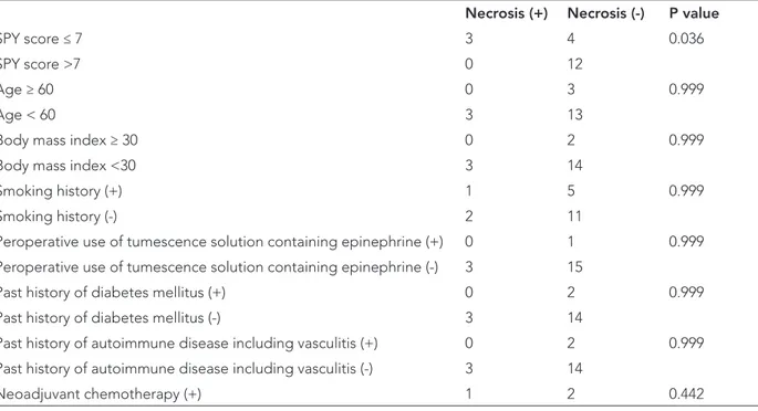
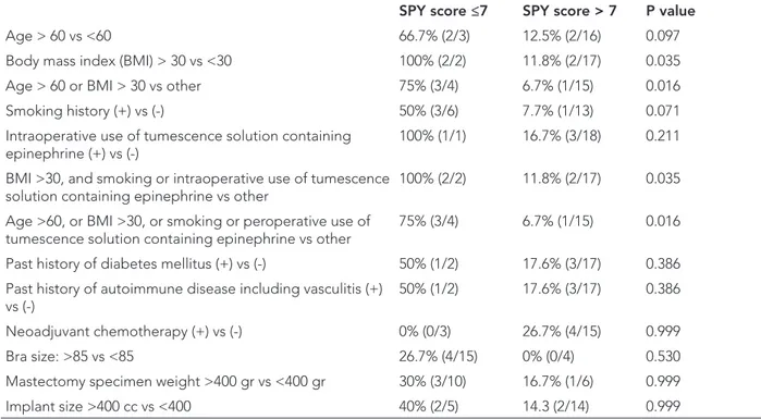
Furthermore, the sensitivity, specificity, and accuracy of LA-ICGA in predicting flap necrosis were 43%, 100%, and 79%, respectively. Patients with a low LA-ICGA score <7 were more likely to develop mastectomy flap necrosis in the fourth postoperative week, whereas other factors such as smoking, diabetes mellitus or neoadjuvant che-motherapy were not found statistically significant (Table 1). Nevertheless, patients aged >60, with a smoking his-tory, a BMI>30, or intraoperative use of tumescence solu-tion containing epinephrine were more likely to have an LA-ICGA score ≤7 which did not predict postoperative flap necrosis (Table 2). LA-ICGA was also able to identify the edges or location of necrotic areas in true positive cases (Figure 1 and 2).
DISCUSSION
Mastectomy skin flap necrosis is one of several frequent complications associated with increased morbidity in breast reconstruction. The significance of accurately predicting mastectomy skin flap viability cannot be em-phasized enough, because such predictions will reduce postoperative morbidity. Although preservation of breast skin contributes to the aesthetic outcome, the presence of necrotic skin flaps contributes to delayed healing, complications, and poor outcome. Necrosis is even more critical when prosthetic implants are being used for
re-Table 1: Factors associated with postoperative flap necrosis
Necrosis (+) Necrosis (-) P value
SPY score ≤ 7 3 4 0.036
SPY score >7 0 12
Age ≥ 60 0 3 0.999
Age < 60 3 13
Body mass index ≥ 30 0 2 0.999
Body mass index <30 3 14
Smoking history (+) 1 5 0.999
Smoking history (-) 2 11
Peroperative use of tumescence solution containing epinephrine (+) 0 1 0.999 Peroperative use of tumescence solution containing epinephrine (-) 3 15
Past history of diabetes mellitus (+) 0 2 0.999
Past history of diabetes mellitus (-) 3 14
Past history of autoimmune disease including vasculitis (+) 0 2 0.999
Past history of autoimmune disease including vasculitis (-) 3 14
construction. Although the safest option is often to re-sect mastectomy skin, this often removes skin that would likely have survived. We had quantified perfusion scores with the current LA-ICGA and assessed whether SPY-Q software does indeed correlate with the areas, which would have necrosis clinically.
Currently, the standard approach to examine mastec-tomy flap viability is via clinician assessment intra-oper-atively. Before closure, the surgeon must assess tissue viability and excise the skin based on the likelihood of necrosis in the postoperative period. With this clinically guided approach, the literature reports mastectomy skin flap necrosis rates of approximately 10-15% (13,14). In the current study, a 15.8% rate of necrosis was observed, which is consistent with the literature. The high rate of flap necrosis after nipple or skin-sparing mastectomies highlights the limitation of current clinical assessment methods used for predicting flap necrosis.
Recently, SPY angiography has been developed as a new tool to evaluate skin perfusion in real-time. The device was designed to assess the patency of bypass anastomo-ses in cardiac surgery (15,16) as well as to evaluate free tissue transfers or microsurgical perforator anastomoses (17,18). A few studies have been published to assess the ability of LA-ICGA to predict flap necrosis in mastecto-mies. Komorowska-Timek and Gurtner in a case series of 24 breasts showed that incorporating SPY values into
Table 2: Factors associated with false positivity in the study patients who have <7 SPY-Q score who did not develop
postoperative necrosis.
SPY score ≤7 SPY score > 7 P value
Age > 60 vs <60 66.7% (2/3) 12.5% (2/16) 0.097
Body mass index (BMI) > 30 vs <30 100% (2/2) 11.8% (2/17) 0.035
Age > 60 or BMI > 30 vs other 75% (3/4) 6.7% (1/15) 0.016
Smoking history (+) vs (-) 50% (3/6) 7.7% (1/13) 0.071
Intraoperative use of tumescence solution containing
epinephrine (+) vs (-) 100% (1/1) 16.7% (3/18) 0.211
BMI >30, and smoking or intraoperative use of tumescence solution containing epinephrine vs other
100% (2/2) 11.8% (2/17) 0.035 Age >60, or BMI >30, or smoking or peroperative use of
tumescence solution containing epinephrine vs other 75% (3/4) 6.7% (1/15) 0.016 Past history of diabetes mellitus (+) vs (-) 50% (1/2) 17.6% (3/17) 0.386 Past history of autoimmune disease including vasculitis (+)
vs (-) 50% (1/2) 17.6% (3/17) 0.386
Neoadjuvant chemotherapy (+) vs (-) 0% (0/3) 26.7% (4/15) 0.999
Bra size: >85 vs <85 26.7% (4/15) 0% (0/4) 0.530
Mastectomy specimen weight >400 gr vs <400 gr 30% (3/10) 16.7% (1/6) 0.999
Implant size >400 cc vs <400 40% (2/5) 14.3 (2/14) 0.999
Figure 1 and 2: Flap necrosis predicted and localized by
SPY-Q numbers ≤7 in a case who underwent immediate reconstruction following NSM.
operational decision-making was associated with a de-crease in the rate of necrosis from 15% to 4% (1). Fur-thermore, Jones et al. used LA-ICGA to inform clinical decisions in 64 cases and found that in five cases where LA-ICGA assessment was ignored, four developed ne-crosis with the last developing blistering in the SPY-pre-dicted distribution (19). These studies did not allow for assessing to what degree LA-ICGA predictions differed from clinical judgment, and whether LA-ICGA over pre-dicted ischemia and also led to the excision of healthy skin not destined for necrosis.
To answer these questions, Newman et al. and Phillips et al. conducted studies where intra-operative excisions were based exclusively on clinician judgment. Newman et al. correlated low LA-ICGA perfusion values to skin ne-crosis in 20 breasts (20). Phillips et al., in a prospective study of 51 tissue expander reconstructions, attempted to quantify the areas predictive of necrosis to provide some framework for interpreting LA-ICGA perfusion val-ues (8). The study, however, used an outdated version of LA-ICGA, which determined perfusion values based on a different scaling system, and has since been replaced by SPY Elite. Recently, Munabi et al. used LA-ICGA prospec-tively to assess the correlation between the LA-ICGA val-ues and skin flap necrosis following both implant-based and autologous breast reconstruction, and assess how those values were affected by certain patients factors (12). They found that by considering an LA-ICGA value of 7 as an indicator of necrosis, the sensitivity and spec-ificity of LA-ICGA were maximized to 88% and 83%, re-spectively. If the cut-off value for necrosis was increased to 13, 100% sensitivity of the device was obtained; how-ever, specificity decreased to 72%. Of all patient factors considered, only temperature and tissue expander vol-ume were found to be statistically significant. All patient temperatures recorded, however, were within reasonable limits of body temperature.
Concerning tissue expander volume, Munabi et al. found that breasts that developed necrosis were statistically more likely to have received a smaller intra-operative fill volume (12). Other researchers have attempted to define threshold values, using the SPY-Q software, which enable reliable prediction of mastectomy skin flap perfusion and necrosis (8,21). Moyer et al. utilized the LA-ICGA perfusion score where the fluorescence was recorded relative to the surrounding well-perfused tissues designated as “100% fluorescent” (21). They showed that a score of less than 33% has a positive predictive value of 88% for necrosis and a negative predictive value of 16% for survival. However, this scoring system was prone to inter-observer variability. In contrast, Phillips et al. and Munabi et al. employed an absolute measurement of tissue fluorescence to de-rive sensitivity and specificity of ICG (8,12). In our study,
LA-ICGA number 7 was used as a cut-off level, and it was discovered that the LA-ICGA had a high specificity (100%) but low sensitivity (43%) with an accuracy rate of 79%, which is acceptable. Since we preferred to use a sili-cone implant rather than a tissue expander, we could not compare the fill-volume used in tissue expanders. Body temperatures in our cases were in reasonable limits and were found to be statistically insignificant.
Demographic factors affecting the LA-ICGA efficiency are variable, and there is little data regarding whether the cost-effectiveness LA-ICGA . Munabi et al. docu-mented that sensitivity and specificity can be confound-ed in smokers and the presence of epinephrine-contain-ing tumescent solution (12). Current evidence indicates that using ICG-based angiography to guide the excision of the hypo-perfused mastectomy skin flap in the oper-ating room results in a significant reduction in necrosis (1,22). Furthermore, in comparison to the historical con-trol, Duggal et al. reported a reduction in the severity of the skin flap necrosis (25% vs. 44.1%) and also the rate of re-operation (6% vs. 14%). These findings translate to a cost saving of USD 614 per patient. However, given the high cost of leasing or purchasing an ICG detector device currently, Kanuri et al. showed that indiscriminate use of ICG in all breast reconstructions is more expensive than the cost related to flap complications by 65% (10). They recommend that reserving the use of FA in high-risk pa-tients—smokers, body mass index (BMI) of greater than 30 kg/m2, and mastectomy weight of greater than 800 g— leads to significant cost-savings. In our study, it was discovered that patients aged>60; patients with a history of smoking; patients with a BMI >30, or patients with in-tra-operative use of tumescence solution containing epi-nephrine were more likely to have a SPY-Q score ≤7 which is not clinically reliable to predict flap necrosis.
In conclusion, SPY-Q numbers of LA-ICGA could be used to predict and locate the necrotic areas in patients under-going reconstruction following NSM. The LA-ICGA as-sessment is a better predictor for necrosis than conven-tional assessment. We may be able to decrease the rate of postoperative flap necrosis by per-operatively debrid-ing the area that demonstrates lower LA-ICGA numbers. LA-ICGA could change operational decision-making by predicting partial necrotic areas, which avoids undergo-ing subsequent excision. There is a need to investigate the accuracy and reliability of ICG angiography in a more extensive series of autologous reconstructions. Further prospective clinical trials, which compare clinical assess-ment versus SPY-Q numbers, are needed.
Ethics Committee Approval: Ethics committee approval was received for this study from the Acıbadem University Ethics Committee.
Informed Consent: Written consent was obtained from the par-ticipants.
Peer Review: Externally peer-reviewed.
Author Contributions: Conception/Design of Study- F.L.B.; Data Acquisition- F.L.B., C.U.; Data Analysis/Interpreta-tion- F.L.B.; Drafting Manuscript- F.L.B.; Critical Revision of Manuscript- C.U.; Final Approval and Accountability- F.L.B., C.U.
Conflict of Interest: Authors declared no conflict of interest. Financial Disclosure: Authors declared no financial support.
Etik Komite Onayı: Etik komite onayı bu çalışma için, Acıbadem Üniversitesi Etik Komitesi’nden alınmıştır.
Bilgilendirilmiş Onam: Katılımcılardan bilgilendirilmiş onam alınmıştır.
Hakem Değerlendirmesi: Dış bağımsız.
Yazar Katkıları: Çalışma Konsepti/Tasarım- F.L.B.; Veri To-plama- F.L.B., C.U.; Veri Analizi/Yorumlama- F.L.B.;Yazı Taslağı- F.L.B.; İçeriğin Eleştirel İncelemesi- C.U.; Son Onay ve Sorumluluk- F.L.B., C.U.
Çıkar Çatışması: Yazarlar çıkar çatışması beyan etmemişlerdir. Finansal Destek: Yazarlar finansal destek beyan etmemişlerdir.
REFERENCES
1. Komorowska-Timek E, Gurtner GC. Intraoperative perfusion mapping with laser-assisted indocyanine green imaging can predict and prevent complications in immediate breast reconstruction. Plast Reconst Surg 2010:125:1065-73.
[CrossRef]
2. Garvey PB, Buchel EW, Pockaj BA, et al. DIEP and pedicled TRAM flaps: a comparison of outcomes. Plast Reconst Surg 2006:117:1711-9. [CrossRef]
3. Pestana IA, Coan B, Erdmann D, et al. Early experience with fluorescent angiography in free-tissue transfer reconstruction. Plast Reconstruct Surg 2009:123:1239-44.
[CrossRef]
4. Newman MI, Samson MC, Tamburrino JF, et al. An investigation of the application of laser-assisted indocyanine green fluorescent dye angiography in pedicle transverse rectus abdominus myocutaneous breast reconstruction. Can J Plast Surg 2011:19:1-5. [CrossRef]
5. Jallali N, Ridha H, Butler PE. Postoperative monitoring of free flaps in UK plastic surgery units. Microsurgery 2005:25:469-72. [CrossRef]
6. Mothes H, Donicke T, Friedel R, et al. Indocyanine-green fluorescence video angiography used clinically to evaluate tissue perfusion in microsurgery. J Trauma 2004:57:1018-24.
[CrossRef]
7. Olivier WA, Hazen A, Levine JP, et al. Reliable assessment of skin flap viability using orthogonal polarization imaging. Plast Reconstruct Surg 2003:112:547-55. [CrossRef]
8. Phillips BT, Lanier ST, Conkling N, et al. Intraoperative perfusion techniques can accurately predict mastectomy skin flap necrosis in breast reconstruction: Results of a prospective trial. Plast Reconstr Surg 2012;129:778-88.
[CrossRef]
9. Newman MI, Samson MC. The application of laser-assisted indocyanine green fluorescent dye angiography in microsurgical breast reconstruction. J Reconstr Microsurg 2009;25:21-6. [CrossRef]
10. Kanuri A, Liu AS, Guo L. Whom should we SPY? A cost analysis of laser-assisted indocyanine green angiography in prevention of mastectomy skin flap necrosis during prosthesis-based breast reconstruction. Plast Reconstr Surg 2014;133:448-54. [CrossRef]
11. Sood M, Glat P. Potential of the SPY intraoperative perfusion assessment system to reduce ischemic complications in immediate postmastectomy breast reconstruction. Ann Surg Innov Res 2013;7(1):9. [CrossRef]
12. Munabi NC, Olorunnipa OB, Goltsman D, et al. The ability of intra-operative perfusion mapping with laser-assisted indocyanine green angiography to predict mastectomy flap necrosis in breast reconstruction: A prospective trial. J Plast Reconstr Aesthet Surg 2014;67:449-55. [CrossRef]
13. Santanelli F, Benedetto L, Sorotos M, et al. Flap survival of skin-sparing mastectomy type IV: a retrospective cohort study of 75 consecutive cases. Ann Surg Oncol 2013;20:981-9. [CrossRef]
14. McCarthy CM, Mehrara BJ, Riedel E, et al. Predicting complications following expander/implant breast reconstruction: an outcomes analysis based on preoperative clinical risk. Plast Reconstr Surg 2008;121(6):1886-92.
[CrossRef]
15. Reuthebuch O, Haussler A, Genoni M, et al. NovadaqSPY: intraoperative quality assessment in off-pump coronary artery bypass grafting. Chest 2004;125(2):418-24. [CrossRef]
16. Desai ND, Miwa S, Kodama D, et al. Improving the quality of coronary bypass surgery with intraoperative angiography: validation of a new technique. J Am Coll Cardiol 2005;46(8): 1521-5. [CrossRef]
17. Francisco BS, Kerr-Valentic MA, Agarwal JP. Laser-assisted indocyanine green angiography and DIEP breast reconstruction. Plast Reconstr Surg 2010;2010:116-8. [CrossRef]
18. Azuma R, Morimoto Y, Masumoto K, et al. Detection of skin perforators by indocyanine green fluorescence nearly infrared angiography. Plast Reconstr Surg 2008;122:1062-7.
[CrossRef]
19. Jones GE, Garcia CA, Murray J, et al. Fluorescent intraoperative tissue angiography for the evaluation of the viability of pedicled TRAM flaps. Plast Reconstr Surg 2009; 124: 53.
20. Newman MI, Samson MC, Tamburrino JF, Swarz KA. Intraoperative laser-assisted indocyanine green angiography for the evaluation of mastectomy flaps in immediate breast reconstruction. J Reconstr Microsurg 2010;26(7):487-92. [CrossRef]
21. Moyer HR, Losken A. Predicting mastectomy skin flap necrosis with indocyanine green angiography: the gray area defined. Plast Reconstr Surg 2012;129:1043-8. [CrossRef]
22. Duggal CS, Madni T, Losken A. An outcome analysis of intraoperative angiography for postmastectomy breast reconstruction. Aesthet Surg J 2014;34(1):61-5. [CrossRef]